. 2022 Dec 29:18:100538.
doi: 10.1016/j.mtbio.2022.100538. eCollection 2023 Feb.

Advanced technologies for molecular diagnosis of cancer: State of pre-clinical tumor-derived exosome liquid biopsies

Affiliations

Advanced technologies for molecular diagnosis of cancer: State of pre-clinical tumor-derived exosome liquid biopsies

Lin Li et al. Mater Today Bio. .

Abstract

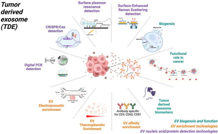
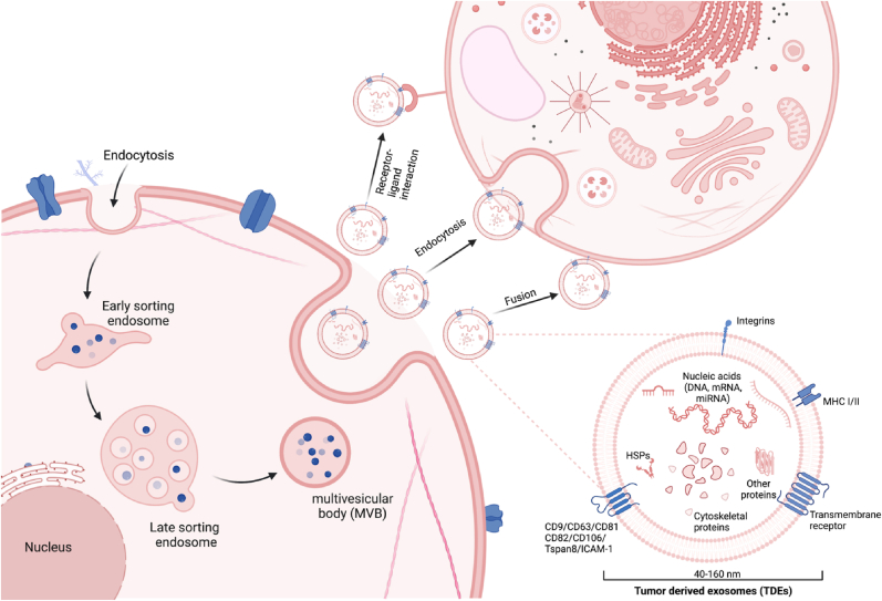
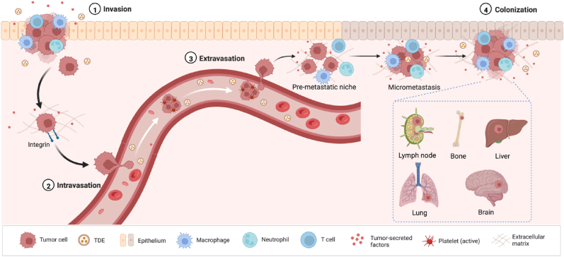
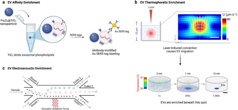
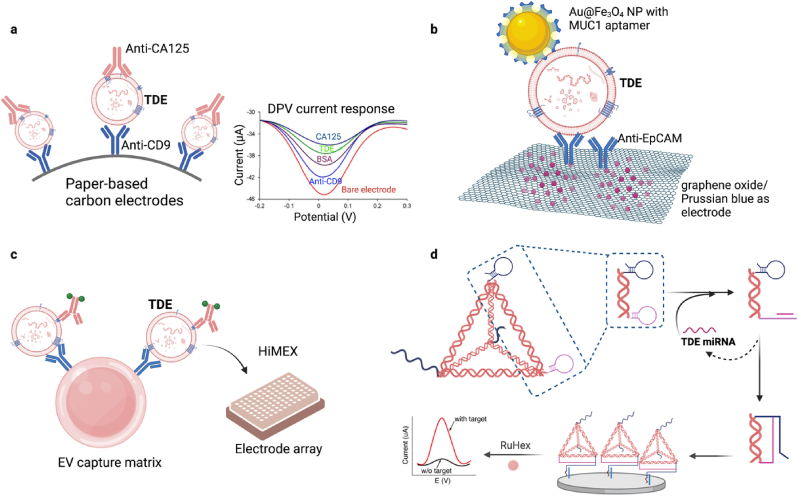
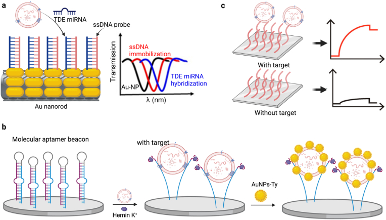
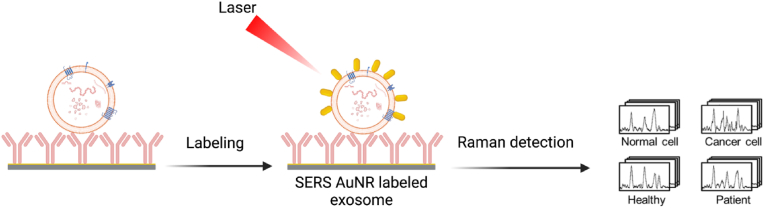
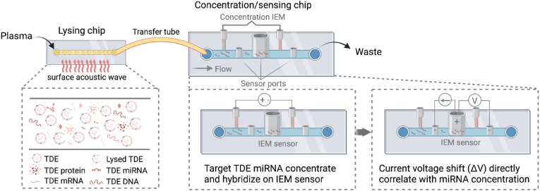
Exosomes are membrane-defined extracellular vesicles (EVs) approximately 40-160 ​nm in diameter that are found in all body fluids including blood, urine, and saliva. They act as important vehicles for intercellular communication between both local and distant cells and can serve as circulating biomarkers for disease diagnosis and prognosis. Exosomes play a key role in tumor metastasis, are abundant in biofluids, and stabilize biomarkers they carry, and thus can improve cancer detection, treatment monitoring, and cancer staging/prognosis. Despite their clinical potential, lack of sensitive/specific biomarkers and sensitive isolation/enrichment and analytical technologies has posed a barrier to clinical translation of exosomes. This review presents a critical overview of technologies now being used to detect tumor-derived exosome (TDE) biomarkers in clinical specimens that have potential for clinical translation.

Keywords: Cancer; Enrichment; Exosome; Isolation; Laboratory developed tests; Molecular diagnosis.


Conflict of interest statement

The authors declare that they have no known competing financial interests or personal relationships that could have appeared to influence the work reported in this paper.

Figures

Image 1
Graphical abstract
Fig. 1
Fig. 1
EV biogenesis, secretion, uptake and composition. EV biogenesis initiates by the inward budding of the early endosome membrane to generate late multivesicular bodies that can secrete mature exosomes into the extracellular space. These EVs can incorporate a complex mixture of factors that derive from multiple cellular compartments. These include lipids, nucleic acids (DNA, mRNA, and miRNA), proteins, and other factors that can play important regulatory roles in cell-to-cell communication events when these factors are internalized by recipient cells following EV endocytosis, fusion or receptor-ligand interactions.
Fig. 2
Fig. 2
Overview of the role of TDEs in the four steps of the metastatic cascade (invasion, intravasation, extravasation, and colonization). TDEs facilitate cancer cell invasion of neighboring tissue through disrupting the extracellular matrix by activating fibroblast proliferation. TDEs transfer factors to cancer cells to initiate an epithelial-mesenchymal transition to form circulating tumor cells (CTCs) during intravasation. These CTCs can then exit the circulation through the capillary bed (extravasation) at pre-metastatic niches (PMNs) prepared by the uptake of circulating TDEs released by the primary tumor. TDEs regulate the colonization of these PMNs by penetrating the vascular endothelium to transfer miRNAs that promote proliferation of micro-metastases and then angiogenesis required to support tumor growth.
Fig. 3
Fig. 3
New methods for EV isolation from liquid biopsies. (a) Affinity enrichment: Fe3O4@TiO2 nanoparticles binding of EV membrane phospholipids for EV enrichment prior to miRNA or protein analysis; (b) Thermophoretic enrichment: laser irradiation forms a region of localized convection that concentrates EVs at the cold lower surface of the microfluidic chamber. (c) Electroacoustic enrichment: electroacoustic energy focused on a biospecimen exerts differential force on its contents based on their size and density to allow EVs to be separated from other components during passage across a microfluidic chamber.
Fig. 4
Fig. 4
Schematic representation of typical electrochemical platforms for TDE detection. (a) TDEs are captured by anti-CD9 antibodies conjugated to paper-based carbon electrodes (PCEs) and then hybridized with an antibody to a cancer biomarker (e.g., CA125), with differential pulse voltammetry (DPV) signal depicting the stepwise attachment of each layer on the PCEs surface. (b) Signal from TDEs captured by spiky Au@Fe3O4 nanoparticles conjugated with an MUC1 aptamer upon their capture on a graphene oxide/Prussian blue electrochemical probe by an EpCAM antibody. (c) TDEs enriched on antibody conjugated magnetic beads are magnetically immobilized on HiMEX electrode arrays that detect electrochemical reactions catalyzed by the enzyme-labeled antibodies detection antibodies bound to these TDEs. (d) Binding of a target miRNA to one of the two hairpins on a multifunctional DNA tetrahedron assisted catalytic hairpin assembly (MDTs-CHA) causes this hairpin to unfold and form a stable complex with the second hairpin and free a sequence that can then bind a capture probe on the surface of a detection electrode, after which incubation of bound MDTs-CHA RuHex produces a signal increase that is proportional to the concentration of the target miRNA.
Fig. 5
Fig. 5
Schematic for the application of surface plasmon resonance (SPR) to the measurement of EV concentration. (a) Binding of a TDE miRNA to a tapered optical fiber laminated with Au nanoparticles and functionalized with single strand DNA (ssDNA) specific for this miRNA causes a shift in the transmission spectrum proportional to target binding; (b) TDE binding to a molecular aptamer beacon conjugated to an SPR sensor exposes a guanine-rich tetramer region causing it to bind hemin and catalyze the deposition of tyramine-coated gold nanoparticles on the exosome surface to markedly enhance SPR signal; (c) Binding of a biotinylated aptamer to a TDE biomarker permits TDE capture and detection on an avidin conjugated SPR biosensor to permit label-free TDE detection.
Fig. 6
Fig. 6
Schematic for the application of surface enhanced Raman spectroscopy (SERS) to quantify EV concentration. EVs are first captured from a specimen by specific binding to target antibodies conjugated on the surface of the slide and then scanned to generate Raman spectra that are analyzed to detected TDE-specific SERS signatures.
Fig. 7
Fig. 7
Schematic of an apta-HCR-CRISPR dual amplification and Cas12a/fluorescent detection approach. In this method EVs are captured from a sample using nanoparticles coated with antibodies against an EV biomarker (CD63, CD81, CD9). Aptamer binding to a biomarker target (nucleolin) exposes a sequence that serves as a template for spontaneous assembly of two hairpin oligonucleotides (H1 and H2) present in excess to promote a hairpin chain reaction. These oligonucleotide concatemers are recognized by a CRISPR/Cas12a complex which then cleaves a quenched fluorescent probe in proportion to their abundance to indirectly quantify the abundance of the EV target molecule.
Fig. 8
Fig. 8
Schematic of an integrated microfluidic platform for TDE-miRNA analysis. This device consists of a lysing chip in which plasma exosomes are lysed by a surface acoustic wave (SAW) after which miRNA from lysed TDEs is concentrated in a region upstream of a positively charged ion-exchange membrane (IEM) by an applied voltage. Following this concentration step, this miRNA is hybridized to a target-specific oligonucleotide conjugated to a negatively charged IEM and miRNA concentration is determined by the change in voltage produced by miRNA binding.
Fig. 9
Fig. 9
Integrated exosome capture, isolation, and detection using the ExoProfile chip platform. (a) Schematic of the ExoProfile chip, which consists of a pneumatic and a fluidic layer and 3D serpentine nanostructures used for in situ exosome phenotyping; (b) Procedure for exosome immunocapture and multiplexed sample analysis, where EV samples are first loaded and captured the 3D nanostructures in the forward direction (blue) before the flow is reversed to (green) load monoclonal detection antibodies contained within the reagent reservoirs on the chip that are blocked during sample loading; (c) Workflow for chip fabrication by microfluidic colloidal self-assembly. Reprinted with permission from Zhang et al. 2019. Copyright 2019 The Royal Society of Chemistry.

Similar articles

Cited by

References

    1. Sung H., Ferlay J., Siegel R.L., Laversanne M., Soerjomataram I., Jemal A., Bray F. Global cancer statistics 2020: GLOBOCAN estimates of incidence and mortality worldwide for 36 cancers in 185 countries. CA A Cancer J. Clin. 2021;71:209–249. doi: 10.3322/caac.21660. - DOI - PubMed
    1. Crosby D., Bhatia S., Brindle K.M., Coussens L.M., Dive C., Emberton M., Esener S., Fitzgerald R.C., Gambhir S.S., Kuhn P., Rebbeck T.R., Balasubramanian S. Early detection of cancer. Science. 2022;375 doi: 10.1126/science.aay9040. - DOI - PubMed
    1. Shyamala K., Girish H.C., Murgod S. Risk of tumor cell seeding through biopsy and aspiration cytology. J. Int. Soc. Prev. Community Dent. 2014;4:5–11. doi: 10.4103/2231-0762.129446. - DOI - PMC - PubMed
    1. Gutman S. Center for Devices and Radiological Health (CDRH); 2004. Evaluation of Automatic Class III Designation CellSearch Epithelial Cell Kit/cell Spotter Analyzer. K031588.
    1. Yu W., Hurley J., Roberts D., Chakrabortty S.K., Enderle D., Noerholm M., Breakefield X.O., Skog J.K. Exosome-based liquid biopsies in cancer: opportunities and challenges. Ann. Oncol. 2021;32:466–477. doi: 10.1016/j.annonc.2021.01.074. - DOI - PMC - PubMed

LinkOut - more resources