Case Reports
. 2022 Dec 22:13:e2021413.
doi: 10.4322/acr.2021.413. eCollection 2023.

Intraoral lipoma with degenerative changes mimicking atypical lipomatous tumor: an immunohistochemical study

Affiliations
Case Reports

Intraoral lipoma with degenerative changes mimicking atypical lipomatous tumor: an immunohistochemical study

Evânio Vilela Silva et al. Autops Case Rep. .

Abstract

Lipomas are mesenchymal neoplasms relatively uncommon in the oral cavity. Lipomas can exhibit histopathological features mimicking atypical lipomatous tumors (ALT) or dysplastic lipoma (DL) in the presence of degenerative changes. Relevantly, immunohistochemistry assists in the correct diagnosis. Herein, we present the case of a 54-year-old male with a sessile nodule located on the dorsum of the tongue. The histopathological analysis showed a diffuse, non-circumscribed adipocytic proliferation constituted by cells of variable size containing cytoplasmic vacuoles and displaced nuclei, some resembling lipoblasts supported by fibrous connective tissue stroma. By immunohistochemistry, tumor cells were positive for vimentin, S100, FASN, CD10, and p16. Rb expression was intact. Moreover, CD34, p53, MDM2, and CDK4 were negative. After 2-year of follow-up, no alteration or recurrence was observed. In conclusion, MDM2, CDK4, p53, and Rb immunomarkers can be used reliably to differentiate benign lipoma with degenerative changes from ALT and DL.

Keywords: Immunohistochemistry; Lipoma; Liposarcoma; Mouth.


Conflict of interest statement

Conflict of interest: All authors declare that they have no conflict of interest.

Figures
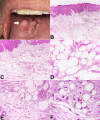
Figure 1
Figure 1. A - Clinical presentation of the lesion showing a sessile nodule located on dorsal region of the tongue (arrow); B to F - Photomicrographs of the surgical specimen with: B and C - diffuse, non-circumscribed adipocytic proliferation with variable cell size (H&E, B x4; C x10); D - note variably sized adipocytes supported by scarce fibrous stroma, mimicking dysplastic lipoma (H&E; x40); E - At the periphery, ring-like cells and vacuolated cells with displaced, slightly hyperchromatic nuclei, some of them with lipoblast-like features, were visualized (H&E, x40); F - Close-up view highlighting cellular details. Note uni- or multivacuolated lipoblast-like cells and nuclei with sharply outlined vacuoles, notably Lochkern change (arrow) (H&E, x100).
Figure 2
Figure 2. Photomicrographs of the surgical specimen. Immunohistochemical analysis with: A - positivity for S100 (x40); B - CD34 highlighted the stromal vessels (x40); C - FASN evidenced adipocytes varying in size and shape (x40); D - negative reaction to MDM2 (x40).
Figure 3
Figure 3. Photomicrographs of the surgical specimen. Immunohistochemical analysis with A - Tumor cells were negative to CDK4 (x40), excluding the possibility of ALT; B - Rb expression was intact (x40); C - CD10 and p16 expression were positive, including the stromal cells (x40); D - CD68 and CD163 evidenced numerous stromal histiocytes (x40).
Figure 4
Figure 4. Photomicrograph of the surgical specimen. Rare tumor cells were Ki-67 positive (x10).

References

    1. Studart-Soares EC, Costa FWG, Sousa FB, Alves APNN, Osterne RLV. Oral lipomas in a Brazilian population: a 10-year study and analysis of 450 cases reported in the literature. Med Oral Patol Oral Cir Bucal. 2010;15(5):e691–6. doi: 10.4317/medoral.15.e691. - DOI - PubMed
    1. Manor E, Sion-Vardy N, Joshua BZ, Bodner L. Oral lipoma: analysis of 58 new cases and review of the literature. Ann Diagn Pathol. 2011;15(4):257–261. doi: 10.1016/j.anndiagpath.2011.01.003. - DOI - PubMed
    1. Santos JL, Ocamoto EA, Almeida LY, Teixeira LR, Ribeiro-Silva A, León JE. Low-fat plexiform spindle cell lipoma with prominent myxoid stroma: an unusual oral presentation and immunohistochemical analysis. J Craniofac Surg. 2017;28(2):e158–60. doi: 10.1097/SCS.0000000000003348. - DOI - PubMed
    1. Rocha AFL, Miotto LN, Ferrisse TM, et al. Low-fat and fat-free spindle cell lipomas in the oral cavity: immunohistochemical analysis and review of the literature. J Cutan Pathol. 2019;46(10):778–783. doi: 10.1111/cup.13512. - DOI - PubMed
    1. Furlong MA, Fanburg-Smith JC, Childers EL. Lipoma of the oral and maxillofacial region: site and subclassification of 125 cases. Oral Surg Oral Med Oral Pathol Oral Radiol Endod. 2004;98(4):441–450. doi: 10.1016/j.tripleo.2004.02.071. - DOI - PubMed

Publication types