Review
. 2022 Aug 19:11:960.
doi: 10.12688/f1000research.123245.1. eCollection 2022.

The mechanistic role of neutrophil lymphocyte ratio perturbations in the leading non communicable lifestyle diseases

Affiliations
Review

The mechanistic role of neutrophil lymphocyte ratio perturbations in the leading non communicable lifestyle diseases

Monalisa Biswas et al. F1000Res. .

Abstract

Inflammation plays a critical role in the development and progression of chronic diseases like type 2 diabetes mellitus, coronary artery disease, and chronic obstructive pulmonary disease. Inflammatory responses are indispensable for pathogen control and tissue repair, but they also cause collateral damage. A chronically activated immune system and the resultant immune dysregulation mediated inflammatory surge may cause multiple negative effects, requiring tight regulation and dampening of the immune response to minimize host injury. While chronic diseases are characterized by systemic inflammation, the mechanistic relationship of neutrophils and lymphocytes to inflammation and its correlation with the clinical outcomes is yet to be elucidated. The neutrophil to lymphocyte ratio (NLR) is an easy-to-measure laboratory marker used to assess systemic inflammation. Understanding the mechanisms of NLR perturbations in chronic diseases is crucial for risk stratification, early intervention, and finding novel therapeutic targets. We investigated the correlation between NLR and prevalent chronic conditions as a measure of systemic inflammation. In addition to predicting the risk of impending chronic conditions, NLR may also provide insight into their progression. This review summarizes the mechanisms of NLR perturbations at cellular and molecular levels, and the key inflammatory signaling pathways involved in the progression of chronic diseases. We have also explored preclinical studies investigating these pathways and the effect of quelling inflammation in chronic disease as reported by a few in vitro, in vivo studies, and clinical trials.

Keywords: Neutrophil-lymphocyte ratio; chronic obstructive pulmonary disease; coronary artery disease; inflammation; non communicable diseases; type 2 diabetes mellitus.


Conflict of interest statement

No competing interests were disclosed.

Figures

Figure 1.
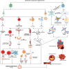
Figure 1.. Role of neutrophils in Type 2 Diabetes Mellitus (T2DM).
A) Hyperglycemia leads to enhanced stimulation of TNF alpha secretion which promotes neutrophil generation and subsequent neutrophil activation. B) Hyperglycemia increases intracellular cAMP which leads to increased generation of inflammatory mediators and reactive oxygen species (ROS). C1) Hyperglycemia activates the polyol pathway and leads to increased sorbitol generation. C2) Decreased glutamine has been implicated in hyperglycemia. B + C1 + C2 leads to exaggerated utilization and thus depletion of cellular NADPH. D1, D2) Hyperglycemia triggers the formation of advanced glycation end products (AGEs) and the release of acute-phase proteins (APPs) which in turn causes increased neutrophil activation. Exaggerated neutrophil activation and decreased cellular NADPH concentrations in the background of an inflammatory microenvironment, leads to dysfunctional/hyperactivated neutrophil which manifest increased degranulation, resistance to apoptosis, and dysfunctional chemotactic abilities. This cascade results in widespread systemic inflammation leading to micro and macrovascular complications of diabetes mellitus. Note: ↑ indicates increased levels while ↓ refers to decreased levels/concentrations. This figure is an original figure produced by the authors for this review article and has been filed for copyright with the Copyright Office, Government of India.
Figure 2.
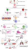
Figure 2.. Role of neutrophils in coronary artery disease (CAD).
Hyperlipidaemia and oxidative damage create a chronic low-grade inflammatory microenvironment that activates the release of granulocyte colony-stimulating factor (GCSF) (A) and pro-inflammatory mediators (B) (increase in IL6, TNF alpha, CXCL1 and decrease in CXCL 12). GCSF triggers the proliferation of neutrophils (A1), while IL6, TNF alpha, and CXCL1 retards neutrophil apoptosis (B1) which leads to an increase in neutrophil count followed by increased neutrophil activation (C) and degranulation (D). These in turn lead to the heightened NET formation (F) and platelet activation (G). Decreased CXCL12 triggering enhanced neutrophil mobilization (B2 a,b) along with activation of macrophage cascade accelerates foam cell formation (E). NETosis and foam cell formation lead to increased secretion of pro-inflammatory proteases, cytokines, and chemokines. The entire sequence of events finally culminates in plaque rupture, and thrombosis which leads to myocardial infarction (MI). Note: ↑ indicates increased levels while ↓ refers to decreased levels/concentrations. This figure is an original figure produced by the authors for this review article and has been filed for copyright with the Copyright Office, Government of India.
Figure 3.
Figure 3.. Role of lymphocytes in coronary artery disease (CAD).
CD4 + T lymphocytes play a vital role in coronary artery disease. Hyperlipidemia and oxidative damage create a chronic low-grade inflammatory microenvironment which stimulates the increased differentiation of CD4 + T cells to Th1 (a.i) and Th17 (a.ii) phenotypes and decreased production of the regulatory Treg cells (c). Further, chronic inflammation triggers the generation of unusual CD4 +CD28 NULL T cells (b). b.i) These are permanently differentiated cytotoxic cells. b.ii) These cells have heightened expression of CX3CR1 which results in increased expression of the anti-apoptotic BCL2 protein. b.iii) These cells express altered Killer Immunoglobulin Receptors (KIRs) which show increased reactivity to self-antigens. c) Perturbation of the Th17 and Treg cell equilibrium further exaggerates the proinflammatory microenvironment. d) B lymphocytes in CAD are shown to manifest increased differentiation of the proinflammatory B2 phenotype (d.i) along with increased expression of BAFF (d.ii). BAFF further aids the B2 cell maturation and increases their resistance to apoptosis. a+b+c+d) Increased inflammatory and oxidative damage of the endothelial membrane through immune cell recruitment accelerates foam cell generation and plaque maturation. The entire sequence of events finally culminates in thrombosis which leads to myocardial infarction (MI). Note: ↑ indicates increased levels while ↓ refers to decreased levels/concentrations. This figure is an original figure produced by the authors for this review article and has been filed for copyright with the Copyright Office, Government of India.
Figure 4.
Figure 4.. Role of neutrophils in chronic obstructive pulmonary disorder (COPD).
Exposure to allergens, respiratory tract infections (RTIs), and cigarette smoking generate an inflammatory microenvironment that leads to immune cell recruitment. Increased immune cell recruitment leads to (a) increased TNF alpha secretion which stimulates neutrophil production, (b) macrophage recruitment and activation which triggers an increased release of inflammatory mediators. (c) Exaggerated release of inflammatory mediators also triggers increased neutrophil priming and results in (d) increased neutrophil activation which leads to increased neutrophil degranulation and increased resistance to apoptosis (e) along with the generation of neutrophils with altered phenotypes. These further result in compressed/collapsed airways and defective mucociliary pathogen clearance (f). e+f) Further intensifies sterile lung inflammation as well as damage due to colonization by opportunistic pathogenic microorganisms which culminates in widespread lung damage in acute exacerbations or chronic long-term COPD. Note: ↑ indicates increased levels while ↓ refers to decreased levels/concentrations. This figure is an original figure produced by the authors for this review article and has been filed for copyright with the Copyright Office, Government of India.
Figure 5.
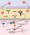
Figure 5.. Mechanism of steroid resistance in COPD.
In normal circumstances, after the transport of steroids in the cytoplasm, the steroid molecule forms a complex with the glucocorticoid receptor and heat shock proteins, HSP 70 and HSP 90. This complex is then translocated through the nuclear member where the Glucocorticoid receptor (GR)-Steroid complex is released. The released complex interacts with histone deacetylases (HDAC) and NF kB (Nuclear Factor kappa-light-chain-enhancer of activated B cells), forming another complex thus preventing the binding of NF kB to the promoter region and derepression of translation of inflammatory genes. In numerous inflammatory conditions like COPD as well as a few cancers, where steroids are one of the prime treatment modalities, a significant number of patients are observed to exhibit resistance to corticosteroid treatment. One of the major reasons for steroid resistance is the increased expression of P-gp (P glycoprotein). The other mechanisms include decreased expression of the glucocorticoid receptors, HSP 90, and HDAC. These perturbations limit the formation of the receptor steroid complex, transport of the complex to the nucleus, and the binding of the complex to NF kB. Thus, NF kB freely binds with the promoter region of the gene which undergoes derepression triggering the expression of inflammatory genes thereby rendering decreased efficacy of the prescribed steroid therapy. Note: ↑ indicates increased levels while ↓ refers to decreased levels/concentration. This figure is an original figure produced by the authors for this review article and has been filed for copyright with the Copyright Office, Government of India.
Figure 6.
Figure 6.. Role of lymphocytes in chronic obstructive pulmonary disease (COPD).
T lymphocytes have been reported to play a vital role in the pathogenesis and progression of COPD. Exposure to allergens, respiratory tract infections, and cigarette smoking generate an inflammatory microenvironment that triggers a) increased activation of the CD8 + T cells (a1) and increased differentiation of the unusual CD8 +CD28 NULL T cells (a2) and b) the increased differentiation of CD4 + cells to Th17 cells, decreased generation/differentiation of the immunoregulatory Treg phenotype (b1) and increased resistance to apoptosis (b2). a2) CD8 +CD28 NULL T cells are characterized by exaggerated secretion of inflammatory mediators. a.2.i) These are permanently differentiated cytotoxic cells. a.2.ii) These cells have heightened expression of anti-apoptotic proteins, making the cells resistant to apoptosis (prolonged survival). a.2.iii) These cells manifest an increased rate of cell division. a.2.iv) These cells show resistance to the action of steroids. b1) Perturbation of the Th17 and Treg cell equilibrium along with b2) the resistance of these cells to apoptosis further exaggerates the proinflammatory microenvironment. a+b) Induces an inflammatory storm via exaggerated release of proinflammatory chemokines, cytokines, prostaglandins, reactive oxygen species, and cytotoxic enzymes like granzymes, perforins, and proteases. These promote alveolar tissue damage and intensify sterile lung inflammation as well as damage due to colonization by pathogenic microorganisms which culminates in widespread damage triggered compression/collapse of airways, characteristic of acute exacerbations or chronic long-term COPD. Note: ↑ indicates increased levels while ↓ refers to decreased levels/concentrations. This figure is an original figure produced by the authors for this review article and has been filed for copyright with the Copyright Office, Government of India.

References

    1. Pawelec G, Goldeck D, Derhovanessian E: Inflammation, ageing and chronic disease. Curr. Opin. Immunol. 2014;29:23–28. 10.1016/j.coi.2014.03.007 - DOI - PubMed
    1. Tsoupras A, Lordan R, Zabetakis I: Inflammation, not Cholesterol, Is a Cause of Chronic Disease. Nutrients. 2018;10(5):604. 10.3390/nu10050604 - DOI - PMC - PubMed
    1. Hansson GK, Libby P: The immune response in atherosclerosis: a double-edged sword. Nat. Rev. Immunol. 2006;6:508–519. 10.1038/nri1882 - DOI - PubMed
    1. World Health Organisation: Noncommunicable diseases. Geneva: WHO;1948. 2021 Apr 12 [cited 2022 Apr 15]; [about 5 screens]. Reference Source
    1. Zahorec R: Neutrophil-to-lymphocyte ratio, past, present and future perspectives. Bratisl. Lek. Listy. 2021;122(7):474–488. 10.4149/BLL_2021_078 - DOI - PubMed

Publication types