Review
. 2022 Dec 23:10:1035513.
doi: 10.3389/fcell.2022.1035513. eCollection 2022.

Understanding laterality disorders and the left-right organizer: Insights from zebrafish

Affiliations
Review

Understanding laterality disorders and the left-right organizer: Insights from zebrafish

Kadeen Forrest et al. Front Cell Dev Biol. .

Abstract

Vital internal organs display a left-right (LR) asymmetric arrangement that is established during embryonic development. Disruption of this LR asymmetry-or laterality-can result in congenital organ malformations. Situs inversus totalis (SIT) is a complete concordant reversal of internal organs that results in a low occurrence of clinical consequences. Situs ambiguous, which gives rise to Heterotaxy syndrome (HTX), is characterized by discordant development and arrangement of organs that is associated with a wide range of birth defects. The leading cause of health problems in HTX patients is a congenital heart malformation. Mutations identified in patients with laterality disorders implicate motile cilia in establishing LR asymmetry. However, the cellular and molecular mechanisms underlying SIT and HTX are not fully understood. In several vertebrates, including mouse, frog and zebrafish, motile cilia located in a "left-right organizer" (LRO) trigger conserved signaling pathways that guide asymmetric organ development. Perturbation of LRO formation and/or function in animal models recapitulates organ malformations observed in SIT and HTX patients. This provides an opportunity to use these models to investigate the embryological origins of laterality disorders. The zebrafish embryo has emerged as an important model for investigating the earliest steps of LRO development. Here, we discuss clinical characteristics of human laterality disorders, and highlight experimental results from zebrafish that provide insights into LRO biology and advance our understanding of human laterality disorders.

Keywords: birth defects; cilia; heterotaxy syndrome; left-right asymmetry; left-right organizer; organ laterality; situs inversus; zebrafish.


Conflict of interest statement

The authors declare that the research was conducted in the absence of any commercial or financial relationships that could be construed as a potential conflict of interest.

Figures

FIGURE 1
FIGURE 1
Human laterality and laterality disorders. (A) Diagram of situs solitus, which refers to the typical asymmetric development and arrangement of internal organs along the left-right body axis. Dashed line indicates the body midline. (B) Diagram of situs inversus totalis, which is a complete mirror-image reversal of organ laterality. (C) Diagram of one example of situs ambiguous or heterotaxy (other arrangement). Situs ambiguous is associated with a broad range of birth defects of the cardiovascular and gastrointestinal systems.
FIGURE 2
FIGURE 2
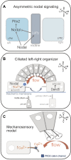
Mechanisms of vertebrate LR patterning. (A) Diagram representing asymmetric Nodal (TGF-β) signaling in left lateral plate mesoderm (LPM). Nodal activates its own transcription as well as the Nodal antagonist Lefty in the embryonic midline that restricts Nodal to the left side, and the transcription factor Pitx2 that can regulate asymmetric morphogenesis of organs such as the heart and gut. (B) Diagram of the mouse left-right organizer (referred to as the node/PNC). Epithelial pit cells with motile cilia generate a leftward fluid flow (red arrow) in a cavity covered by Reichert’s membrane. Return flow (blue arrow) occurs away from the ciliated epithelium. Leftward flow is sensed in larger crown cells that have immotile primary cilia. In left-sided crown cells, flow induces increased Ca2+ fluxes and degradation of the Nodal inhibitor Dand5. Loss of Dand5 increases Nodal expression in left crown cells, which can then activate asymmetric Nodal expression in left LPM. (C) Diagram of the mechanosensory cilia model for sensing flow. It is proposed that posteriorly tilted motile cilia on pit cells generate a leftward flow that induces bending of mechanosensory primary cilia in left-side crown cells to open the stretch-activated cation channel PKD2 in the ciliary membrane that initiates asymmetric Ca2+ signaling. A = anterior, P = posterior, L = left, R = right, D = dorsal, V = ventral.
FIGURE 3
FIGURE 3
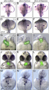
Asymmetric markers and laterality defects in the zebrafish embryo. (A–F) Dorsal views of RNA in situ hybridizations of spaw expression at the 18-somite stage (A–C) or pitx2c expression at the 20-somite stage (D–F). Typical asymmetric expression (situs solitus) occurs in left lateral plate mesoderm [arrowhead in (A,D)]. Defects in LR patterning include right-sided [situs inversus; arrowhead in (B,E)] or bilateral [situs ambiguous; arrowheads in (C,F)] expression. (G–L) Visualization of heart jogging at 26 hpf (G–I) and heart looping at 48 hpf (J–L) in living transgenic Tg(myl7:GFP) (Huang et al., 2003) embryos that express GFP (green) specifically in cardiomyocytes. GFP fluorescence images are superimposed on brightfield images of the embryo. The forming heart tube normally migrates (‘jogs’) to left of the midline [arrow in (G)]. LR patterning defects can result in reversed (arrow in H) or midline [arrow in (I)] jogging. Next, as in other vertebrates, the heart typically loops to the right [situs solitus; (J)]. Heart looping defects can include reversed [situs inversus; (K)] and midline [situs ambiguous; (L)] looping. v = ventricle, a = atrium. (M–O) Dorsal view of RNA in situ hybridizations of foxa3 expression in the gastrointestinal tract at 48 hpf. The marker foxa3 labels the gut tube, left-sided liver (Li) and right-sided pancreas (Pa) in embryos with situs solitus (M). Examples of laterality defects of the gut include reversed orientation [situs inversus; (N)] and left-sided liver [situs ambiguous; (O)]. Dashed lines indicate the midline. A = anterior, P = posterior, L = left, R = right, D = dorsal, V = ventral.
FIGURE 4
FIGURE 4
Kupffer’s vesicle is the zebrafish left-right organizer. (A–B) Live images of zebrafish embryos at the 6-somite stage (∼12 hpf). Side view (A) and dorsal view (B) of the fluid-filled Kupffer’s vesicle (KV) lumen in the tailbud. KV is adjacent to the posterior end of the midline notochord. (C) Higher magnification of fluorescent immunostaining of KV (boxed region in B) using aPKC antibodies to outline epithelial KV cells (red) and acetylated Tubulin antibodies to label cilia (green). Cilia project b from each KV cell into the lumen. A = anterior, P = posterior, L = left, R = right, D = dorsal, V = ventral.
FIGURE 5
FIGURE 5
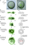
Steps of Kupffer’s vesicle morphogenesis. (A) Live images of Tg (sox17:GFP) (Chung and Stainier 2008) embryos that express GFP (green) in dorsal forerunner cells (DFCs) and Kupffer’s vesicle (KV) show the location of these cells in the zebrafish embryo during epiboly and early somitogenesis. The GFP signal is superimposed on a brightfield image of the embryo. A = anterior, P = posterior, L = left, R = right, D = dorsal, V = ventral. The orientation of the axes shown in (A) applies to all images (A–F). (B–F) High-resolution snapshots of DFCs and KV cells in live Tg(sox17:EGFP-CAAX) (Dasgupta et al., 2018) embryos expressing membrane-localized GFP (shown using an inverted thalium lookup table) alongside simplified diagrams of cell morphologies that represent specific steps of DFC/KV development. Examples of mechanisms that regulate each step are listed (see main text for details). (B) Approximately 20–30 mesenchymal DFCs are specified at the 50% epiboly stage. (C) DFCs move towards the posterior of the embryo and proliferate between the 50–80% epiboly stages (Supplementary Movie S5). (D) At the end of epiboly, DFCs undergo a mesenchymal-to-epithelial transition, and cluster to form a rosette-like structure (Supplementary Movie S6). (E) During early somite stages, a fluid-filled lumen expands (Supplementary Movie S6) and cilia elongate into the lumen. At the 2-somite stage anterior and posterior KV cells have similar morphologies. (F) Between the 4 and 6 somite stages KV cells undergo asymmetric cell shape changes along the anterior-posterior axis, termed KV remodeling (Supplementary Movie S7). This allows tight packing of cilia in the anterior region of KV to drive strong right-to-left fluid flow (red arow). More widely spaced cilia in the posterior region mediate a slower left-to-right flow (blue arrow).

References

    1. Ablooglu A. J., Tkachenko E., Kang J., Shattil S. J. (2010). Integrin alphaV is necessary for gastrulation movements that regulate vertebrate body asymmetry. Development 137, 3449–3458. 10.1242/dev.045310 - DOI - PMC - PubMed
    1. Adams D. S., Robinson K. R., Fukumoto T., Yuan S., Albertson R. C., Yelick P., et al. (2006). Early, H+-V-ATPase-dependent proton flux is necessary for consistent left-right patterning of non-mammalian vertebrates. Development 133, 1657–1671. 10.1242/dev.02341 - DOI - PMC - PubMed
    1. Afzelius B. A. (1976). A human syndrome caused by immotile cilia. Science 193, 317–319. 10.1126/science.1084576 - DOI - PubMed
    1. Agarwal R., Varghese R., Jesudian V., Moses J. (2021). 'The heterotaxy syndrome: Associated congenital heart defects and management. Indian J. Thorac. Cardiovasc Surg. 37, 67–81. 10.1007/s12055-020-00935-y - DOI - PMC - PubMed
    1. Albertson R. C., Yelick P. C. (2005). Roles for fgf8 signaling in left-right patterning of the visceral organs and craniofacial skeleton. Dev. Biol. 283, 310–321. 10.1016/j.ydbio.2005.04.025 - DOI - PubMed

LinkOut - more resources