

Correlations between Visual Performance and Chorioretinal Variables after Vitrectomy for the Idiopathic Macular Hole
- PMID: 36620525
- PMCID: PMC9812597
- DOI: 10.1155/2022/6641956
Correlations between Visual Performance and Chorioretinal Variables after Vitrectomy for the Idiopathic Macular Hole
Abstract
Purpose: To investigate the relationships between visual function and the retinal and choroid microstructure in idiopathic macular hole patients after surgery.
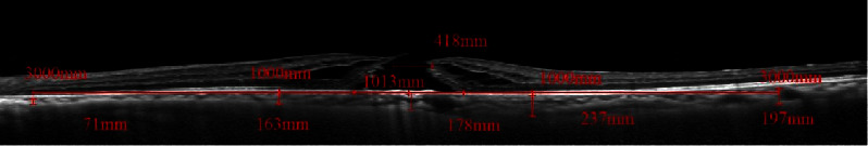
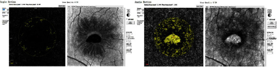
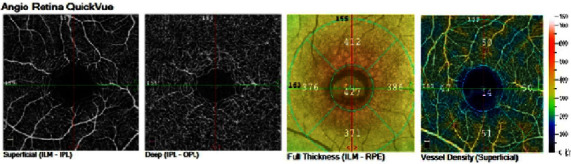
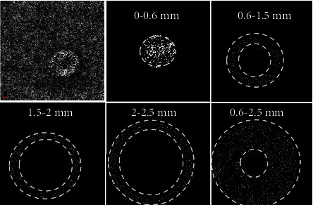
Methods: A prospective study investigated changes in the fundus structure and visual function in 16 macular hole patients before and after surgery. Patients' best-corrected visual acuity (BCVA) and retinal sensitivity were measured by an EDTRS visual chart and microperimetry (MP1), respectively. The thickness of the retina and the blood supply to the retina and superficial choroid were detected by OCTA, and the choroidal capillary blood flow density was analysed with MATLAB. The thickness of the choroid and the aperture size of the macular hole were detected by Heidelberg OCT.
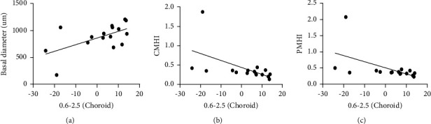
Results: Compared with before surgery and one month after surgery, the BCVA (3 months: 0.47 ± 0.27, before: 1.02 ± 0.22, 1 month: 0.66 ± 0.27, and P < 0.05) and the central sensitivity of the retina (3 months: 14.88 ± 2.87 dB, before: 8.76 ± 3.27 dB, 1 month: 12.22 ± 3.30 dB, and P < 0.05) were significantly improved three months after surgery. The change in BCVA was significantly correlated with the basal diameter (r = 0.677 and P = 0.004), the minimum diameter (r = 0.585 and P = 0.017), the macular hole cystoid height area index (r = -0.618 and P = 0.011), the central macular hole index (r = -0.727 and P = 0.001), the peripheral macular hole index (r = -0.758 and P = 0.001), the central tractional hole index (r = -0.717 and P = 0.002), the peripheral tractional hole index (r = -0.725 and P = 0.001), and changes in the peripheral blood vessel density of the choroid capillary layer (r = 0.585 and P = 0.0017). The change in central retinal sensitivity was correlated with the change in the superficial foveal avascular zone (FAZ; r = 0.520 and P = 0.039), change in the retinal superficial peripheral blood flow density (r = -0.503 and P = 0.047), change in the deep FAZ (r = 0.599 and P = 0.014), and change in the retinal deep peripheral blood flow density (r = -0.601 and P = 0.014).
Conclusions: The morphology of the macular hole as well as changes to the retinal and choroidal microstructure contributes to the recovery of visual function after surgery.
Copyright © 2022 Minfeng Chen et al.
Conflict of interest statement
The authors declare that they have no conflicts of interest.
Figures
Similar articles
-
[Choroidal blood flow and visual function in idiopathic macular hole].Zhonghua Yan Ke Za Zhi. 2022 Jun 11;58(6):412-419. doi: 10.3760/cma.j.cn112142-20210918-00436. Zhonghua Yan Ke Za Zhi. 2022. PMID: 35692022 Chinese.
-
Optical coherence tomography angiography features in patients with idiopathic full-thickness macular hole, before and after surgical treatment.Clin Interv Aging. 2019 Mar 8;14:505-514. doi: 10.2147/CIA.S189417. eCollection 2019. Clin Interv Aging. 2019. PMID: 30880931 Free PMC article.
-
Evaluating the Quantitative Foveal Avascular Zone and Retino-Choroidal Vessel Density Using Optical Coherence Tomography Angiography in a Healthy Indian Population.Cureus. 2022 Aug 4;14(8):e27669. doi: 10.7759/cureus.27669. eCollection 2022 Aug. Cureus. 2022. PMID: 36072178 Free PMC article.
-
[New examination methods for macular disorders--application of diagnosis and treatment].Nippon Ganka Gakkai Zasshi. 2000 Dec;104(12):899-942. Nippon Ganka Gakkai Zasshi. 2000. PMID: 11193944 Review. Japanese.
-
[Relationship between vitrectomy and the morphology and function of the retina].Nippon Ganka Gakkai Zasshi. 2003 Dec;107(12):836-64; discussion 865. Nippon Ganka Gakkai Zasshi. 2003. PMID: 14733133 Review. Japanese.
Cited by
-
Changes in Outcomes of Macular Optical Coherence Tomography Angiography Following Surgery for Optic Disc Pit Maculopathy.Diagnostics (Basel). 2024 Apr 23;14(9):874. doi: 10.3390/diagnostics14090874. Diagnostics (Basel). 2024. PMID: 38732289 Free PMC article.
-
Bibliometric analysis of ophthalmic OCT and OCT angiography research trends over the past 20 years.Int Ophthalmol. 2024 Sep 9;44(1):374. doi: 10.1007/s10792-024-03292-6. Int Ophthalmol. 2024. PMID: 39251539
-
Artificial intelligence in predicting macular hole surgery outcomes: a focus on optical coherence tomography parameters.BMC Ophthalmol. 2025 Jul 22;25(1):419. doi: 10.1186/s12886-025-04256-9. BMC Ophthalmol. 2025. PMID: 40696275 Free PMC article.
References
-
- Johnson M. W., Van Newkirk M. R., Meyer K. A. Perifoveal vitreous detachment is the primary pathogenic event in idiopathic macular hole formation. Archives of Ophthalmology . 2001;119(2):215–222. - PubMed
LinkOut - more resources
Full Text Sources
Miscellaneous
