. 2023 Jan;12(1):22-32.
doi: 10.1302/2046-3758.121.BJR-2022-0263.R1.

Femoral impingement in maximal hip flexion is anterior-inferior distal to the cam deformity in femoroacetabular impingement patients with femoral retroversion : implications for hip arthroscopy

Affiliations

Femoral impingement in maximal hip flexion is anterior-inferior distal to the cam deformity in femoroacetabular impingement patients with femoral retroversion : implications for hip arthroscopy

Adam Boschung et al. Bone Joint Res. 2023 Jan.

Abstract

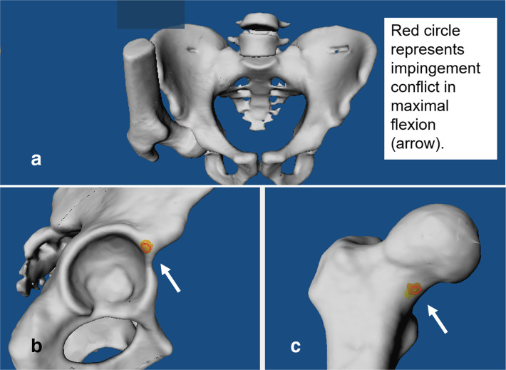
Aims: Femoroacetabular impingement (FAI) patients report exacerbation of hip pain in deep flexion. However, the exact impingement location in deep flexion is unknown. The aim was to investigate impingement-free maximal flexion, impingement location, and if cam deformity causes hip impingement in flexion in FAI patients.

Methods: A retrospective study involving 24 patients (37 hips) with FAI and femoral retroversion (femoral version (FV) < 5° per Murphy method) was performed. All patients were symptomatic (mean age 28 years (SD 9)) and had anterior hip/groin pain and a positive anterior impingement test. Cam- and pincer-type subgroups were analyzed. Patients were compared to an asymptomatic control group (26 hips). All patients underwent pelvic CT scans to generate personalized CT-based 3D models and validated software for patient-specific impingement simulation (equidistant method).

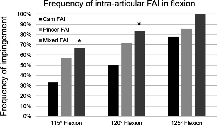
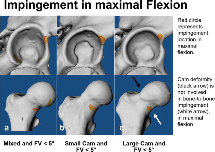
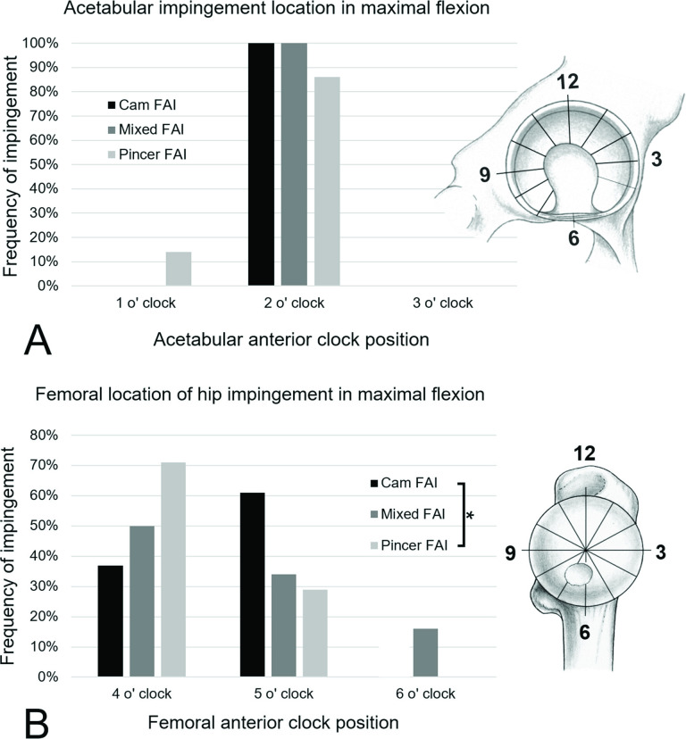
Results: Mean impingement-free flexion of patients with mixed-type FAI (110° (SD 8°)) and patients with pincer-type FAI (112° (SD 8°)) was significantly (p < 0.001) lower compared to the control group (125° (SD 13°)). The frequency of extra-articular subspine impingement was significantly (p < 0.001) increased in patients with pincer-type FAI (57%) compared to cam-type FAI (22%) in 125° flexion. Bony impingement in maximal flexion was located anterior-inferior at femoral four and five o'clock position in patients with cam-type FAI (63% (10 of 16 hips) and 37% (6 of 10 hips)), and did not involve the cam deformity. The cam deformity did not cause impingement in maximal flexion.

Conclusion: Femoral impingement in maximal flexion was located anterior-inferior distal to the cam deformity. This differs to previous studies, a finding which could be important for FAI patients in order to avoid exacerbation of hip pain in deep flexion (e.g. during squats) and for hip arthroscopy (hip-preservation surgery) for planning of bone resection. Hip impingement in flexion has implications for daily activities (e.g. putting on shoes), sports, and sex.Cite this article: Bone Joint Res 2023;12(1):22-32.

Keywords: CT scans; Extra-articular hip impingement; FAI; Femoroacetabular impingement; Hip arthroscopy; Hip preservation surgery; Subspine impingement; cam deformities; cam-type femoroacetabular impingement; femoral retroversion; femoroacetabular impingement; flexion; hip arthroscopy; hip flexion; hip pain; hips.


Figures

Fig. 1
Fig. 1
a) CT-based 3D model of the pelvis and proximal femur is shown of a patient with cam-type femoroacetabular impingement. Impingement simulation in maximal flexion shows b) acetabular and c) femoral intra-articular impingement conflict (red circle, arrow).
Fig. 2
Fig. 2
Location of acetabular (top) and femoral (below) impingement for the three study groups (a) mixed-type femoroacetabular impingement (FAI), b) pincer-type FAI, and c) cam-type FAI) of patients with decreased femoral version (FV < 5°) is shown for 125° of flexion.
Fig. 3
Fig. 3
Frequency of intra-articular hip impingement for 115°, 120°, and 125° of flexion is shown comparing three groups (mixed-type femoroacetabular impingement (FAI), pincer-type FAI, and cam-type FAI) of patients with decreased femoral version < 5°. The asterisk indicates significant difference compared to cam-type FAI.
Fig. 4
Fig. 4
Location of acetabular (top) and femoral (below) impingement in maximal flexion is shown for three patients with decreased femoral version (FV < 5°): a) a patient with mixed-type femoroacetabular impingement, b) a patient with a small cam deformity, and c) a patient with a large cam deformity.
Fig. 5
Fig. 5
Location of a) acetabular and b) femoral hip impingement in maximal flexion is shown for the three study groups. Clock face positions were used: 3 o’clock represents anterior, 12 o’clock represents superior, and 6 o’clock represents inferior. The asterisk indicates significant difference between cam-type and pincer-type femoroacetabular impingement (FAI).

Similar articles

Cited by

References

    1. Griffin DR, Dickenson EJ, O’Donnell J, et al. . The Warwick Agreement on femoroacetabular impingement syndrome (FAI syndrome): an international consensus statement. Br J Sports Med. 2016;50(19):1169–1176. 10.1136/bjsports-2016-096743 - DOI - PubMed
    1. Schmaranzer F, Todorski IAS, Lerch TD, Schwab J, Cullmann-Bastian J, Tannast M. Intra-articular lesions: Imaging and surgical correlation. Semin Musculoskelet Radiol. 2017;21(5):487–506. 10.1055/s-0037-1606133 - DOI - PubMed
    1. Clohisy JC, Knaus ER, Hunt DM, Lesher JM, Harris-Hayes M, Prather H. Clinical presentation of patients with symptomatic anterior hip impingement. Clin Orthop Relat Res. 2009;467(3):638–644. 10.1007/s11999-008-0680-y - DOI - PMC - PubMed
    1. Ng KCG, Lamontagne M, Adamczyk AP, Rakhra KS, Rahkra KS, Beaulé PE. Patient-specific anatomical and functional parameters provide new insights into the pathomechanism of cam FAI. Clin Orthop Relat Res. 2015;473(4):1289–1296. 10.1007/s11999-014-3797-1 - DOI - PMC - PubMed
    1. Diamond LE, Dobson FL, Bennell KL, Wrigley TV, Hodges PW, Hinman RS. Physical impairments and activity limitations in people with femoroacetabular impingement: a systematic review. Br J Sports Med. 2015;49(4):230–242. 10.1136/bjsports-2013-093340 - DOI - PubMed