. 2023 Feb;299(2):102873.
doi: 10.1016/j.jbc.2023.102873. Epub 2023 Jan 5.

Natural and synthetic 2-oxoglutarate derivatives are substrates for oncogenic variants of human isocitrate dehydrogenase 1 and 2

Affiliations

Natural and synthetic 2-oxoglutarate derivatives are substrates for oncogenic variants of human isocitrate dehydrogenase 1 and 2

Xiao Liu et al. J Biol Chem. 2023 Feb.

Abstract

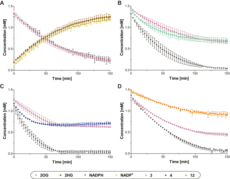
Variants of isocitrate dehydrogenase (IDH) 1 and 2 (IDH1/2) alter metabolism in cancer cells by catalyzing the NADPH-dependent reduction of 2-oxoglutarate (2OG) to (2R)-hydroxyglutarate. However, it is unclear how derivatives of 2OG can affect cancer cell metabolism. Here, we used synthetic C3- and C4-alkylated 2OG derivatives to investigate the substrate selectivities of the most common cancer-associated IDH1 variant (R132H IDH1), of two cancer-associated IDH2 variants (R172K IDH2, R140Q IDH2), and of WT IDH1/2. Absorbance-based, NMR, and electrochemical assays were employed to monitor WT IDH1/2 and IDH1/2 variant-catalyzed 2OG derivative turnover in the presence and absence of 2OG. Our results reveal that 2OG derivatives can serve as substrates of the investigated IDH1/2 variants, but not of WT IDH1/2, and have the potential to act as 2OG-competitive inhibitors. Kinetic parameters reveal that some 2OG derivatives, including the natural product 3-methyl-2OG, are equally or even more efficient IDH1/2 variant substrates than 2OG. Furthermore, NMR and mass spectrometry studies confirmed IDH1/2 variant-catalyzed production of alcohols in the cases of the 3-methyl-, 3-butyl-, and 3-benzyl-substituted 2OG derivatives; a crystal structure of 3-butyl-2OG with an IDH1 variant (R132C/S280F IDH1) reveals active site binding. The combined results highlight the potential for (i) IDH1/2 variant-catalyzed reduction of 2-oxoacids other than 2OG in cells, (ii) modulation of IDH1/2 variant activity by 2-oxoacid natural products, including some present in common foods, (iii) inhibition of IDH1/2 variants via active site binding rather than the established allosteric mode of inhibition, and (iv) possible use of IDH1/2 variants as biocatalysts.

Keywords: (R)-2-hydroxyglutarate; 2-oxoacids; 2-oxoglutarate; 2HG; 2OG; IDH; acute myeloid leukemia; alternative substrates; cancer metabolism-related IDH mutations; epigenetics; isocitrate dehydrogenase; α-ketoacids; α-ketoglutarate.


Conflict of interest statement

Conflict of interest The authors declare that they have no conflict of interest with the contents of this article.

Figures

Figure 1
Figure 1
Isocitrate dehydrogenase catalysis.A, WT IDH1-3 catalyze the Mg(II)-dependent conversion of isocitrate to 2OG and CO2 coupled with conversion of the cosubstrate NADP+ (or NAD+) reduction to NADPH (or NADH); (B) cancer-associated IDH variants, e.g. R132H IDH1, R132C IDH1, R172K IDH2, and R140Q IDH2, catalyze the stereospecific reduction of 2OG to (R)-2-hydroxyglutarate (2HG) coupled with conversion of NADPH to NADP+. Note that WT IDH1/2 do not efficiently catalyze the reduction of 2OG to 2HG (3, 5); (C) potential reaction of cancer-associated IDH1/2 variants with 2OG derivatives; (D) view of the R132H IDH1 active site in a closed conformation in complex with Ca(II) (green), 2OG (teal), and NADP+ (yellow), the R132H variation is in magenta; two R132H IDH1 molecules (gray and lavender) interact with each other and form the catalytically active dimer (PDB ID: 3INM (54)); (E) view of the R140Q IDH2 active site in complex with NADP+ (yellow) in an open conformation; the R140Q variation is in magenta; two R140Q IDH2 molecules (gray and lavender) interact with each other and form the catalytically active dimer (PDB ID: 5SVO (75)); (F) view of the R172K IDH2 active site in complex with NADP+ (yellow) in a closed conformation; the R172K variation, which corresponds to R132 in IDH1, is in magenta; two R172K IDH2 molecules (gray and lavender) interact with each other and form the catalytically active dimer (PDB ID: 5SVN (75)). 2OG, 2-oxoglutarate; IDH, isocitrate dehydrogenase.
Figure 2
Figure 2
Kinetic parameters for the IDH1/2 variant-catalyzed reduction of 2OG derivatives.A and B, the R132H IDH1 variant catalyzes the reduction of (A) 3-methyl-2OG (1) giving 2-hydroxy-3-methylglutarate (33) and (b) 3-butyl-2OG (4) giving 3-butyl-2-hydroxyglutarate (34) as confirmed by NMR and/or MS analyses (Fig. S1); CK, Kmapp- values of R132H IDH1 (orange circles), R172K IDH2 (blue triangles), and R140Q IDH2 (black boxes) for (C) 2OG, (D) 1, (E) 2, (F) 3, (G) 4, (H) 7, (I) 12, (J) 21, and (K) 23. IDH1/2 variant assays were performed as described in the Experimental procedures section, results are a mean of two independent runs, each composed of technical duplicates (n = 2; mean ± SD). 2OG, 2-oxoglutarate.
Figure 3
Figure 3
1H NMR and electrochemical studies indicate that R132H IDH1 preferentially catalyzes the reduction of one enantiomer from racemic mixtures of C3/C4-substituted 2OG derivatives.1H NMR time course monitoring the R132H IDH1-catalyzed reduction of (A and B) 2OG, (C) 1, (D) 4, (E) 7, and (F) 12 in the presence of equimolar amounts of NADPH (1.5 mM) (A) or a twofold excess of NADPH (3.0 mM) and a fourfold greater R132H IDH1 concentration (2.0 μM) (BF). The time scales were normalized to the end of the first of 50 subsequent NMR experiments after the addition of R132H IDH1 to the reaction mixture (t = 0 min), by which time low levels of conversion were manifest; (G) cyclic voltammograms for a stationary porous electrode containing ‘electroactive’ R132H IDH1 (55) in the presence of 2OG and racemic 12. The concentration of 12 depletes at the electrode-solution interface faster than 2OG at similar currents (R132H IDH1 faradaic ‘coupled’ current (55) decreased by −43% for 12 compared to −10% for 2OG during each scan); (H) Michaelis–Menten curves showing the simulated R132H IDH1 activity based on experimental Kmapp- and vmaxapp-values for 2OG (blue), racemic 12 (black), and (predicted) enantiopure 12 (red). The initial and final concentrations indicate the expected activity/current for each substrate based on the conditions used in panel G if 500 μM of each substrate was consumed (locally) at the electrode-solution interface during each scan. The predicted changes in catalytic rate (enantiopure 12: −46%; racemic 12: −22%; 2OG: −9%) suggest that only one enantiomer of 12 is (at least) an efficient substrate for R132H IDH1. ENADP(H)0 and E2OG/2HG0 denote formal potentials for the NADP+/NADPH and 2OG/2HG couples, respectively (55). 2OG, 2-oxoglutarate; 2HG, 2-hydroxyglutarate.
Figure 4
Figure 4
1H NMR competition assays provide evidence that 3-butyl–substituted 2OG (4) is a better R132H IDH1 substrate than 2OG under the tested conditions.1H NMR time course monitoring the R132H IDH1-catalyzed reduction of (A) 2OG alone (1.5 mM; note that Figure 3A and panel (A) in this figure are identical, panel (A) is shown to help the direct comparison with panels BD) and equimolar mixtures of 2OG and (B) 3, (C) 4, or (D) 12 in the presence of equimolar NADPH (1.5 mM). The time scales were normalized to the end of the first of 50 subsequent NMR experiments after the addition of R132H IDH1 to the reaction mixture (t = 0 min), by which time low levels of conversion were manifest. Results are a mean of two independent duplicates (n = 2; mean ± SD). 2OG, 2-oxoglutarate.
Figure 5
Figure 5
Crystallographic analysis with R132C/S280F IDH1 reveals the active site–binding mode of 3-butyl-2OG (4). Color code: R132C/S280F IDH1: monomer 1: wheat, monomer 2: green; carbon-backbone of (3R)-butyl-2OG [(3R)-4] in yellow, (3S)-butyl-2OG [(3S)-4] in teal; NADPH in gray; oxygen: red; nitrogen: blue. A, ribbon view of R132C/S280F IDH1:Ca:NADPH:4 (PDB ID: 8BAY, 2.35 Å resolution) showing the dimer formed by chain A (wheat) and chain B (green); B, view of the metal and substrate-binding site revealing the Ca(II)-binding residues Aps252 (monomer 2, green, helix α9), Asp275 and Asp279 (monomer 1, wheat, α10) and the 4-binding residues Thr77 and Ser94; (C) 2mFo-DFc electron density map contoured to 1.0 σ around 4 and Ca(II) in complex with R132C/S280F IDH1 and NADPH; (D) superimposition of the active site views from chain A of the R132C/S280F IDH1:Ca:NADPH:4 structure (PDB ID: 8BAY, 2.35 Å resolution) and the reported R132C/S280F IDH1:Ca(II):NADPH:2OG structure (cyan; PDB ID: 7PJM (53)) reveal a similar binding mode and conformation for 2OG and 4. 2OG, 2-oxoglutarate.
Figure 6
Figure 6
2OG derivative 11 inhibits R172K IDH2 more efficiently than R132H IDH1 and R140Q IDH2. Representative dose-response curves used to determine IC50 values for (A) NOG and (B) 11 for R132H IDH1 (orange circles), R172K IDH2 (blue triangles), and R140Q IDH2 (black boxes). Hill coefficients (76) of the inhibition curves were in the range of the expected value of −1 for substrate-competitive IDH1/2 variant inhibitors. 2OG, 2-oxoglutarate; NOG, N-oxalylglycine.

References

    1. Xu X., Zhao J., Xu Z., Peng B., Huang Q., Arnold E., et al. Structures of human cytosolic NADP-dependent isocitrate dehydrogenase reveal a novel self-regulatory mechanism of activity. J. Biol. Chem. 2004;279:33946–33957. - PubMed
    1. Liu Y., Lang F., Chou F.-J., Zaghloul K.A., Yang C. Isocitrate dehydrogenase mutations in glioma: genetics, biochemistry, and clinical indications. Biomedicines. 2020;8:294. - PMC - PubMed
    1. Dang L., Su S.-S.M. Isocitrate dehydrogenase mutation and (R)-2-hydroxyglutarate: from basic discovery to therapeutics development. Annu. Rev. Biochem. 2017;86:305–331. - PubMed
    1. Yang H., Ye D., Guan K.-L., Xiong Y. IDH1 and IDH2 mutations in tumorigenesis: mechanistic insights and clinical perspectives. Clin. Cancer Res. 2012;18:5562–5571. - PMC - PubMed
    1. Losman J.-A., Kaelin W.G., Jr. What a difference a hydroxyl makes: mutant IDH, (R)-2-hydroxyglutarate, and cancer. Genes Dev. 2013;27:836–852. - PMC - PubMed

Publication types

MeSH terms

Substances

LinkOut - more resources