. 2023 Feb;299(2):102869.
doi: 10.1016/j.jbc.2023.102869. Epub 2023 Jan 5.

RanBP9 controls the oligomeric state of CTLH complex assemblies

Affiliations

RanBP9 controls the oligomeric state of CTLH complex assemblies

Pia Maria van Gen Hassend et al. J Biol Chem. 2023 Feb.

Abstract

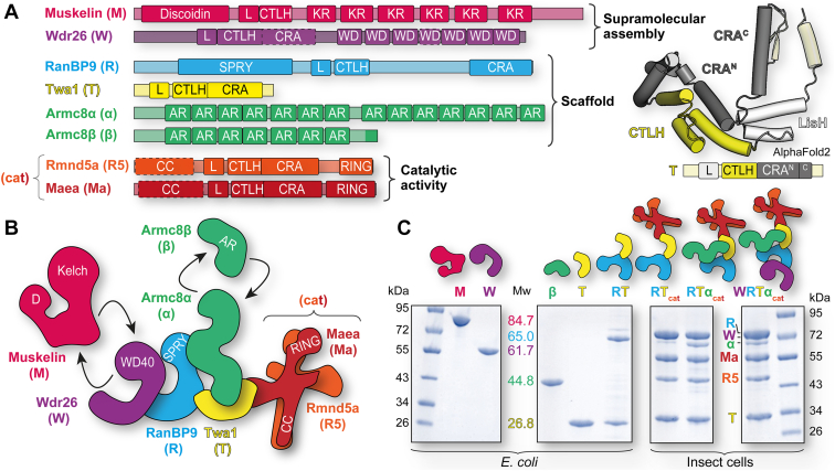
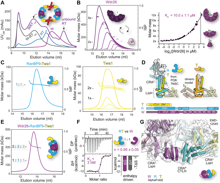
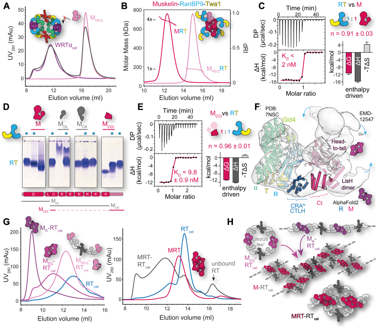
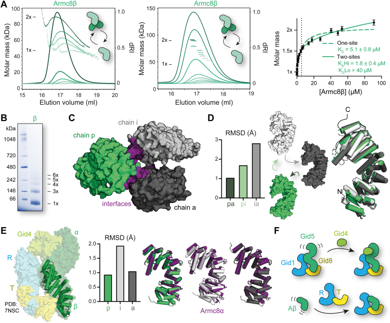
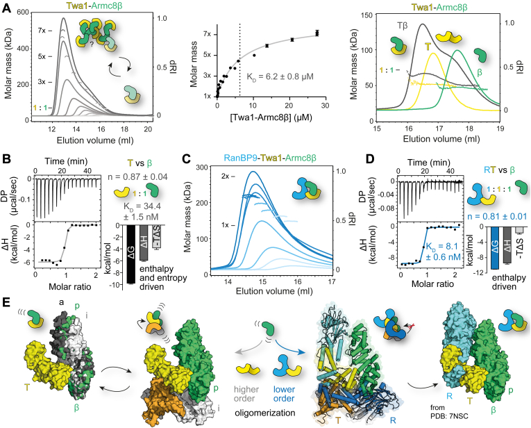
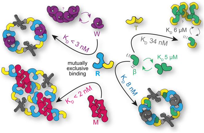
The CTLH (C-terminal to lissencephaly-1 homology motif) complex is a multisubunit RING E3 ligase with poorly defined substrate specificity and flexible subunit composition. Two key subunits, muskelin and Wdr26, specify two alternative CTLH complexes that differ in quaternary structure, thereby allowing the E3 ligase to presumably target different substrates. With the aid of different biophysical and biochemical techniques, we characterized CTLH complex assembly pathways, focusing not only on Wdr26 and muskelin but also on RanBP9, Twa1, and Armc8β subunits, which are critical to establish the scaffold of this E3 ligase. We demonstrate that the ability of muskelin to tetramerize and the assembly of Wdr26 into dimers define mutually exclusive oligomerization modules that compete with nanomolar affinity for RanBP9 binding. The remaining scaffolding subunits, Armc8β and Twa1, strongly interact with each other and with RanBP9, again with nanomolar affinity. Our data demonstrate that RanBP9 organizes subunit assembly and prevents higher order oligomerization of dimeric Wdr26 and the Armc8β-Twa1 heterodimer through its tight binding. Combined, our studies define alternative assembly pathways of the CTLH complex and elucidate the role of RanBP9 in governing differential oligomeric assemblies, thereby advancing our mechanistic understanding of CTLH complex architectures.

Keywords: GID–CTLH complex; ITC; RING E3 ligase; SEC–MALS; X-ray crystallography.


Conflict of interest statement

Conflict of interest The authors declare that they have no conflicts of interest with the contents of this article.

Figures

Figure 1
Figure 1
Modular reconstitution of CTLH complexes.A, domain architecture of CTLH subunits based on the InterPro database and additional assignments based on AlphaFold2 predictions (dotted lines). Folding of the recurring LisH (L), CTLH, and CRA domain sequence is illustrated by the Twa1 AlphaFold2 model. B, model of the CTLH complex as defined earlier (6). Unique domains are highlighted, and arrows represent alternative subunits. C, SD-PAGE analyses of purified individual proteins and different CTLH subcomplexes after expression in either Escherichia coli or insect cells. Theoretical molecular weights (colored numerals with units in kilodalton) of the monomeric proteins as reference for the SEC–MALS analyses are indicated. AR, armadillo repeat; CC, coiled-coil region; CRA, CT-11–RanBPM; CTLH, C-terminal to lissencephaly-1 homology motif; D, discoidin; KR, kelch repeat; L, lissencephaly-1 homology; RING, really interesting new gene; SPRY, SPla and the RYanodine receptor; SEC-MALS, multiangle light scattering coupled to analytical size-exclusion chromatography; WD, WD40 repeat.
Figure 2
Figure 2
Tight binding to RanBP9 prevents higher order oligomerization of dimeric Wdr26.A, Wdr26-mediated CTLH complex assembly. Analytical SEC documenting assembly of the WRTcat complex from Wdr26 (W) and the coexpressed RTcat complex compared with the coexpressed WRTcat complex. The model indicates the assembly of this complex based on published data (6). See Fig. S1A for SDS-PAGE analysis of fractions of the WRTcat elution profile. B, self-association of Wdr26 analyzed with SEC–MALS at various protein concentrations (see Fig. S2 for more details). At the elution peak represented by the differential refractive index (dRI) profile signal, molar masses are depicted (left). Theoretical molar masses of a dimeric (2×) and octameric (8×) Wdr26 assembly are indicated. The best-fit value of the dissociation constant (KD) and its standard error (SE) are reported. C, SEC–MALS analysis of the RT complex and Twa1. D, based on the cryo-EM structure of RT (derived from PDB entry: 7NSC), a model of a Twa1 homodimer utilizing the same binding mode was constructed by superimposing Twa1 with RanBP9. Schematic representations of the domains of T, R, and W illustrate similarities in their architecture. E, molar mass determination of the WRT complex by SEC–MALS. F, isothermal titration calorimetry (ITC) studies to determine binding of the RT complex to Wdr26. The differential power (DP) (upper panel) was integrated over time, and the released heat (ΔH) plotted against the molar ratio of the RT complex (lower panel) and fitted with a one-site binding model. KD values (KD = 3.4 ± 3.5 nM) and the signature binding plot were derived from three measurements in which the interaction was not too tight to still permit reliable data analysis of a total of six measurements. The error bars reflect the SE of the change in enthalpy (ΔH), entropy (ΔT), and the Gibbs free energy (ΔG). G, model illustrating the Wdr26–RanBP9 interaction. The AlphaFold2 predicted structures of Wdr26, RanBP9, and Twa1 were fitted into the cryo-EM map (EMD-12545) of the human SA module. CTLH, C-terminal to lissencephaly-1 homology motif; L, LisH; MALS, multiangle light scattering; PDB, Protein Data Bank; RTcat, RanBP9–Twa1–Rmnd5a–Maea complex; SEC, size-exclusion chromatography; W, Wdr26; WD, WD40 repeat.
Figure 3
Figure 3
Tight binding to RanBP9 preserves the tetrameric assembly of muskelin.A, mutually exclusive binding of Wdr26 and muskelin to the CTLH complex. UV280 elution profiles of an aSEC run documenting the assembly of the WRTαcat complex with the monomeric muskelin mutant MREQ (N144R, F184E, and L196E). For comparison, see Fig. S5. B, SEC–MALS analysis of the MRT and MREQRT complexes. C, ITC analysis for the binding of RT to muskelin. Similar to Figure 2F, the dissociation constant (KD = 1.7 ± 6.7 nM) and signature binding plot could be derived for only three of six measurements. D, native agarose gel electrophoreses (NAGE) of differently truncated muskelin constructs in a complex assembly with the RT complex and as individual proteins. Domains that are included in the construct are depicted as follows: discoidin (D), LisH (L), CTLH (C), Kelch repeats (K), and C-terminal module (Ct). Analysis of further constructs by NAGE are shown in Fig. S6. E, ITC study of binding of the RT complex to a MCCt construct comprised of a fusion of the N-terminal part of the CTLH domain with the C-terminal module of muskelin. F, model of how the C-terminal module of muskelin binds to RanBP9. The high-resolution cryo-EM structure of RTα–Gid4 (PDB entry: 7NSC) and the AlphaFold2 predictions of muskelin and RanBP9 were fitted into the 10 Å cryo-EM map (EMD-12547). Muskelin dimerization sites are denoted, and blue arrows display further binding sites of RanBP9. G, complex assembly of different muskelin variants (MREQ, MR, and MEQ) displayed in shades of purple (left) and the MRT complex (right) with the RTcat complex as analyzed by aSEC. See Fig. S1C for SDS-PAGE analysis of selected fractions. H, model illustrating possible assemblies from (G). aSEC, analytical size-exclusion chromatography; CTLH, C-terminal to lissencephaly-1 homology motif; ITC, isothermal titration calorimetry; LisH, lissencephaly-1 homology; M, muskelin; SEC-MALS, multiangle light scattering coupled to aSEC; PDB, Protein Data Bank; RTcat, RanBP9–Twa1–Rmnd5a–Maea complex; W, Wdr26; α, Armc8α.
Figure 4
Figure 4
Dynamic oligomerization and conformational flexibility of Armc8β.A, SEC–MALS analysis of the Armc8β self-association (similar to Fig. 2B). B, blue native PAGE analysis of Armc8β. C, the crystal structure of Armc8β (PDB entry: 8A1I) reveals three chains in the asymmetric unit: chains p (green), i (gray), and a (black). D, overlay of the three chains in ribbon representation reveals conformational changes. Rms deviations of the different chains toward each other are summarized in the bar chart. E, comparison of the p chain (ribbon) of Armc8β with Armc8α as observed in the cryo-EM structure of the RTα–Gid4 complex (PDB entry: 7NSC). Superposition of the three different chains of Armc8β with Armc8α (purple) in ribbon representation illustrates the rms deviations depicted in the bar chart. F, model of how complex binding rigidifies the flexibility of Armc8. SEC-MALS, multiangle light scattering coupled to analytical size-exclusion chromatography; PDB, Protein Data Bank.
Figure 5
Figure 5
Tight binding to RanBP9 prevents higher order oligomerization of the Twa1–Armc8β complex.A, SEC–MALS analysis of the Tβ complex self-association conducted as in Figures 2B and 4A. Full molar mass peak profiles are shown in Fig. S1B. In addition, assembly and molecular weight analysis of the binary complex at low concentrations are depicted (right). B, ITC binding studies of Twa1 to Armc8β displayed as for Figure 3C. The affinity constant KD and signature binding plot parameters including their standard errors were derived from 18 measurements. C, molecular weight determination of RTβ complex self-association with SEC–MALS at different concentrations was limited by the instability of the complex at higher concentrations. See Fig. S1C for full molar mass profiles. D, ITC analysis of the RT complex binding to Aβ derived from seven measurements. E, model of how the oligomerization of the binary Tβ complex is prevented upon addition of RanBP9. To demonstrate this the Twa1-Armc8α module from the RTα–Gid4 complex (PDB entry: 7NSC) was superimposed with the three Armc8β conformations observed in the crystal structure. ITC, isothermal titration calorimetry; SEC-MALS, multiangle light scattering coupled to analytical size exclusion chromatography; PDB, Protein Data Bank.
Figure 6
Figure 6
Tight binding to RanBP9 governs differential oligomerization of CTLH complex assemblies. CTLH, C-terminal to lissencephaly-1 homology motif; M, muskelin; R, RanBP9; T, Twa1; W, Wdr26; β, Armc8β.

References

    1. Sherpa D., Chrustowicz J., Schulman B.A. How the ends signal the end: regulation by E3 ubiquitin ligases recognizing protein termini. Mol. Cell. 2022;82:1424–1438. - PMC - PubMed
    1. Deshaies R.J., Joazeiro C.A.P. RING domain E3 ubiquitin ligases. Annu. Rev. Biochem. 2009;78:399–434. - PubMed
    1. Hershko A., Ciechanover A. The ubiquitin system. Annu. Rev. Biochem. 1998;67:425–479. - PubMed
    1. Zheng N., Shabek N. Ubiquitin ligases: structure, function, and regulation. Annu. Rev. Biochem. 2017;86:129–157. - PubMed
    1. Kobayashi N., Yang J., Ueda A., Suzuki T., Tomaru K., Takeno M., et al. RanBPM, Muskelin, p48EMLP, p44CTLH, and the armadillo-repeat proteins ARMC8alpha and ARMC8beta are components of the CTLH complex. Gene. 2007;396:236–247. - PubMed

Publication types

MeSH terms

Substances

LinkOut - more resources