. 2023 Feb;299(2):102877.
doi: 10.1016/j.jbc.2023.102877. Epub 2023 Jan 6.

Membrane skeleton hyperstability due to a novel alternatively spliced 4.1R can account for ellipsoidal camelid red cells with decreased deformability

Affiliations

Membrane skeleton hyperstability due to a novel alternatively spliced 4.1R can account for ellipsoidal camelid red cells with decreased deformability

Yuqi Chen et al. J Biol Chem. 2023 Feb.

Abstract

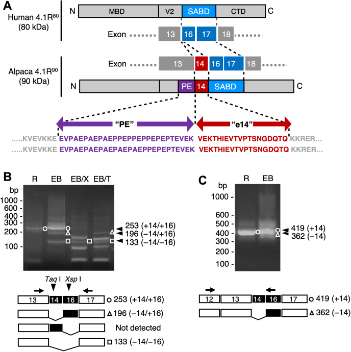
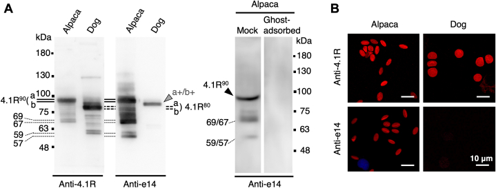
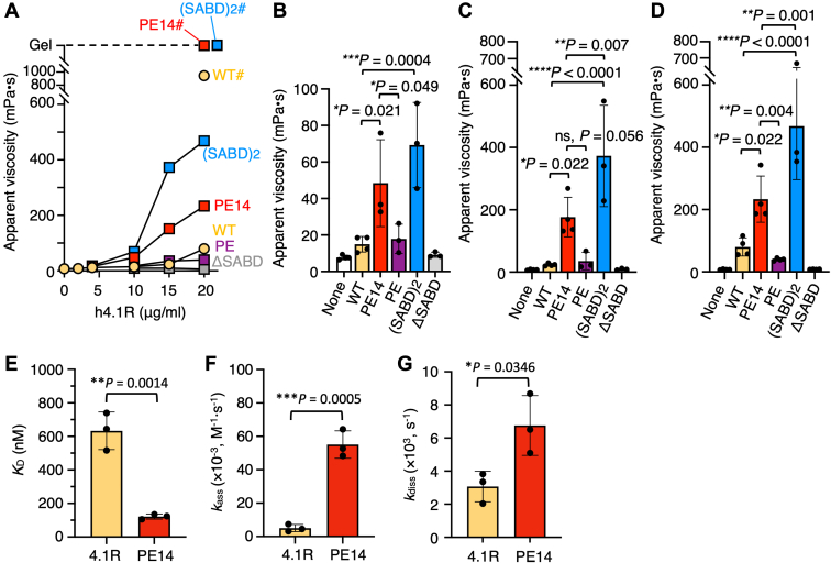
The red blood cells (RBCs) of vertebrates have evolved into two basic shapes, with nucleated nonmammalian RBCs having a biconvex ellipsoidal shape and anuclear mammalian RBCs having a biconcave disk shape. In contrast, camelid RBCs are flat ellipsoids with reduced membrane deformability, suggesting altered membrane skeletal organization. However, the mechanisms responsible for their elliptocytic shape and reduced deformability have not been determined. We here showed that in alpaca RBCs, protein 4.1R, a major component of the membrane skeleton, contains an alternatively spliced exon 14-derived cassette (e14) not observed in the highly conserved 80 kDa 4.1R of other highly deformable biconcave mammalian RBCs. The inclusion of this exon, along with the preceding unordered proline- and glutamic acid-rich peptide (PE), results in a larger and unique 90 kDa camelid 4.1R. Human 4.1R containing e14 and PE, but not PE alone, showed markedly increased ability to form a spectrin-actin-4.1R ternary complex in viscosity assays. A similar facilitated ternary complex was formed by human 4.1R possessing a duplication of the spectrin-actin-binding domain, one of the mutations known to cause human hereditary elliptocytosis. The e14- and PE-containing mutant also exhibited an increased binding affinity to β-spectrin compared with WT 4.1R. Taken together, these findings indicate that 4.1R protein with the e14 cassette results in the formation and maintenance of a hyperstable membrane skeleton, resulting in rigid red ellipsoidal cells in camelid species, and suggest that membrane structure is evolutionarily regulated by alternative splicing of exons in the 4.1R gene.

Keywords: alternative splicing; camelid; elliptocyte; erythrocyte; hereditary elliptocytosis; membrane protein; membrane structure; plasma membrane; protein 4.1R; spectrin.


Conflict of interest statement

Conflict of interest The authors declare that they have no conflicts of interest with the contents of this article.

Figures

Figure 1
Figure 1
Characterization of the ellipticity of alpaca RBCs and the 90 kDa 4.1R in alpaca RBC membranes.A, Left panel. A new methylene blue–stained smear of freshly obtained alpaca peripheral blood, showing a younger aggregate reticulocyte (arrow) and punctate reticulocytes (arrowhead). Right panel. Ellipticity of aggregate (R1) and punctate (R2) reticulocytes and erythrocytes (E). Data are expressed as the mean ± S.D. (n = 20). ∗∗∗∗p < 0.0001 by one-way ANOVA with Tukey’s multiple comparison test. Bar represents 10 μm. B, alpaca RBCs washed in PBS (left) and ellipsoid Triton shell after solubilization of membranes in PBS containing 1% Triton X-100 on ice for 30 min (right). Bars, 10 μm. C, 8% SDS-PAGE gel of membrane proteins from human, canine, bovine, and alpaca RBCs stained with Coomassie brilliant blue (CBB, 7 μg/lane) and immunoblotted with anti-canine 4.1R antibody (IB, 3 μg/lane). The migration of alpaca 4.1R (4.1R90, a and b) was slower than that of human, canine, and bovine 4.1R proteins (4.1R80, a and b). D, membrane ghosts (Gh) from human and alpaca RBCs were incubated in 1% Triton X-100 solution on ice for 1 h followed by centrifugation at 100,000g for 30 min to separate the supernatant (Sup) and precipitate (Prec). Samples equivalent to ghosts containing 2 μg of proteins were subjected to SDS-PAGE and immunoblotting to detect spectrin (Spectrin), band 3 (Band 3), 4.1R (4.1R), and β-actin (β-Actin). The migrating positions of size markers are shown in kDa. RBC, red blood cell.
Figure 2
Figure 2
Involvement of the exon 14–derived sequence in alpaca 4.1R90cDNA.A, cDNA and amino acid sequence analyses of alpaca 4.1R90, focusing on the regions derived from exons 13–17, in comparison with the relevant structures of human 4.1R80. Structural domains of 4.1R80 were assigned as described (35): CTD, C-terminal domain; MBD, membrane-binding domain; SABD, spectrin–actin-binding domain; V2, variable region 2. The C-terminal region of V2, which is encoded by exon 13, of alpaca 4.1R90 contains a unique amino acid sequence “PE” (shown in purple letters) with two allelic differences. The exon 14–derived sequence “e14” is flanked by the PE and SABD sequences and is shown in red. The complete amino acid sequence and an example of LC-MS/MS analysis of alpaca 4.1R90 are shown in the Supporting Information (Fig. S2 and Table S1). B, cDNA fragments encompassing exons 13–17 that were PCR amplified from alpaca reticulocyte (R) and day 6 erythroblasts (EB) using the primers Vic41.5′e13PE.F and Vic41.e17.KKHHASI.R. The arrows show the positions of the primers. The cDNA fragments obtained include a 253 bp fragment containing nucleotides derived from exons 14 and 16 (○, +14/+16), a 196 bp fragment with an exon 16 sequence (△, −14/+16), and a 133 bp fragment lacking sequences from exons 14 and 16 (□, −14/−16). Because exons 14 and 16 contain unique restriction sites for Taq I and Xsp I, respectively (arrowheads), digestion with Xsp I (EB/X) or Taq I (EB/T) confirmed that the 253 bp and 196 bp fragments contain exon 14–derived sequence, whereas only 253 bp fragment contains exon 16–derived nucleotides. A cDNA fragment containing exon 14–derived nucleotides alone was not detected. C, cDNA fragments encompassing exons 12–16 PCR amplified from reticulocyte (R) and erythroblasts (EB) using the primers Vic41.e12.TRQASA.F and Vic41.seq.e16.R2 (arrows). The 419 bp fragment containing the exon 14 sequence (○, +14) and the 362 bp fragment without this sequence (△, −14) are indicated. SABD, spectrin–actin-binding domain.
Figure 3
Figure 3
Immunoblotting and immunofluorescent detection of the exon 14–derived sequence in alpaca 4.1R90.A, immunoblotting analysis of 4.1R proteins from alpaca and dog RBC membranes using anti-canine 4.1R (Anti-4.1R) and anti-e14 (Anti-e14) antibodies. Alpaca 4.1R90 reacted with both antibodies, whereas dog 4.1R80 reacted only with anti-4.1R antibody. The anti-e14 antibody reacted weakly with proteins slightly larger than 4.1R80 (a+/b+) in dog membranes, indicating the presence of a 4.1R80 isoform having the e14 sequence at a very low level. Each lane contained 3 μg (alpaca) and 2 μg (dog) membrane proteins (left panels). The right panel shows the immunoblotting of alpaca RBC membranes with anti-e14 antibody previously incubated with (Ghost-adsorbed) or without (Mock) an excess amount of alpaca RBC membranes, as described in the Experimental procedures. The migrating positions of size markers are shown in kDa. B, immunofluorescent detection of 4.1R protein in alpaca and dog RBCs showing the involvement of the e14 sequence in alpaca RBC membranes. Bars represent 10 μm. RBC, red blood cell.
Figure 4
Figure 4
GST-fused h4.1R mutants used in the gelation assay.A, schemes of h4.1R WT and its mutants used in gelation assays. Mutants included PE14, h4.1R[PE14]; PE, h4.1R[PE]; (SABD)2, h4.1R(SABD)2; and ΔSABD, h4.1RΔSABD. Assignment of structural domains is described in the legend for Figure 2. B, Coomassie blue–stained SDS-gel of the purified recombinants. Human RBC membrane proteins are also shown (Ghost), with 4.1R80 (4.1R80) indicated. The apparent molecular masses of GST-fused recombinants and size markers are shown in kDa (right panel). GST, glutathione-S-transferase; RBC, red blood cell; SABD, spectrin–actin-binding domain.
Figure 5
Figure 5
Effects of the PE14 sequence on spectrin–actin–4.1R ternary complex formation and 4.1R binding to the N-terminal region of β-spectrin.A, results of falling ball viscometry assays, showing the apparent viscosity due to the spectrin–actin–4.1R complex formation. Each reaction contained 10 μg/ml spectrin tetramers, 250 μg/ml F-actin, and appropriate concentrations (0–20 μg/ml) of GST-fused h4.1R WT or the h4.1R mutants shown in Figure 3. Data shown are the means of three independent experiments. The viscosities obtained for the WT (WT#) and the PE14 (PE14#) and (SABD)2 ((SABD)2#) recombinants in the presence of 20 μg/ml spectrin tetramers are also shown. PE14# and (SABD)2# reactions showed complete gelation of the solutions (Gel). B–D, detailed data and comparisons for reactions containing 10 (B), 15 (C), and 20 μg/ml (D) h4.1R recombinants. None indicates that the reaction mixture contained only spectrin and F-actin. Data are expressed as the mean ± S.D. (n = 3–4). p-values are calculated by one-way ANOVA with Dunnett’s multiple comparison test. E–G, results of surface plasmon resonance assays, showing the binding affinities of h4.1R WT (WT) and h4.1R[PE14] (PE14) to the N-terminal fragment of β-spectrin. KD values (E) are calculated from the measurements of kass (F) and kdiss (G). Data are expressed as the mean ± S.D. (n = 3). p-values calculated by unpaired t tests. GST, glutathione-S-transferase; SABD, spectrin–actin-binding domain.

References

    1. Lazarides E. From genes to structural morphogenesis: the genesis and epigenesis of a red blood cell. Cell. 1987;51:345–356. - PubMed
    1. Lazarides E., Woods C. Biogenesis of the red blood cell membrane-skeleton and the control of erythroid morphogenesis. Annu. Rev. Cell Biol. 1989;5:427–452. - PubMed
    1. Fowler V.M. Chapter two–the human erythrocyte plasma membrane: a Rosetta stone for decoding membrane-cytoskeleton structure. Curr. Top. Membr. 2013;72:39–88. - PubMed
    1. Gallagher P.G. Hereditary elliptocytosis: spectrin and protein 4.1R. Sem Hematol. 2004;41:142–164. - PubMed
    1. Gallagher P.G. In: Hematology: Basic Principles and Practice. 7th Ed. Hoffman R., Benz E.J. Jr., Silberstein L.E., Heslop H.E., Weitz J.I., Anastasi J., et al., editors. Elsevier; Philadelphia, PA: 2017. Red blood cell membrane disorder.

Publication types

LinkOut - more resources