. 2022 Dec 8;6(2):e202201533.
doi: 10.26508/lsa.202201533. Print 2023 Feb.

SLAMF6 compartmentalization enhances T cell functions

Affiliations

SLAMF6 compartmentalization enhances T cell functions

Yevgeniya Gartshteyn et al. Life Sci Alliance. .

Abstract

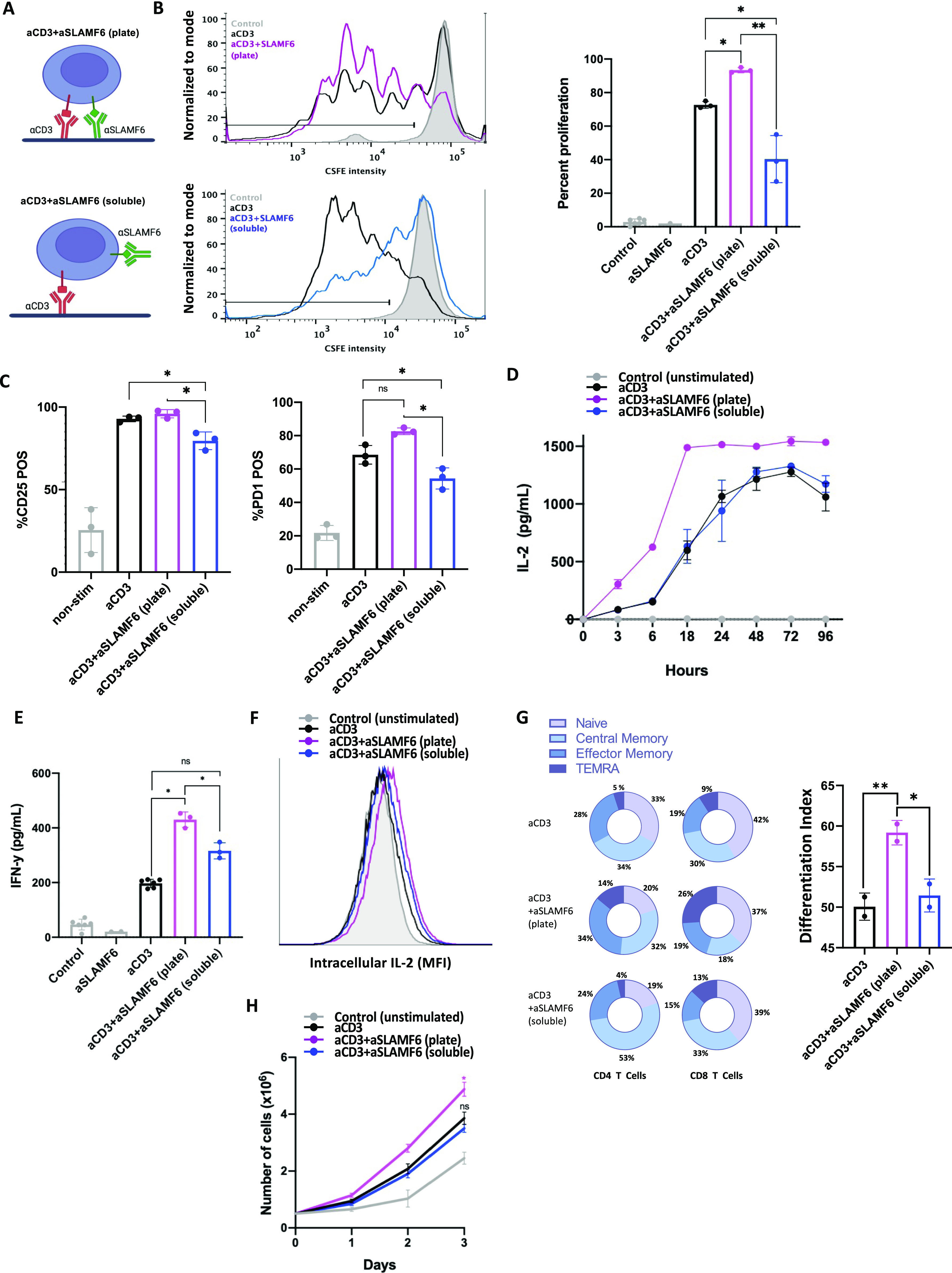
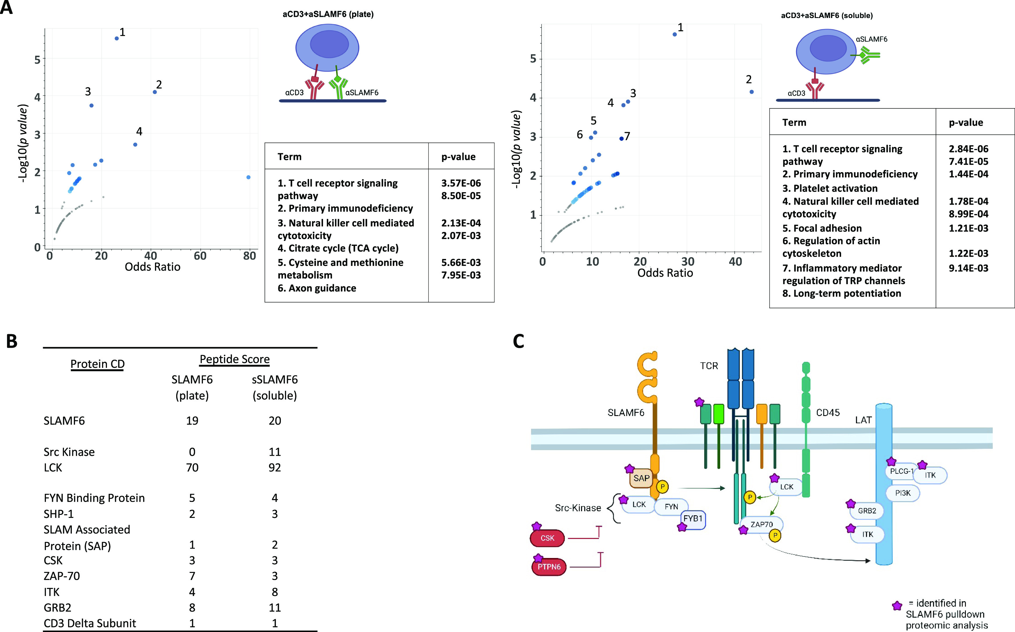
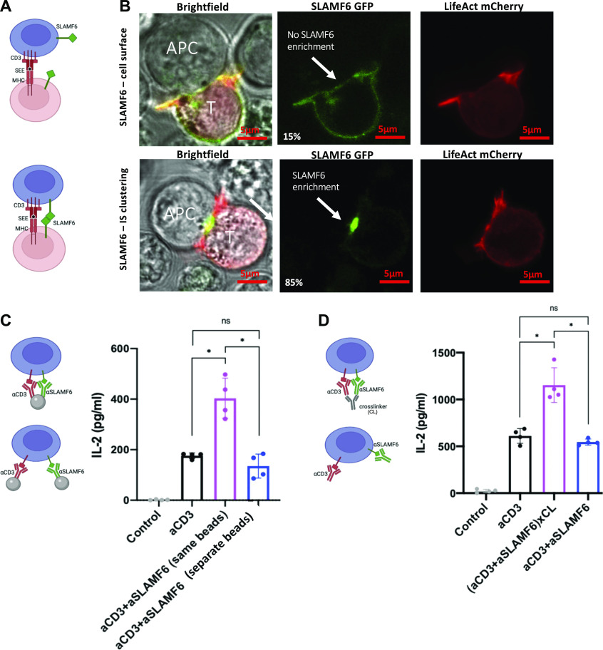
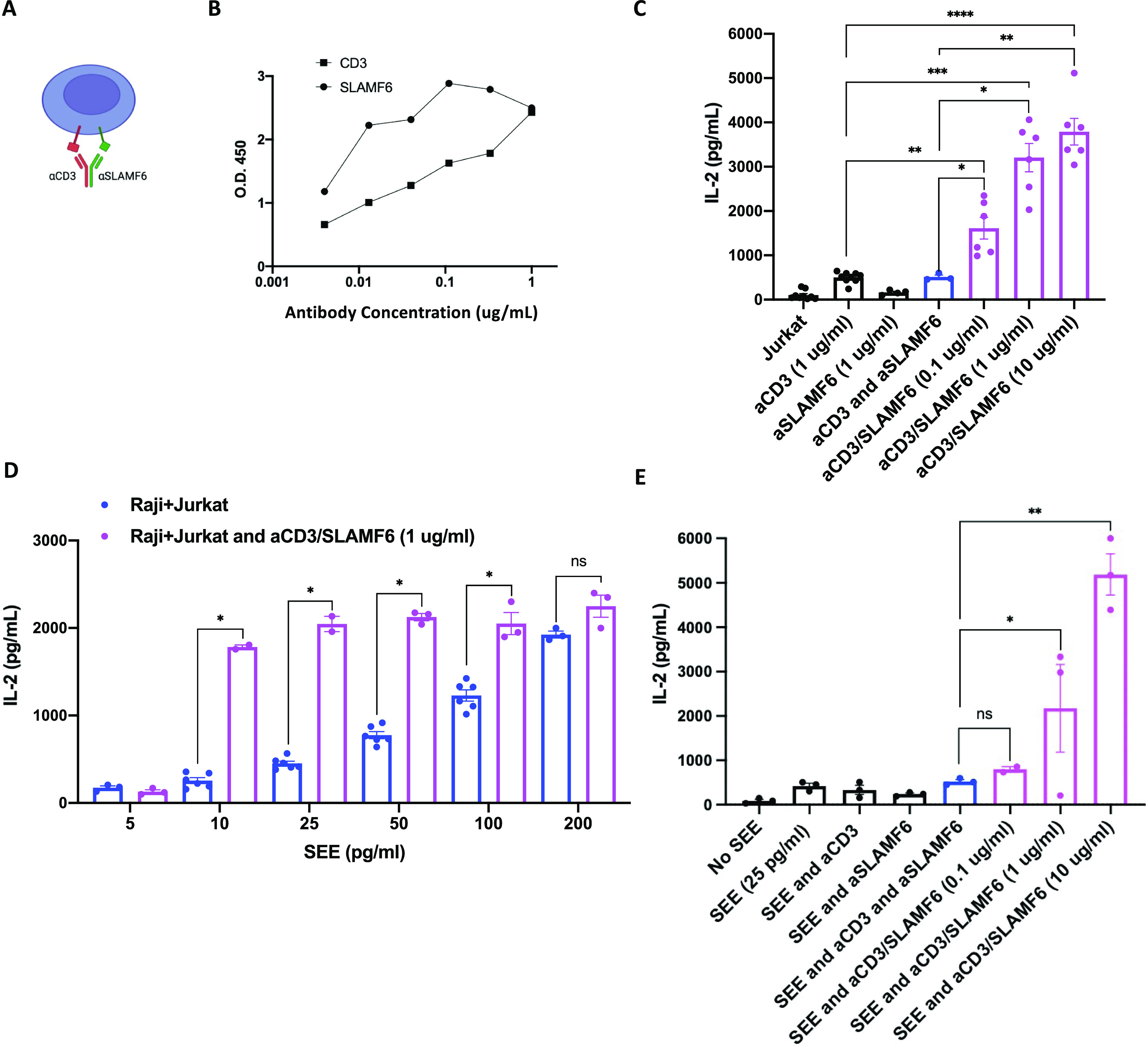
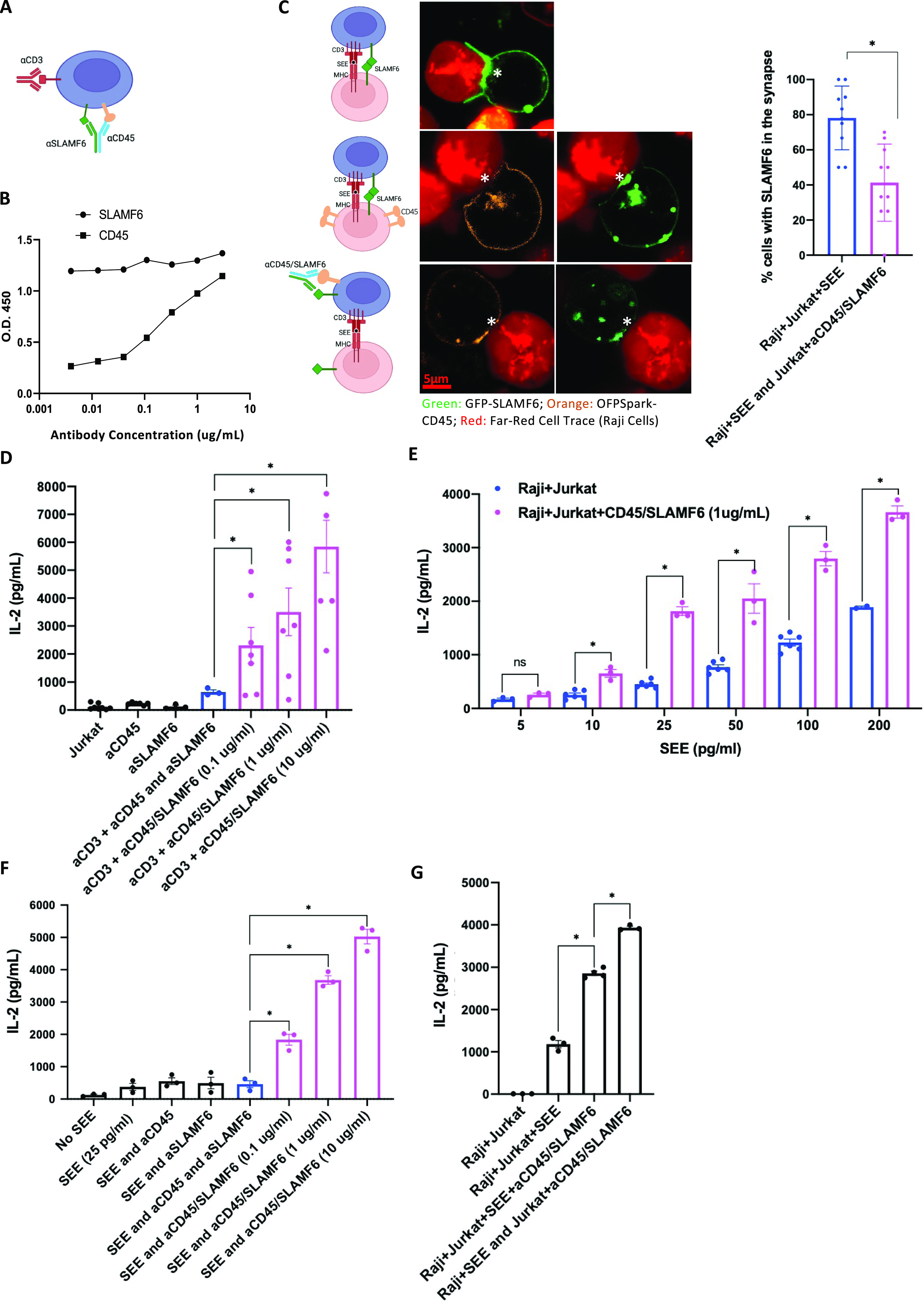
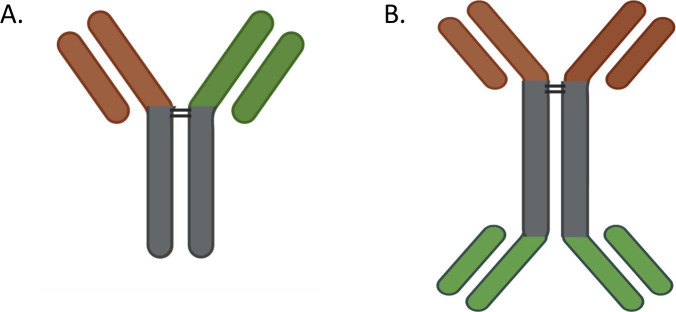
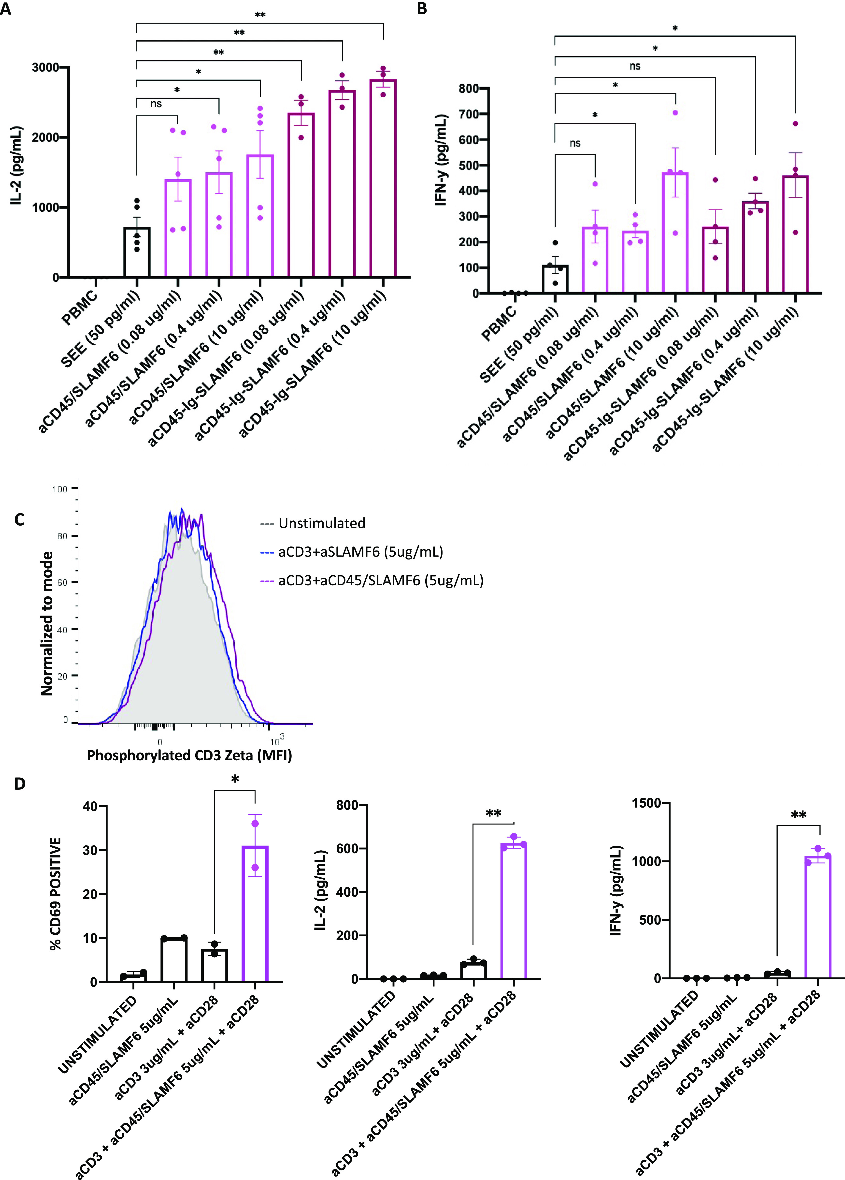
Signaling lymphocyte activation molecule family member 6 (SLAMF6) is a T cell co-receptor. Previously, we showed that SLAMF6 clustering was required for T cell activation. To better understand the relationship between SLAMF6 location and function and to evaluate the role of SLAMF6 as a therapeutic target, we investigated how its compartmentalization on the cell surface affects T cell functions. We used biochemical and co-culture assays to show that T cell activity is enhanced when SLAMF6 colocalizes with the CD3 complex. Mechanistically, co-immunoprecipitation analysis revealed the SLAMF6-interacting proteins to be those essential for signaling downstream of T cell receptor, suggesting the two receptors share downstream signaling pathways. Bispecific anti-CD3/SLAMF6 antibodies, designed to promote SLAMF6 clustering with CD3, enhanced T cell activation. Meanwhile, anti-CD45/SLAMF6 antibodies inhibited SLAMF6 clustering with T cell receptor, likely because of the steric hindrance, but nevertheless enhanced T cell activation. We conclude that SLAMF6 bispecific antibodies have a role in modulating T cell responses, and future work will evaluate the therapeutic potential in tumor models.


Conflict of interest statement

The authors declare that they have no conflict of interest.

Figures

Figure 1.
Figure 1.. Separation of SLAMF6 and CD3 inhibits T cell proliferation.
(A) A schematic representation of the assays used to study SLAMF6 compartmentation. T cells were stimulated with either immobilized anti-CD3 (αCD3) and anti-SLAMF6 (αSLAMF6) on a plate surface (top) or immobilized αCD3 but soluble αSLAMF6 (bottom). (B) Freshly isolated primary CD3+ T cells were stained with CFSE then cultured in the presence of plate-coated αCD3 or plate-coated αCD3 + αSLAMF6 or plate-coated αCD3 + soluble αSLAMF6. After 120 h, the cells were assayed for FITC fluorescence for three independent experiments (n = 3). The data were analyzed for percent (%) of proliferating cells as depicted (black line); plate-coated αCD3 + αSLAMF6 resulted in greater proliferation as compared with αCD3 alone, whereas addition of soluble αSLAMF6 inhibited proliferation. (C) CD25 and PD-1 expression at 120 h was analyzed using flow cytometry. (D, E) Freshly isolated primary CD3 T cells were cultured in the presence of plate-coated αCD3 + αSLAMF6 or plate-coated αCD3 + soluble αSLAMF6. (D, E) The supernatant was harvested and IL-2 levels at different time intervals over 96 h and (E) IFN-y levels at 48 h were analyzed by ELISA. (F) Jurkat T cells were stimulated in the presence of brefeldin for 6 h, after which time intracellular IL-2 was analyzed by flow cytometry. This experiment was repeated twice (n = 2). (G) Freshly isolated primary CD3 T cells were cultured as above for 120 h. Cell differentiation was analyzed based on cell surface expression of CD45RA and CCR7. A weighed T cell maturation index was calculated as (1*Naïve + 2*Central Memory + 3*Effector Memory + 4*Terminal Effector Memory)/4. This experiment was repeated twice with the average value shown here. (H) Cell number was assessed by automated cell counting every 24 h. The experiment was done in triplicate (n = 3). *P ≤ 0.05 for an unpaired t test.
Figure S1.
Figure S1.. Gating strategy for flow cytometry.
(A, B) T cells were identified on the FSC–SSC plot followed by (B) selection of single cells. (C) Live cells were identified as those that did not take up UV Zombie dye. (D) CD4 and CD8 T cell populations were identified. For all subsequent markers, FMOs were used to identify the negative populations.
Figure 2.
Figure 2.. SLAMF6 and TCR signaling complexes share key mediators.
V5-SLAMF6-expressing Jurkat T cells were stimulated with plate-coated αCD3 + αSLAMF6 (plate) or plate-coated αCD3 + soluble αSLAMF6 (soluble) for 15 min. The cells were then lysed and the lysate mixed with V5-coupled agarose beads to enrich for V5-tagged SLAMF6 immunoprecipitation. Pull-down lysate proteins were separated by electrophoresis and submitted for mass spectrometry analysis. (A) Protein enrichment pathway analysis for the plate versus soluble stimulation conditions was performed. (B) Proteins interacting with SLAMF6 were identified, listed by peptide-spectrum match score. (C) A schematic demonstrating that the SLAMF6-interacting proteins identified in the immunoprecipitation (marked by a purple star) are known to be essential for proximal TCR signal transduction, emphasizing the interconnection between the two receptors and their signaling interactome. Three independent experimental repeats were performed. (n = 3).
Figure S2.
Figure S2.. Protein–protein interactions downstream of SLAMF6 activation differ based on stimulation condition.
V5-SLAMF6-expressing Jurkat T cells were stimulated with plate-coated αCD3 + αSLAMF6 (plate) or plate-coated αCD3 + soluble αSLAMF6 (soluble) for 15 min. The cells were then lysed and the lysate mixed with V5-coupled agarose beads to enrich for V5-tagged SLAMF6 immunoprecipitation. Pull-down lysate proteins were separated by electrophoresis and submitted for mass spectrometry analysis. (A, B) Protein–protein interaction identified prediction models of kinases involved downstream of SLAMF6 signaling in the plate (A) and soluble (B) conditions. Kinases that were validated in the pull-down are shown in red.
Figure 3.
Figure 3.. SLAMF6 clustering in the immunologic synapse (IS) enhances cytokine secretion.
(A) A schematic representation of SLAMF6 in the absence (top) or presence (bottom) of homophilic receptor ligation in a T-B cell co-culture. (B) Jurkat T cells were transfected via nucleofection with LifeAct mCherry and SLAMF6 GFP, then co-cultured with Raji B Cell APCs. Images are representative of at least 30 cells from a least two independent experiments. Scale bar is 5 μm. Actin clearance was defined as a mature IS. (C) Jurkat T cells were treated with αCD3-conjugated beads and αSLAMF6-conjugated beads versus αCD3 + αSLAMF6–conjugated beads (both antibodies on the same bead). After 24 h, the supernatant was harvested and IL-2 levels were analyzed by ELISA for at least three independent experiments (n = 3). (D) Jurkat T cells were treated with αCD3 + αSLAMF6 or αCD3 + αSLAMF6 + cross-linker for 24 h, after which the supernatant was harvested and IL-2 levels were analyzed by ELISA for at least three independent experiments (n = 3). *P < 0.05 for an unpaired t test.
Figure 4.
Figure 4.. Anti-CD3/SLAMF6–bispecific antibody clusters SLAMF6 to the CD3 and augments T cell activation.
(A) A schematic representation of anti-CD3/SLAMF6–bispecific antibody (aCD3/SLAMF6) binding and clustering the two receptors together. (B) aCD3/SLAMF6 antibody binding was validated using an ELISA assay: αCD3 binding was assessed against immobilized SLAMF6 KO Jurkat T cells, whereas anti-SLAMF6 binding was assessed against immobilized Raji B cells. (C) Jurkat T cells were treated with αCD3, αCD3 + soluble αSLAMF6, or aCD3/SLAMF6 at three different concentrations of the bispecific antibody: 0.1, 1, and 10 µg/ml. After 24 h, IL-2 levels were analyzed by ELISA for at least two independent experiments (n = 2). (D) Raji B cells were preloaded with increasing concentrations of SEE and co-cultured with Jurkat T cells in the absence (blue) or presence (magenta) of 1 µg/ml of aCD3/SLAMF6 antibody. IL-2 levels were analyzed for at least two independent experiments (n = 2). (E) Raji B cells were preloaded with SEE and co-cultured with Jurkat T cells at increasing concentrations of the αCD3/SLAMF6 antibody. IL-2 levels were analyzed for at least two independent experiments (n = 2). *P < 0.05, **P < 0.01, ***P < 0.001 for an unpaired t test.
Figure 5.
Figure 5.. Anti-CD45/SLAMF6–bispecific antibody inhibits SLAMF6 enrichment in the synapse but still augments T cell activation.
(A) A schematic representation of anti-CD45/SLAMF6 (CD45/SLAMF6) binding to inhibit SLAMF6 clustering with CD3 in the IS. (B) αCD45/SLAMF6 antibody binding was quantified using an ELISA assay: αCD45 and αSLAMF6 binding was assessed against immobilized, recombinant ectodomains of CD45 and SLAMF6, respectively. (C) Jurkat T cells were transfected with GFP-tagged SLAMF6, and Raji B cells were stained with LifeAct Far Red and preloaded with SEE (2 ng/ml). Jurkat T cells were then co-cultured with Raji B cells for 30 min. Synapse formation with enrichment of SLAMF6 in the IS was visualized (top row). To visualize the distribution of CD45, we next transfected the Jurkat T cells with OFPSpark-tagged CD45 and GFP-tagged SLAMF6. In the Jurkat T–Raji B co-cultures, exclusion of CD45 was coupled with enrichment of SLAMF6 in the IS (middle row). Finally, we pretreated Jurkat T cells with anti-CD45/SLAMF6 10 µg/ml for 15 min (bottom image). Exclusion of CD45 was now associated with a lack of enrichment of SLAMF6 in the IS (bottom row). Images are representative of at least 40 cell conjugates per each experimental condition from two independent experiments. Scale bar is 5 μm. Percent of cell conjugates with SLAMF6 enrichment in the IS was quantified; results are summarized in the bar graph. (D) Jurkat T cells were treated with αCD3 and αCD45/SLAMF6 at three different concentrations: 0.1, 1, and 10 µg/ml of the bispecific antibody. After 24 h, IL-2 levels were analyzed by ELISA. (E) Raji B cells were preloaded with different concentrations of SEE and co-cultured with Jurkat T cells in the absence (blue) or presence (magenta) of 1 µg/ml of αCD45/SLAMF6 antibody. IL-2 levels were analyzed for at least three independent experiments (n = 3). (F) Raji B cells were preloaded with SEE and co-cultured with T cells at increasing concentrations of αCD45/SLAMF6 antibody. IL-2 levels were analyzed for at least three independent experiments (n = 3). (G) Raji B cells and Jurkat T cells were co-cultured either in the presence of, or after T cell pretreatment with, αCD45/SLAMF6. Specifically, in the first experimental condition, αCD45/SLAMF6 was added as Jurkat T–Raji B conjugates formed (supporting in trans antibody ligation), whereas in the second experimental condition, Jurkat T cells were pretreated with αCD4/SLAMF6 for 30 min, washed, and subsequently co-cultured with the Raji B cells, supporting in cis antibody ligation on T cells before addition of the B cells. IL-2 levels were analyzed for at least three independent experiments (n = 3). *P < 0.05 for an unpaired t test.
Figure S3.
Figure S3.. Electrophoresis confirming purity of synthesized antibodies.
Bispecific antibodies, anti-CD3/SLAMF6 and anti-CD45/SLAMF6, were supplemented with 1 M HEPS buffer, and purity was determined by running PAGE gel against 1 µg of BSA as a control.
Figure S4.
Figure S4.. Bispecific antibodies.
(A, B) Schematic of (A) monovalent and (B) bivalent bispecific antibodies.
Figure 6.
Figure 6.. Anti-CD45-SLAMF6 antibody enhances T cell response in primary human cell–based assay.
PBMCs were treated with SEE and three different concentrations of either the monovalent (αCD45/SLAMF6) or the bivalent (αCD45-Ig-SLAMF6) anti-CD45/SLAMF6–bispecific antibody. (A, B) After 24 h, (A) IL-2 and (B) IFN-γ levels were measured by ELISA. The results of at least three independent experiments (n = 3) are shown. (C, D) Primary human CD3-positive T cells were isolated from whole blood and cultured with anti-CD3 in the presence αCD45/SLAMF6. (C) After 5 min, the cells were analyzed for phosphorylation of CD3 ζ chain using flow cytometry. (D) After 24 h, the cells were analyzed for CD69 expression, and the supernatant was analyzed for IL-2 and IFN-y release. *P < 0.05, **P < 0.01 for an unpaired t test.

References

    1. Eisenberg G, Engelstein R, Geiger-Maor A, Hajaj E, Merims S, Frankenburg S, Uzana R, Rutenberg A, Machlenkin A, Frei G, et al. (2018) Soluble SLAMF6 receptor induces strong CD8(+) T-cell effector function and improves anti-melanoma activity in vivo. Cancer Immunol Res 6: 127–138. 10.1158/2326-6066.cir-17-0383 - DOI - PubMed
    1. Schenkel JM, Herbst RH, Canner D, Li A, Hillman M, Shanahan SL, Gibbons G, Smith OC, Kim JY, Westcott P, et al. (2021) Conventional type I dendritic cells maintain a reservoir of proliferative tumor-antigen specific TCF-1(+) CD8(+) T cells in tumor-draining lymph nodes. Immunity 54: 2338–2353.e6. 10.1016/j.immuni.2021.08.026 - DOI - PMC - PubMed
    1. Yigit B, Wang N, Ten Hacken E, Chen SS, Bhan AK, Suarez-Fueyo A, Katsuyama E, Tsokos GC, Chiorazzi N, Wu CJ, et al. (2019) SLAMF6 as a regulator of exhausted CD8(+) T cells in cancer. Cancer Immunol Res 7: 1485–1496. 10.1158/2326-6066.cir-18-0664 - DOI - PubMed
    1. Hajaj E, Eisenberg G, Klein S, Frankenburg S, Merims S, Ben David I, Eisenhaure T, Henrickson SE, Villani AC, Hacohen N, et al. (2020) SLAMF6 deficiency augments tumor killing and skews toward an effector phenotype revealing it as a novel T cell checkpoint. Elife 9: e52539. 10.7554/elife.52539 - DOI - PMC - PubMed
    1. Valdez PA, Wang H, Seshasayee D, van Lookeren Campagne M, Gurney A, Lee WP, Grewal IS (2004) NTB-A, a new activating receptor in T cells that regulates autoimmune disease. J Biol Chem 279: 18662–18669. 10.1074/jbc.m312313200 - DOI - PubMed

Publication types

MeSH terms

Substances