. 2023 Jan 17;120(3):e2215071120.
doi: 10.1073/pnas.2215071120. Epub 2023 Jan 9.

Repeat-associated non-AUG translation induces cytoplasmic aggregation of CAG repeat-containing RNAs

Affiliations

Repeat-associated non-AUG translation induces cytoplasmic aggregation of CAG repeat-containing RNAs

Michael R Das et al. Proc Natl Acad Sci U S A. .

Abstract

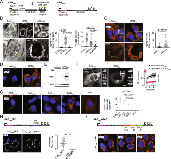
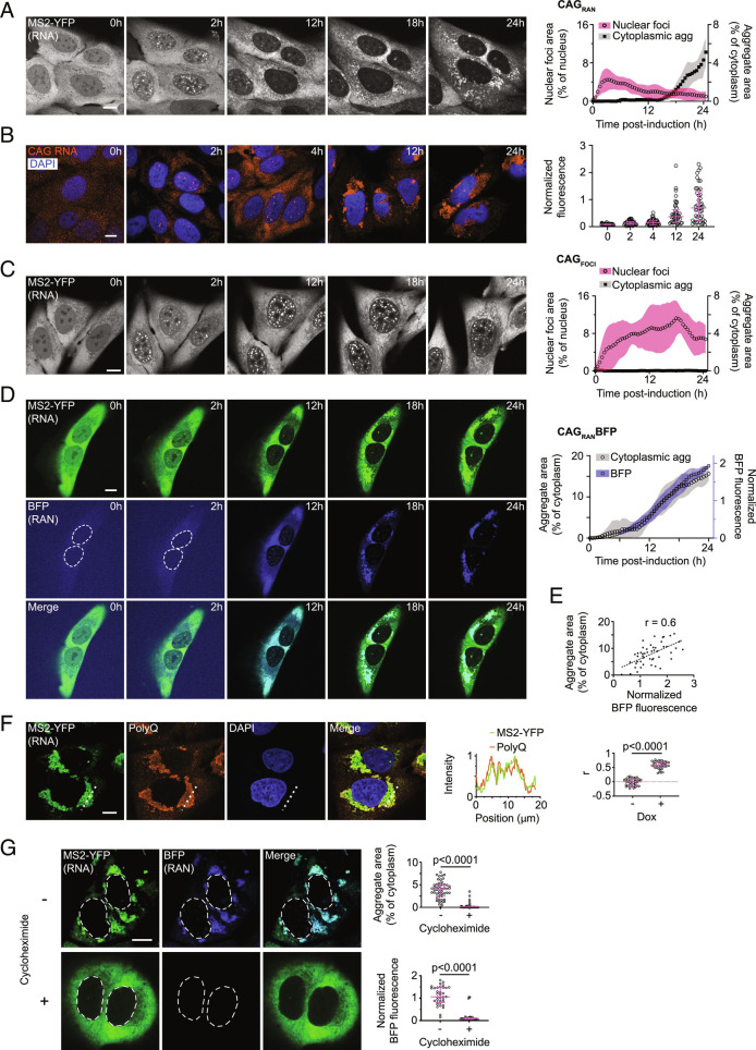
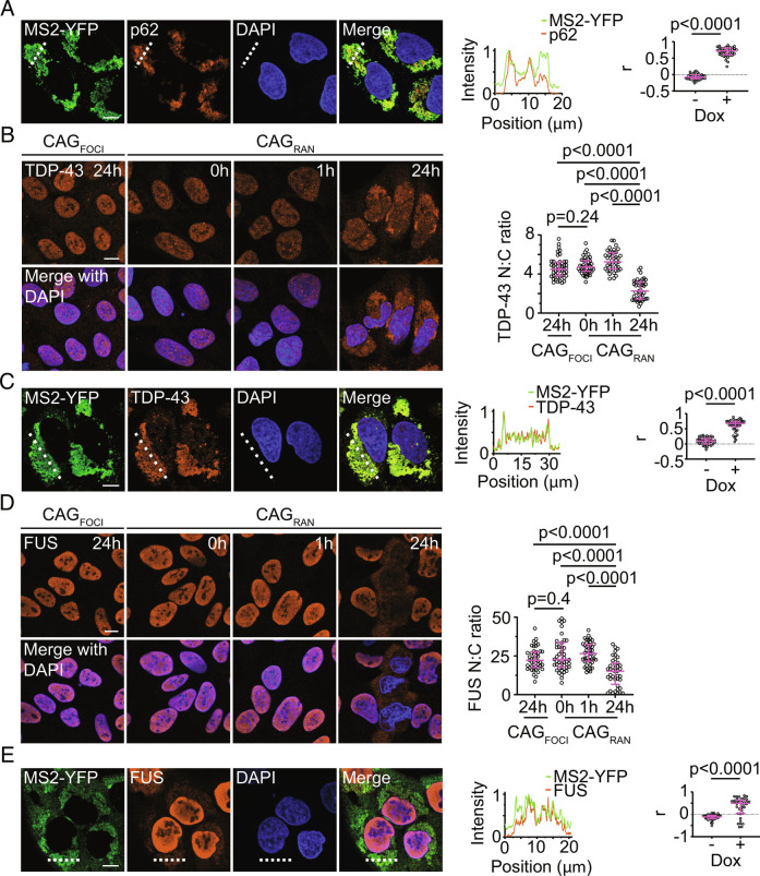
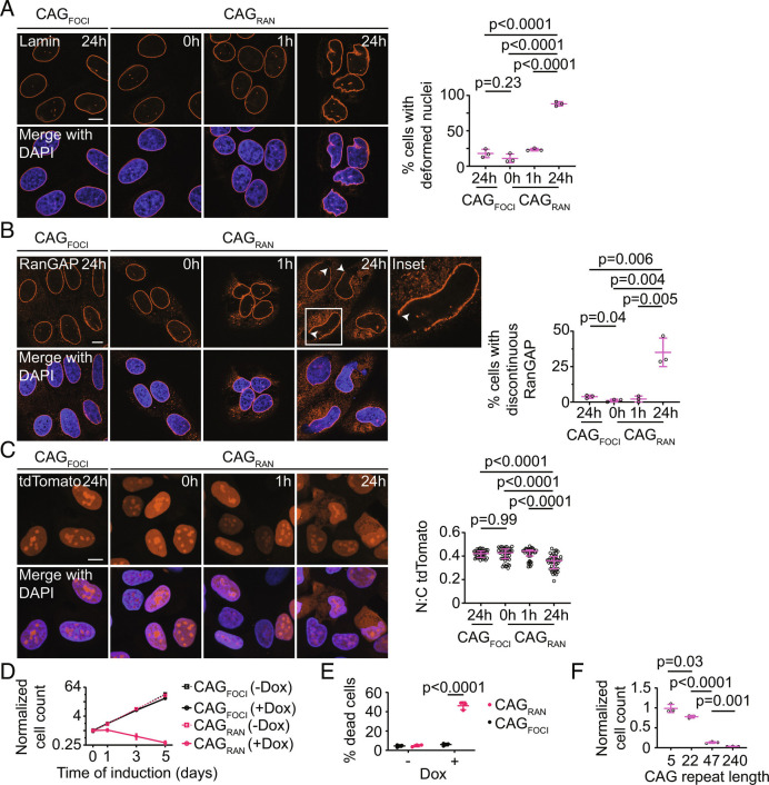
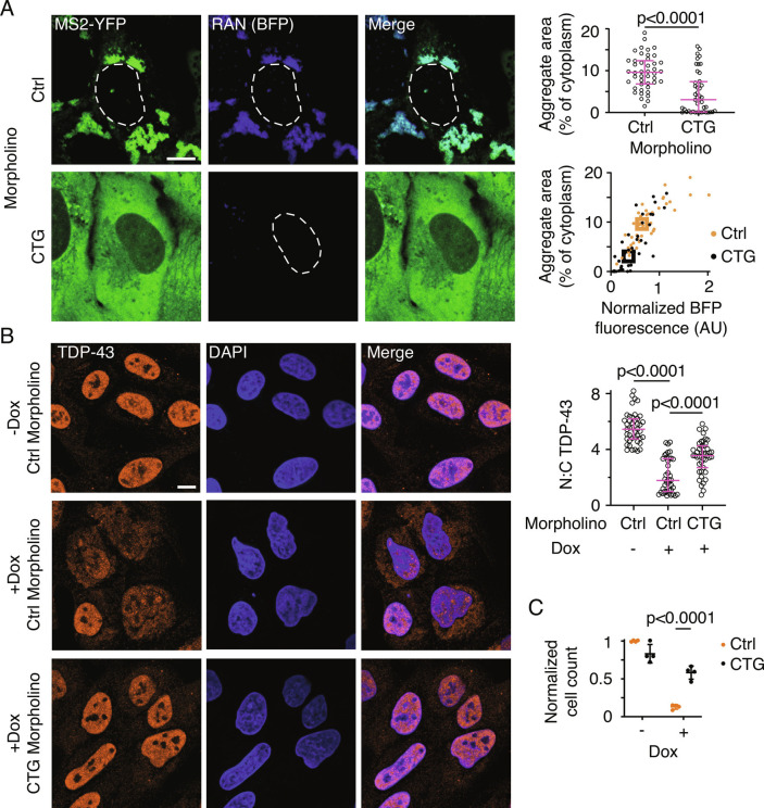
CAG trinucleotide repeat expansions cause several neurodegenerative diseases, including Huntington's disease and spinocerebellar ataxia. RNAs with expanded CAG repeats contribute to disease in two unusual ways. First, these repeat-containing RNAs may agglomerate in the nucleus as foci that sequester several RNA-binding proteins. Second, these RNAs may undergo aberrant repeat-associated non-AUG (RAN) translation in multiple frames and produce aggregation-prone proteins. The relationship between RAN translation and RNA foci, and their relative contributions to cellular dysfunction, are unclear. Here, we show that CAG repeat-containing RNAs that undergo RAN translation first accumulate at nuclear foci and, over time, are exported to the cytoplasm. In the cytoplasm, these RNAs are initially dispersed but, upon RAN translation, aggregate with the RAN translation products. These RNA-RAN protein agglomerates sequester various RNA-binding proteins and are associated with the disruption of nucleocytoplasmic transport and cell death. In contrast, RNA accumulation at nuclear foci alone does not produce discernable defects in nucleocytoplasmic transport or cell viability. Inhibition of RAN translation prevents cytoplasmic RNA aggregation and alleviates cell toxicity. Our findings demonstrate that RAN translation-induced RNA-protein aggregation correlates with the key pathological hallmarks observed in disease and suggest that cytoplasmic RNA aggregation may be an underappreciated phenomenon in CAG trinucleotide repeat expansion disorders.

Keywords: RAN translation; RNA aggregation; RNA localization; Repeat expansion diseases.


Conflict of interest statement

The authors declare no competing interest.

Figures

Fig. 1.
Fig. 1.
Sequence context modulates the localization and RAN translation of CAG repeat-containing RNAs. A, Schematics for CAGRAN and CAGFOCI constructs. B and C, Representative micrographs of cells expressing the indicated constructs imaged using MS2–YFP (B, Left) or via FISH probes against the MS2 tag (C, Left), and corresponding quantification of the aggregate and foci areas (B and C, Right). Boxes show regions used for Insets. D and E, Immunofluorescence micrographs (D) and immunoblot (E) for indicated samples using polyglutamine (polyQ) antibody. F, Representative micrographs before (pre) and at the indicated timepoints after photobleaching for nuclear foci of CAGFOCI (Left) and cytoplasmic inclusions of CAGRAN (Middle) and corresponding fluorescence recovery after photobleaching (FRAP) trajectories (Right). G, Fluorescence micrographs of cells transfected with the indicated constructs and stained using a FISH probe against the CAG repeats (Left) and corresponding quantification of the percentage of cells with cytoplasmic RNA aggregates (Right). H, Schematic for CAGRANBFP construct. Asterisk denotes the location of stop codons in CAGRANSTOP-BFP construct (Top). Representative fluorescence micrographs 24 h post-induction (Bottom, Left), and quantification of BFP fluorescence, normalized to cell area (Bottom, Right). I, Schematic for CAGRAN3xTag (Top) and representative immunofluorescence micrographs 24 h post-induction staining for the indicated tags (Bottom). Micrographs in F are representative of ≥20 independent photobleaching events; all other micrographs are representative of ≥40 cells across ≥2 independent experiments. Each data point in B, C, and H represents a single cell. Error bars denote median ± interquartile range. Data in G are summarized as mean ± SD over 3 independent experiments. Significance values were calculated using Mann–Whitney tests in B, C, F, and H and using Student’s t test in G. Micrographs were independently scaled in B, C, F, and G. Where indicated, cell nuclei are stained with DAPI. (Scale bars, 10 µm.)
Fig. 2.
Fig. 2.
Cytoplasmic RNA aggregation follows accumulation at foci and coincides with RAN translation. A, Representative micrographs of cells expressing CAGRAN at the stated timepoints after induction (Left) and corresponding quantification (Right) of nuclear (circles, pink shading, left y-axis) and cytoplasmic (squares, gray shading, right y-axis) inclusions. B, Similar to A, imaged via FISH probes against the CAG repeats (Left) and corresponding quantification of the FISH signal (Right). C, Similar to A except for cells expressing CAGFOCI. D, Representative micrographs of cells expressing CAGRANBFP at the stated timepoints after induction imaged in MS2–YFP (Top) and BFP (Middle) channels, and their overlay (Bottom). Plot to the right shows the cytoplasmic aggregate area (circles, gray shading) and BFP fluorescence (squares, blue shading). E, Quantification of BFP fluorescence in relation to cytoplasmic RNA aggregate area. Dashed line represents a linear regression fit. F, Representative immunofluorescence micrographs showing CAGRAN RNA and polyQ-containing proteins. Intensity profile of MS2–YFP and polyQ signal along the dashed line in the micrographs (Middle). Correlation between MS2–YFP and polyQ-immunofluorescence signals (Right). G, Micrographs of cells expressing CAGRANBFP without (−) or with 10 µg/mL cycloheximide (+) treatment for 14 h (Left). Corresponding quantification of RNA aggregate area (Right, Top) and of BFP fluorescence (Right, Bottom). In A, C, and D, each data point is the mean of ≥4 cells and is a rolling average of three successive timepoints, shaded regions depict SD, and data are representative of ≥4 experiments with ≥25 cells. Each data point in B, E, F, and G represents a single cell and micrographs are representative of ≥2 independent experiments with ≥40 cells. Fluorescence in B, D, E, and G is normalized to cell area. Data in B, F, and G are summarized as median ± interquartile range and significance values were calculated by Mann–Whitney tests. Images in B and D and MS2–YFP images in G are independently scaled. Where indicated, cell nuclei are stained with DAPI. (Scale bars, 10 µm.)
Fig. 3.
Fig. 3.
RNA–RAN protein aggregates recruit nuclear RNA-binding proteins. A, Representative immunofluorescence micrographs showing colocalization between CAGRAN RNA and p62 (Left), and intensity profile of the two channels along the dashed line (Middle). Correlation between MS2–YFP and p62 signals with (+) and without (−) RNA induction (Right). B, Representative immunofluorescence micrographs showing TDP-43 in cells expressing CAGFOCI or CAGRAN for the stated times (Left). Quantification of the ratio of nuclear to cytoplasmic TDP-43 (Right). C, Similar to A except staining for TDP-43 instead of p62. D, Similar to B except staining for FUS instead of TDP-43. E, Similar to A except staining for FUS instead of p62. Cell nuclei are stained with DAPI. Data are representative of ≥2 independent experiments with ≥40 cells. Each data point represents a single cell and data are summarized as median ± interquartile range. Significance values were calculated by Mann–Whitney tests. (Scale bars, 10 µm.)
Fig. 4.
Fig. 4.
RNA–RAN protein aggregates disrupt nucleocytoplasmic transport and induce cell death. A and B, Representative immunofluorescence micrographs showing Lamin (A) or RanGAP1 (B) in cells expressing CAGRAN or CAGFOCI for the stated times (Left). Quantification of cells with deformed nuclei (A, Right) and discontinuous RanGAP1 staining (B, Right). Each data point represents an independent experiment (≥3 experiments) evaluating ≥40 cells. C, Representative micrographs of NLS-tdTomato-NES nuclear transport reporter in fixed cells expressing CAGRAN or CAGFOCI for the stated times (Left), and corresponding quantification of the nuclear to cytoplasmic tdTomato fluorescence (Right). Each data point represents a single cell and data are summarized as the median ± interquartile range, n ≥ 40 cells. Significance values were calculated by Mann–Whitney U tests. D, Quantification of cell populations expressing CAGRAN or CAGFOCI over 5 d, normalized to population size at day 0 (immediately prior to induction). Each data point is the mean of three biological replicates, and error bars denote SD. E, Quantification of cell death caused by expression of CAGRAN or CAGFOCI over 3 d, as measured by trypan blue staining. F, Quantification of cell populations expressing CAGRAN with varying number of CAG repeats 5 d post-induction. Cell counts are normalized to uninduced controls. Each data point in E and F represents a separate biological replicate, n ≥ 3. In A, B, E, and F, data are summarized as mean ± SD and significance values were calculated by Student’s t tests. Where indicated, cell nuclei are stained with DAPI. (Scale bars, 10 µm.)
Fig. 5.
Fig. 5.
Inhibition of RAN translation prevents cell toxicity and nuclear transport defects. A, Representative micrographs of cells expressing CAGRANBFP for 24 h and treated with 50 µM of a control (Ctrl) or 8×CTG morpholino (CTG) for 48 h (Left). MS2–YFP images are independently scaled. Quantification of aggregate area (Right, Top) and in relation to BFP fluorescence (Right, Bottom). Data are representative of ≥3 independent experiments and ≥40 cells. B, Representative immunofluorescence micrographs showing TDP-43 localization in cells treated with indicated morpholinos for 48 h, with (+Dox) or without (−Dox) CAGRAN induction (Left), and corresponding quantification of the ratio of nuclear to cytoplasmic TDP-43 (Right). Cell nuclei are stained with DAPI. Data are representative of ≥40 cells. C, Quantification of cell survival 4 d post CAGRAN induction after treatment with the indicated morpholinos. Each data point represents a separate biological replicate, n = 4. Each data point in A and B represents a single cell; bar graph data are summarized as median ± interquartile range and boxes in the scatterplot in A represent the median values for each population. Significance values in A and B were calculated by Mann–Whitney U tests. Data in C are summarized as mean ± SD and the significance value was calculated by Student’s t test. (Scale bars, 10 µm.)

References

    1. Malik I., Kelley C. P., Wang E. T., Todd P. K., Molecular mechanisms underlying nucleotide repeat expansion disorders. Nat. Rev. Mol. Cell Biol. 22, 589–607 (2021). - PMC - PubMed
    1. Paulson H., “Repeat expansion diseases” in Handbook of Clinical Neurology, (Elsevier, 2018), pp. 105–123. - PMC - PubMed
    1. Zu T., et al. , Non-ATG-initiated translation directed by microsatellite expansions. Proc. Natl. Acad. Sci. U.S.A. 108, 260–265 (2011). - PMC - PubMed
    1. Jiang H., Mankodi A., Swanson M. S., Moxley R. T., Thornton C. A., Myotonic dystrophy type 1 is associated with nuclear foci of mutant RNA, sequestration of muscleblind proteins and deregulated alternative splicing in neurons. Hum. Mol. Genet. 13, 3079–3088 (2004). - PubMed
    1. Wojciechowska M., Krzyzosiak W. J., Cellular toxicity of expanded RNA repeats: Focus on RNA foci. Hum. Mol. Genet. 20, 3811–3821 (2011). - PMC - PubMed

Publication types

MeSH terms