. 2023 Apr;51(4):427-435.
doi: 10.1124/dmd.122.000975. Epub 2023 Jan 9.

Relevance of Human Aldoketoreductases and Microbial β-Glucuronidases in Testosterone Disposition

Affiliations

Relevance of Human Aldoketoreductases and Microbial β-Glucuronidases in Testosterone Disposition

Abdul Basit et al. Drug Metab Dispos. 2023 Apr.

Abstract

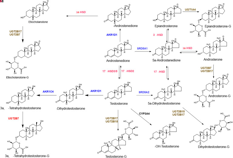
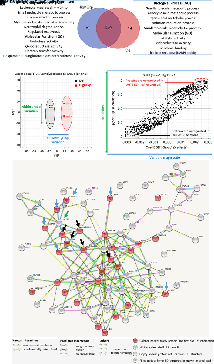
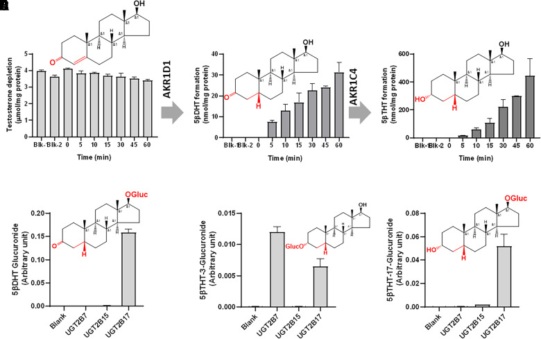
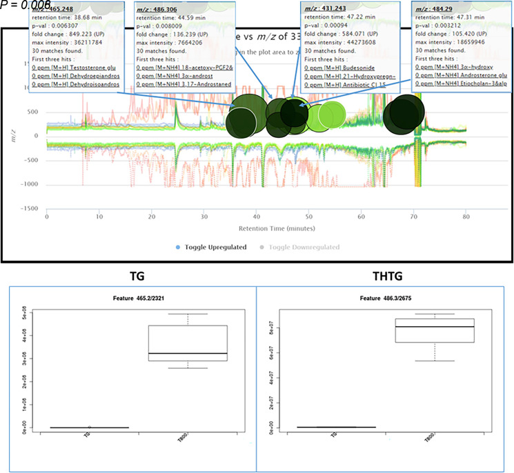
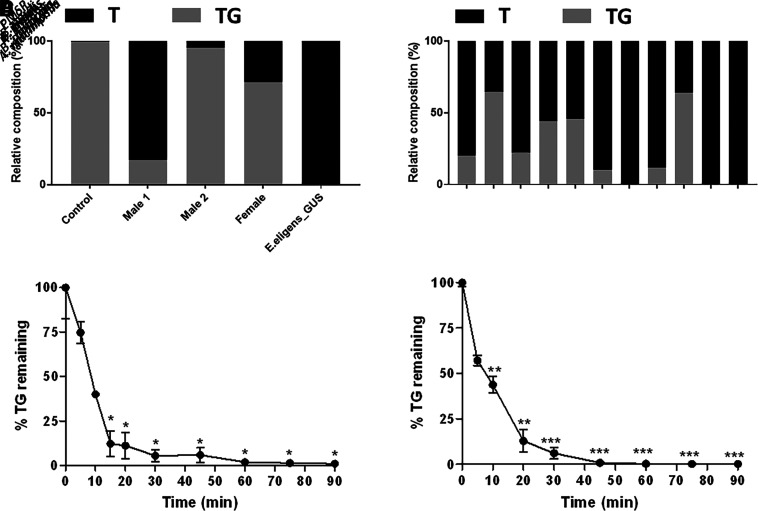
Testosterone exhibits high variability in pharmacokinetics and glucuronidation after oral administration. Although testosterone metabolism has been studied for decades, the impact of UGT2B17 gene deletion and the role of gut bacterial β-glucuronidases on its disposition are not well characterized. We first performed an exploratory study to investigate the effect of UGT2B17 gene deletion on the global liver proteome, which revealed significant increases in proteins from multiple biological pathways. The most upregulated liver proteins were aldoketoreductases [AKR1D1, AKR1C4, AKR7A3, AKR1A1, and 7-dehydrocholesterol reductase (DHCR7)] and alcohol or aldehyde dehydrogenases (ADH6, ADH1C, ALDH1A1, ALDH9A1, and ALDH5A). In vitro assays revealed that AKR1D1 and AKR1C4 inactivate testosterone to 5β-dihydrotestosterone (5β-DHT) and 3α,5β-tetrahydrotestosterone (3α,5β-THT), respectively. These metabolites also appeared in human hepatocytes treated with testosterone and in human serum collected after oral testosterone dosing in men. Our data also suggest that 5β-DHT and 3α, 5β-THT are then eliminated through glucuronidation by UGT2B7 in UGT2B17 deletion individuals. Second, we evaluated the potential reactivation of testosterone glucuronide (TG) after its secretion into the intestinal lumen. Incubation of TG with purified gut microbial β-glucuronidase enzymes and with human fecal extracts confirmed testosterone reactivation into testosterone by gut bacterial enzymes. Both testosterone metabolic switching and variable testosterone activation by gut microbial enzymes are important mechanisms for explaining the disposition of orally administered testosterone and appear essential to unraveling the molecular mechanisms underlying UGT2B17-associated pathophysiological conditions. SIGNIFICANCE STATEMENT: This study investigated the association of UGT2B17 gene deletion and gut bacterial β-glucuronidases with testosterone disposition in vitro. The experiments revealed upregulation of AKR1D1 and AKR1C4 in UGT2B17 deletion individuals, and the role of these enzymes to inactivate testosterone to 5β-dihydrotestosterone and 3α, 5β-tetrahydrotestosterone, respectively. Key gut bacterial species responsible for testosterone glucuronide activation were identified. These data are important for explaining the disposition of exogenously administered testosterone and appear essential to unraveling the molecular mechanisms underlying UGT2B17-associated pathophysiological conditions.


Figures

Fig. 1.
Fig. 1.
Testosterone metabolism scheme in the gene deletion and the high expressers of UGT2B17.
Fig. 2.
Fig. 2.
Untargeted proteomics data of the postmitochondrial (S9) fractions isolated from the liver tissue of the UGT2B17 deletion subjects (Del) versus UGT2B17 high expressers (HighExp). The Venn diagram represents the number of proteins identified in both the groups and the biological processes and molecular functions associated with significantly upregulated proteins (A). OPLS-DA analysis on the liver proteome shows clustering of individual samples between the deletion and the high-expresser groups (B). The S-plot from OPLS-DA model highlights the differentially expressed proteins in both the groups (C). Protein interactome analysis by STRING analysis of the upregulated proteins in UGT2B17 deletion (D). Significantly upregulated proteins in UGT2B17 gene deletion individuals are associated with aldoketoreductases [AKR1D1, AKR1C4, AKR1A1, AKR7A3, CBR4, and dehydrocholesterol reductase (DHCR)-7; indicated by blue arrows], aldehyde dehydrogenase pathways [aldehyde dehydrogenase (ALDH)-1A1, ALDH5A1, ALDH9A1, ADH1C, and ADH6; indicated by black arrows], and UGT2B7 (indicated by green arrow). The protein interactome of the downregulated proteins is presented in Supplemental Fig. 1.
Fig. 3.
Fig. 3.
In vitro testosterone metabolism by aldoketoreductases in liver cytosol samples from the UGT2B17 deletion subjects (A–C) and in the recombinant UGTs (D–F). Testosterone to 5β-DHT and 5β-THT conversion with time by AKR1D1 and AKR1C4 in the human liver cytosol obtained from the UGT2B17 deletion subjects (A–C). Blk-1 and Blk-2 samples do not conatin NADPH and the enzyme, respectively. In vitro glucuronidation of 5β-DHT and 5β-THT using the recombinant human UGT2B enzymes (D–F). Structures of testosterone, 5β-DHT, 3α,5β-THT, 5β-DHT-glucuronide, 3α,5β-THT-3-glucuronide, and 3α,5β-THT-17-glucuronide are shown in (A–F), respectively.
Fig. 4.
Fig. 4.
Metabolic cloud plot representing differentially elevated metabolites in the serum after oral testosterone dosing in men. The cloud plot represents 16 elevated features in human serum sample after oral 800 mg testosterone dose (T800) as compared with the predose (T0) sample (Supplemental Table 4), with P < 0.01, fold change > 35, and m/z range 200–600. Visualization represents P value by color intensity (more intense means lower P value) and the fold-change by the radius of the circle. The whisker-box plots show the elevated levels (mass spectrometry peak intensity) of TG (M+H, m/z 465.2482) and 3α,5β-THT-glucuronide (THTG; M+NH4, m/z 486.3060) after oral 800 mg testosterone administration (T800) as compared with the predose sample (T0).
Fig. 5.
Fig. 5.
In vitro bacterial GUS activity toward testosterone glucuronide deconjugation. GUS activity toward testosterone glucuronide in fecal samples at 1 µM TG (A). Deconjugation of TG to testosterone by GUS enzymes from various gut bacterial species at 20 µM TG concentration (B). TG disappearance with time in the presence of GUS enzymes from E. coli (C) and E. eligens (D) at 10 µM TG concentration. The experiments were performed in triplicates, where percent coefficient of variation (%CV) was within 30% as indicated in (C) and (D). TG disappearance rate [0 minutes versus individual timepoints; (C) and (D)] was compared using ANOVA followed by Dunnett’s T3 multiple comparisons test, with *P < 0.05; **P < 0.01; ***P < 0.001.

References

    1. Amory JK, Bremner WJ (2005) Oral testosterone in oil plus dutasteride in men: a pharmacokinetic study. J Clin Endocrinol Metab 90:2610–2617. - PubMed
    1. Balhara A, Basit A, Argikar UA, Dumouchel JL, Singh S, Prasad B (2021) Comparative Proteomics Analysis of the Postmitochondrial Supernatant Fraction of Human Lens-Free Whole Eye and Liver. Drug Metab Dispos 49:592–600. - PubMed
    1. Barski OA, Tipparaju SM, Bhatnagar A (2008) The aldo-keto reductase superfamily and its role in drug metabolism and detoxification. Drug Metab Rev 40:553–624. - PMC - PubMed
    1. Basit A, Amory JK, Prasad B (2018) Effect of Dose and 5α-Reductase Inhibition on the Circulating Testosterone Metabolite Profile of Men Administered Oral Testosterone. Clin Transl Sci 11:513–522. - PMC - PubMed
    1. Bhasin S, Cunningham GR, Hayes FJ, Matsumoto AM, Snyder PJ, Swerdloff RS, Montori VM (2006) Testosterone therapy in adult men with androgen deficiency syndromes: an endocrine society clinical practice guideline. J Clin Endocrinol Metab 91:1995–2010. - PubMed

Publication types