. 2023 Jan 9;14(1):116.
doi: 10.1038/s41467-022-35741-0.

Apremilast prevents blistering in human epidermis and stabilizes keratinocyte adhesion in pemphigus

Affiliations

Apremilast prevents blistering in human epidermis and stabilizes keratinocyte adhesion in pemphigus

Anna M Sigmund et al. Nat Commun. .

Erratum in

Abstract

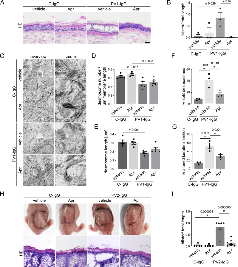
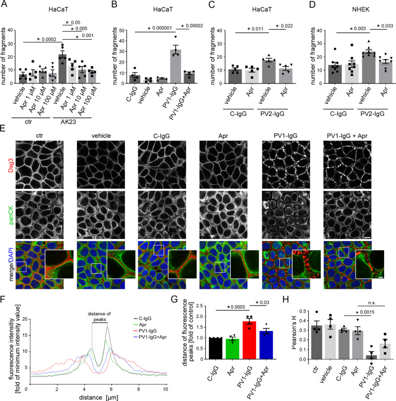
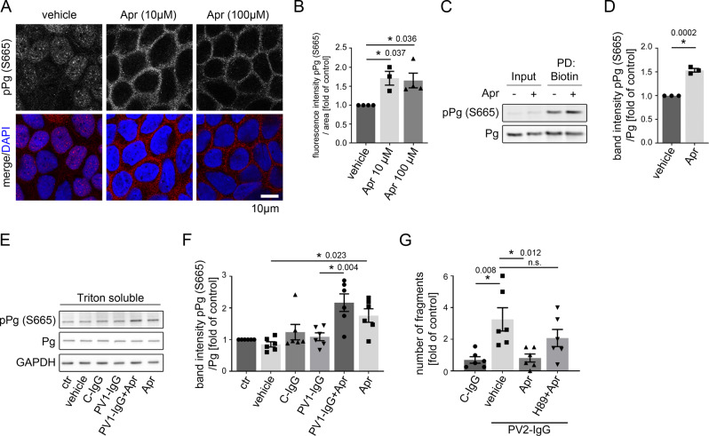
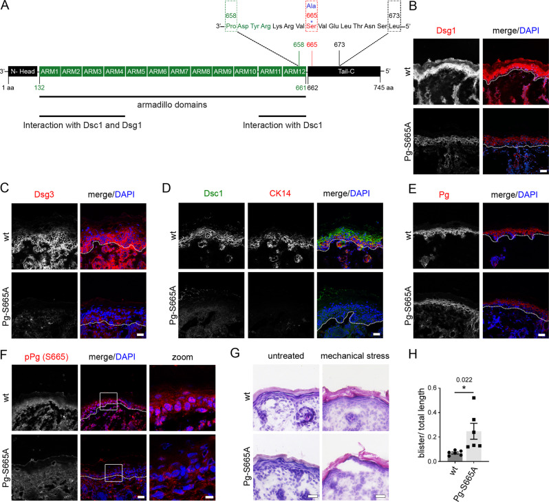
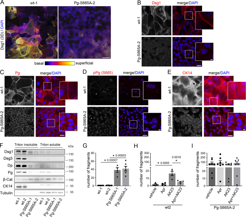
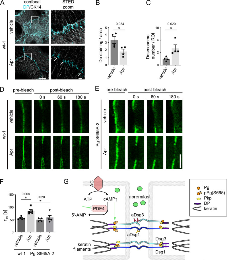
Pemphigus vulgaris is a life-threatening blistering skin disease caused by autoantibodies destabilizing desmosomal adhesion. Current therapies focus on suppression of autoantibody formation and thus treatments directly stabilizing keratinocyte adhesion would fulfill an unmet medical need. We here demonstrate that apremilast, a phosphodiesterase 4 inhibitor used in psoriasis, prevents skin blistering in pemphigus vulgaris. Apremilast abrogates pemphigus autoantibody-induced loss of keratinocyte cohesion in ex-vivo human epidermis, cultured keratinocytes in vitro and in vivo in mice. In parallel, apremilast inhibits keratin retraction as well as desmosome splitting, induces phosphorylation of plakoglobin at serine 665 and desmoplakin assembly into desmosomal plaques. We established a plakoglobin phospho-deficient mouse model that reveals fragile epidermis with altered organization of keratin filaments and desmosomal cadherins. In keratinocytes derived from these mice, intercellular adhesion is impaired and not rescued by apremilast. These data identify an unreported mechanism of desmosome regulation and propose that apremilast stabilizes keratinocyte adhesion and is protective in pemphigus.


Conflict of interest statement

The authors declare no competing interests.

Figures

Fig. 1
Fig. 1. Apremilast is protective in an ex vivo pemphigus skin model and in vivo in a neonatal pemphigus mouse model.
A HE staining of human ex vivo skin samples from healthy humans after injection of vehicle (DMSO) or apremilast (Apr) prior to injection of PV1-IgG or control (C)-IgG. Apremilast (100 µM) prevented PV1-IgG-induced intraepidermal blister formation. Scale bar = 25 µm. Representative of n = 3. B Quantification of blister length revealed a significant decrease in blister length after apremilast treatment (n = 3). C Electron microscopic analysis of desmosome ultrastructure of ex vivo skin samples. Scale bar (overview) = 500 nm. Zoom in areas marked with white rectangles. Scale bar (zoom) = 200 nm. Representative of n = 5. Quantification revealed no rescue of PV1-IgG-induced reduction in number (D, n = 5) and length (E, n = 5) of desmosomes but showed improvement of PV1-IgG-induced formation of split desmosomes (F, n = 3) and altered keratin network insertion (G, n = 3) by apremilast. H Macroscopic and microscopic phenotype of epidermis of neonatal pemphigus mouse model. c-IgG-injected samples revealed a normal appearance of the epidermis. PV2-IgG induced suprabasal blistering which was abrogated by apremilast (1 µM) treatment. Scale bar = 25 µm. Representative of n > 5. I Quantification of blistered epidermis. Apremilast diminished PV-IgG-induced suprabasal epidermal blister formation. (C-IgG samples: n = 5; PV-IgG+vehicle: n = 6, PV-IgG+Apr: n = 7). Columns indicate mean value ± SEM, *P < 0.05 (exact values are depicted in the figure). One-way ANOVA with Bonferroni correction. pemphigus vulgaris (PV). Source data are provided as a Source Data file.
Fig. 2
Fig. 2. Apremilast ameliorates PV-IgG-induced loss of intercellular adhesion and keratin filament alterations.
AD Dispase-based keratinocyte dissociation assay of human keratinocytes. 1 h pre-incubation of apremilast attenuated anti-Dsg3 antibody (AK23) in a dose-dependent fashion (A, n = 5). 100 µM apremilast also ameliorated PV1-IgG- (B, n = 4) and PV2-IgG- (C, n = 5) induced loss of cell cohesion in HaCaT cells. D Primary keratinocytes (NHEK) show same results with PV2-IgG. (n = 8). E Immunostaining of Dsg3 and keratin filaments (panCK) in HaCaTs. Apremilast (100 µM) ameliorated PV1-induced keratin retraction but not Dsg3 depletion and fragmentation of staining. Scale bar = 10 µm. Zoom in areas marked with white rectangles. Scale bar (zoom) = 2.5 µm. Representative of n = 4. F, G Quantification of cytokeratin fluorescence intensity in small areas perpendicular to the respective cell border. F Average of keratin fluorescence intensity measured along 10 µm spanning a cell border under respective conditions. G Apremilast improved PV1-IgG-induced increase in distance of fluorescence peaks as a measure for retraction of the keratin cytoskeleton (n = 4). H Calculation of Pearson’s correlation coefficient. Apremilast ameliorated PV1-IgG induced loss of co-localization between keratin filaments and Dsg3 (n = 4). Columns indicate mean value ± SEM, *P < 0.05 (exact values are depicted in the figure). One-way ANOVA with Bonferroni correction. Source data are provided as a Source Data file.
Fig. 3
Fig. 3. Apremilast restores PV1-IgG-induced retraction of the keratin cytoskeleton but has no effect on Dsg3.
A Immunostaining of Dsg3 in stably expressing CK5-YFP HaCaTs. Apremilast (100 µM) prevented PV-IgG-induced retraction of the keratin cytoskeleton but not Dsg3 depletion and fragmentation of Dsg3 staining. Representative of n = 4. Scale bar = 10 µm. Areas for zoom in marked with white rectangles. Scale bar (zoom) = 2.5 µm. B Quantification of the number of keratin-bearing processes over 10 µm of the membrane. Pretreatment of apremilast restored PV1-IgG-induced decreased number of keratin-bearing cell processes (n = 4). C Topography overview images of atomic force microscopy (AFM) measurements on living HaCaTs using a Dsg3-functionalized tip revealing cell borders bridged by dense filamental structures. Scale bar = 10 µm. Small areas along the cell borders (green rectangles) were chosen for adhesion measurements. In these areas each pixel represents a force-distance-curve. In the adhesion panel each green pixel represents a Dsg3-specific binding event. Scale bar = 1 µm. D, E Quantification of AFM adhesion measurements. D Apremilast (100 µM) had no effect on Dsg3 binding frequency. Additionally, the unbinding force (E) as a measure for the single molecule binding strength was unaltered in cells treated with apremilast (8 cell borders from 4 independent experiments with 900 force-distance curves/ cell border). Bars indicate mean value ±SEM. *P < 0.05 (exact values are depicted in the figure). One-way ANOVA with Bonferroni correction (B), two-tailed Student’s t test (D, E). Source data are provided as a Source Data file.
Fig. 4
Fig. 4. Apremilast increases phosphorylation of Pg at S665.
A Immunostaining of phosphorylated Pg at S665 in HaCaT showed increased phosphorylation of Pg at S665 upon apremilast (10 and 100 µM) treatment. Representative of n = 4. Scale bar = 10 µm. B Quantification confirms significant stronger pPG (S665) staining along keratinocyte cell borders (n = 4). Bars indicate mean value ±SEM. C Biotinylation assay reveal higher levels of pPg (S665) at the cell membrane of HaCaT keratinocytes after apremilast (10 µM) compared to control. Representative of n = 3. D Quantification of the biotin pulldown of the biotinylation assay. Representative Western blot of HaCaT Triton soluble fraction (E) and quantification of band intensity of pPg (S5665) (F) show a significant increased phosphorylation of Pg at S665 after apremilast (100 µM) treatment. GAPDH and total Pg was used as loading control (n = 6). G Inhibition of PKA (H89) inhibited the protective effect of apremilast (10 µM) on PV2-IgG-induced loss of cell adhesion in keratinocyte dissociation assay in HaCaTs (n = 6). Columns indicate mean value ±SEM. *P < 0.05 (exact values are depicted in the figure). Two-tailed Student’s t test (D), One-way ANOVA with Bonferroni correction (B, F, G). Source data are provided as a Source Data file.
Fig. 5
Fig. 5. Phosphorylation of Pg at S665 is important for desmosomal adhesion in murine epidermis.
A Introduction of S665A mutation into the plakoglobin (JUP) locus. The phospho-site of interest is located at S665 in the C-terminal tail region. B, C Immunostaining for Dsg1 (wt: n = 4, Pg-S665A: n = 7) and Dsg3 (n = 3) was diminished in Pg-S665A mice. D Dsc1 and CK14 reveal a differentiation-dependent staining pattern in wt and are almost absent in Pg-S665A mice (n = 5). E Pg is reduced along cell borders in Pg-S665A (n = 6) mice compared to wt (n = 5). F pPg-S665 was present in a dotted fashion along cell borders and in the nuclei in wt (n = 3) but not in Pg-S665A (n = 6) epidermis. White rectangles represent areas for zoom in. Scale bar (zoom) = 8 µm. BF White dotted lanes represent basement membranes. Representatives of n ≥ 3. Scale bar = 25 µm. G HE staining of wt and Pg-S665A neonatal epidermis revealed no significant changes in epidermal morphology or thickness in untreated conditions. In contrast, in Pg-S665A but not in wt mice blisters occurred after exposure to mechanical stress. Representative of n = 6. Scale bar = 25 µm. H Quantification of HE after application of mechanical stress. Columns indicate mean value ±SEM. *P = 0.022. Two-tailed Student’s t test. Source data are provided as a Source Data file.
Fig. 6
Fig. 6. Keratinocytes phospho-deficient at Pg S665 (Pg-S665A) reveal impaired intercellular adhesion accompanied with drastic changes in desmosomal proteins and keratin filament cytoskeleton.
A Color-encoded Z-stack of wt and Pg-S665A keratinocytes revealed multiple layers in wt and only 1-2 layers in Pg-S665A keratinocytes. Representative of n = 3. Scale bar = 50 µm. A, B Dsg1 was expressed primarily in the superficial epidermal layers in a linear fashion along cell borders. In contrast, Dsg1 was drastically reduced and restricted to the basal layer in Pg-S665A keratinocytes. C Pg was reduced and appeared less linearized in Pg-S665A keratinocytes compared to wt. D pPg-S665 was absent in Pg-S665A keratinocytes confirming phospho-deficiency at this site whereas it appeared dotted along cell borders and in the nuclei in wt keratinocytes. E The keratin cytoskeleton showed a dense network spanning the whole cell in wt keratinocytes whereas it was rarefied and drastically altered in Pg-S665A keratinocytes. BE Scale bar = 25 µm. White rectangles depict areas for zoom in. Scale bar (zoom) = 5 µm. Representatives of n = 4 (B), n = 3 (CE). F Triton-based separation revealed a drastic impairment of Dsg1 and Dsg3 expression in the Triton insoluble, desmosome bearing fraction in Pg-S665A keratinocytes. Similar observations were made for Dp and Pg. Importantly, keratin 14 (CK14) was almost absent in Pg-S665A keratinocytes. In contrast, β-catenin was slightly upregulated in the Triton insoluble fraction. Tubulin serves as loading control. Representative of n = 4. G Dissociation assay in wt and Pg-S665A keratinocyte cell lines showed a drastic impairment of intercellular adhesion in Pg-S665A cells (wt-1, Pg-S665A-2: n = 6; wt-2, Pg-S665A-1: n = 4). H Dissociation assay in wt murine keratinocytes revealed a significant fragmentation after 24 h of incubation with the pathogenic monoclonal anti-Dsg3 antibody AK23. Pre-incubation with apremilast (Apr) for 1 h ameliorated AK23-induced loss of intercellular adhesion in wt keratinocytes (n = 4, for vehicle: n = 3). In contrast, in Pg-S665A keratinocytes (I), which already showed drastic impairment of intercellular adhesion under resting conditions, AK23 had no additional effect on intercellular adhesion (n = 7). Apremilast did not restore impaired intercellular adhesion neither under basal conditions nor after AK23 incubation. Columns indicate mean value ±SEM. P < 0.05 (exact values are depicted in the figure). One-way ANOVA with Bonferroni correction. Source data are provided as a Source Data file.
Fig. 7
Fig. 7. Apremilast regulates desmoplakin recruitment and Dsg3 membrane availability.
A Representative immunostaining of wt murine keratinocytes incubated with apremilast for 2 h and stained for desmoplakin (Dp) and cytokeratin 14 (CK14). Under control conditions, Dp was present at linear streaks along the cytokeratin filament at cellular contact points. In contrast, after apremilast (10 µM) treatment, Dp was present in desmosomes (white arrows). Scale bar = 10 µm. Zoom in areas marked with white rectangles. Scale bar (zoom) = 1 µm. Representative of n = 4. B, C Quantification of Dp staining. Dp-covered area is significantly reduced after apremilast treatment compared to control cells. In contrast, number of desmosomes/ROI was elevated. D, E FRAP on murine keratinocytes transfected with Dsg3-GFP. D, F In wt murine keratinocytes, apremilast (10 µM) lead to enhanced recovery halftime (τ1/2). Scale bar = 5 µm. E, F In contrast, no changes in τ were present in Pg-S665A keratinocytes (n = 5, for Pg-S665A vehicle: n = 4). G Schematic of mechanisms of protective cAMP signaling in pemphigus. Apremilast prevents PV-IgG-induced loss of intercellular adhesion. Autoantibodies directed against desmogleins induce uncoupling of keratin filaments from the desmosomal plaque. Increase of intercellular cAMP by the phosphodiesterase 4- (PDE4) inhibitor apremilast ameliorates this effect by PKA-dependent phosphorylation of Pg at S665. Columns indicate mean values ±SEM. *P < 0.05 (exact values are depicted in the figure). Two-tailed Student’s t test (B, C), one-way ANOVA with Bonferroni correction (F). Source data are provided as a Source Data file.

References

    1. Broussard JA, Getsios S, Green KJ. Desmosome regulation and signaling in disease. Cell Tissue Res. 2015;360:501–512. - PMC - PubMed
    1. Garrod D, Chidgey M. Desmosome structure, composition and function. Biochim. Biophys. Acta. 2008;1778:572–587. - PubMed
    1. Green, K. J., Jaiganesh, A. & Broussard, J. A. Desmosomes: essential contributors to an integrated intercellular junction network. F1000Res10.12688/f1000research.20942.1 (2019). - PMC - PubMed
    1. Waschke J. Desmogleins as signaling hubs regulating cell cohesion and tissue/organ function in skin and heart - EFEM lecture 2018. Ann. Anat. 2019;226:96–100. - PubMed
    1. Schmidt E, Kasperkiewicz M, Joly P. Pemphigus. Lancet. 2019;394:882–894. - PubMed

Publication types

LinkOut - more resources