. 2023 Jan 9;22(1):4.
doi: 10.1186/s12943-022-01703-9.

Tumor-secreted exosomal miR-141 activates tumor-stroma interactions and controls premetastatic niche formation in ovarian cancer metastasis

Affiliations

Tumor-secreted exosomal miR-141 activates tumor-stroma interactions and controls premetastatic niche formation in ovarian cancer metastasis

Yulan Mo et al. Mol Cancer. .

Abstract

Background: Metastatic colonization is one of the critical steps in tumor metastasis. A pre-metastatic niche is required for metastatic colonization and is determined by tumor-stroma interactions, yet the mechanistic underpinnings remain incompletely understood.

Methods: PCR-based miRNome profiling, qPCR, immunofluorescent analyses evaluated the expression of exosomal miR-141 and cell-to-cell communication. LC-MS/MS proteomic profiling and Dual-Luciferase analyses identified YAP1 as the direct target of miR-141. Human cytokine profiling, ChIP, luciferase reporter assays, and subcellular fractionation analyses confirmed YAP1 in modulating GROα production. A series of in vitro tumorigenic assays, an ex vivo model and Yap1 stromal conditional knockout (cKO) mouse model demonstrated the roles of miR-141/YAP1/GROα/CXCR1/2 signaling cascade. RNAi, CRISPR/Cas9 and CRISPRi systems were used for gene silencing. Blood sera, OvCa tumor tissue samples, and tissue array were included for clinical correlations.

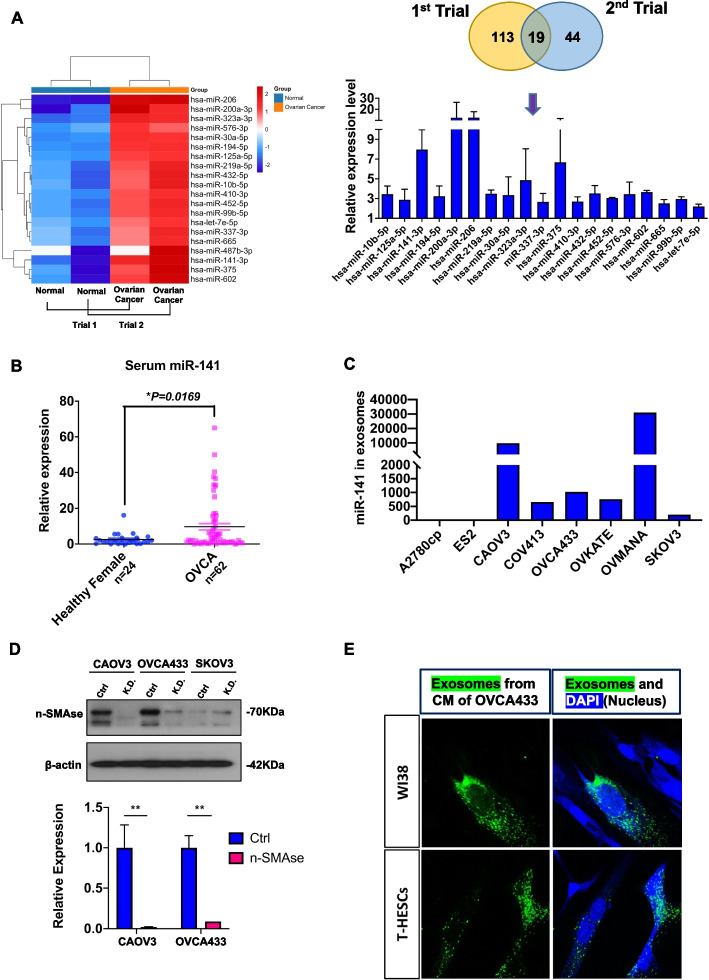
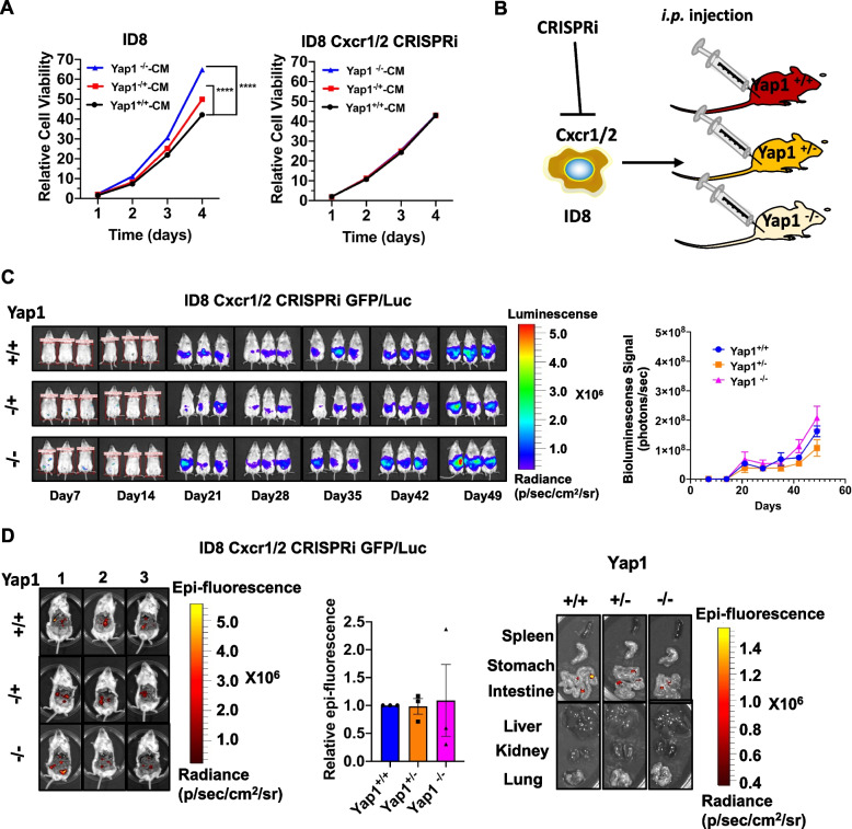
Results: Hsa-miR-141-3p (miR-141), an exosomal miRNA, is highly secreted by ovarian cancer cells and reprograms stromal fibroblasts into proinflammatory cancer-associated fibroblasts (CAFs), facilitating metastatic colonization. A mechanistic study showed that miR-141 targeted YAP1, a critical effector of the Hippo pathway, reducing the nuclear YAP1/TAZ ratio and enhancing GROα production from stromal fibroblasts. Stromal-specific knockout (cKO) of Yap1 in murine models shaped the GROα-enriched microenvironment, facilitating in vivo tumor colonization, but this effect was reversed after Cxcr1/2 depletion in OvCa cells. The YAP1/GROα correlation was demonstrated in clinical samples, highlighting the clinical relevance of this research and providing a potential therapeutic intervention for impeding premetastatic niche formation and metastatic progression of ovarian cancers.

Conclusions: This study uncovers miR-141 as an OvCa-derived exosomal microRNA mediating the tumor-stroma interactions and the formation of tumor-promoting stromal niche through activating YAP1/GROα/CXCRs signaling cascade, providing new insight into therapy for OvCa patients with peritoneal metastases.

Keywords: Hippo/YAP1/pathway; Ovarian cancer; Peritoneal metastases; Tumor-stroma interactions; cancer-associated fibroblasts; miR-141.


Conflict of interest statement

The authors declare that they have no competing interests.

Figures

Fig. 1
Fig. 1
Identification of miR-141 as a secretary miRNA from ovarian cancers. (A) The heatmap indicated the changes in secretary miRNA expression in ovarian cancer patients and normal donors using a miRCURY LNA miRNA PCR Array. N = 2 independent experiments (left). The graph showed the expression of secretory miRNAs compared between ovarian cancer patients and normal donors in the miRCURY LNA miRNA Cancer Focus PCR Panel (right). N = 2 independent experiments. (B) The graphical chart showed the elevated level of exosomal miR-141 in the serum of OvCa patients (n = 62) compared with normal women (n = 24) by qPCR analysis (mean ± SEM, t-test). (C) The graphical chart showed the secretory miR-141 in a panel of ovarian cancer cell lines by qPCR analysis. N = 1 independent experiment. (D) Knockdown of n-SMase2 by a lentiviral shRNAi approach led to the complete suppression of miR-141 exosome production in the conditioned medium of CAOV3 and OVCA433 cells. β-actin and U6 were the internal controls. (n = 3, mean ± SEM, t-test, **P < 0.01) (E) The immunofluorescence microscope showed PHK67 labeled exosomal miR-141 derived from OVCA433 conditioned medium, localized in the cytoplasm of WI-38 and T HESC stromal cells. Representative images are shown in color. Blue, DAPI staining in the nucleus. Green, staining of the exosomes with PKH67. Scale bar = 20 μm
Fig. 2
Fig. 2
MiR-141 reprograms stromal fibroblasts to be oncogenic drivers, and GROα is a major proinflammatory cytokine (A) The XTT cell proliferation assay indicated that stromal cell-conditioned medium (SCCM) from miR-141-transfected WPMY-1 cells (SCCM miR-141) increased the cell proliferation of ES-2 and SKOV3 cells on Day 2 and Day 3, compared with scrambled control medium from WPMY-1 cells (SCCM Ctrl) (n = 5, Mean ± SD, t-test, *P < 0.05, **P < 0.01). N = 1 independent experiment. (B) Transwell cell migration/invasion assays demonstrated that SCCM miR-141 treatment had convincingly induced a higher capacity of migration at 16 h or invasion at 24 h in ES-2 and SKOV3 cells as compared with SCCM Ctrl treatment (n = 6, mean ± SEM, t test, **P < 0.01, ***P < 0.001). Scale bar =100 μm. (C) Human XL cytokine array analysis revealed the number of inflammatory cytokines such as GROα in the stromal cell-conditioned medium derived from WPMY-1 scrambled control cells (SCCM Ctrl) and miR-141 overexpressing cells (SCCM miR-141). N = 2 independent experiments. (D) The XTT cell proliferation assay showed a dose-dependent increase in cell proliferation of ES-2 and OVCA433 cells upon a 4-day incubation with recombinant GROα (50 ng/mL) as compared with the respective untreated control (n = 6, mean ± SEM, two-way ANOVA, **P < 0.01, ***P < 0.001, ****P < 0.0001). N = 3 independent experiments. (E) Transwell cell migration/invasion assays showed that the recombinant GROα (60 ng/mL) remarkably promoted cell migration/invasion capacity in ES-2 and OVCA433 cells after 14 h (migration) and 20 h (invasion) as compared with the respective untreated control (Ctrl) (n = 6, mean ± SEM, t-test, *P < 0.05, **P < 0.01, ***P < 0.001, ****P < 0.0001). Scale bar = 100 μm
Fig. 3
Fig. 3
YAP1 of Hippo signaling is the direct target of miR-141 (A) The schematic drawing showed YAP1 constructs with wild-type (YAP1-WT) and mutant (YAP1-MT) miR-141 binding sites paired with the miR141 sequence (upper). Dual-luciferase assay showed the relative luciferase activities that cotransfection of pmir-YAP1-WT or pmir-YAP1-MT and the miR-141 expressing plasmid pmR-141 (pmR-ZsGreen1 empty vector was used as negative control) in WPMY-1 and HEK293 cell lines (mean ± SEM, t-test, *P < 0.05, **P < 0.01). N = 3 independent experiments. (B) Confirmation of miR-141 as a target of YAP1 by dose-dependent transfection of pmR-Zsgreen1-miR141 (0, 0.5, 1.0, and 2.0 μg) into WPMY-1, T HESC and WI-38 cells by western blot analysis. The relative YAP1 expression (YAP1/β-actin) was quantified by ImageJ software. (C) Graphic charts compared the relative transcription level of GROα between WPMY-1 stromal cells with either shRNA-mediated knockdown of YAP1 (YAP1-KD), CRISPR/Cas9 system-mediated knockout of YAP1 (YAP1low/−) or knockout of TAZ (TAZlow/− #1 and TAZlow/− #2) with the respective control (Ctrl) (upper). QPCR and ELISA analyses showed the relative expression of GROα in WPMY-1 YAP1low/− cells transfected with the YAP1-expressing plasmid (0, 1 and 2 μg) (lower) (mean ± SEM, t-test, *P < 0.05, **P < 0.01, ***P < 0.001, ****P < 0.0001). N = 3 independent experiments. (D) Western blot analysis showed the changes in YAP1/TAZ in the cytoplasm and nucleus in WPMY-1 cells with shRNA-mediated YAP1 knockdown approach (YAP1-KD). β-actin and Lamin A/C were used as the internal controls for the cytosol and nuclear proteins, respectively. (E) Immunofluorescence microscopy showed changes in YAP1/TAZ in the cytoplasm and nucleus in YAP1 knockdown and miR-141-overexpressing WPMY-1 cells, and parental cells were used as a control. Scale bar = 20 μm. (F) The graph showed the relative percent input of ChIP that indicated the interaction between TEAD and the predicted binding sites on GROα in T HESCs cells with TEAD overexpression and vector control (mean ± SEM, t-test, *P < 0.05). (G) Graphic charts show the relative luciferase activity of cotransfected TEAD1, TAZ and pGL3-GROα (N ~ III) in the WPMY-1 cells by dual-luciferase assay (mean ± SEM, one-way ANOVA, *P < 0.05, ***p < 0.001, ****p < 0.0001). N = 3 independent experiments
Fig. 4
Fig. 4
GROα enhances the tumor colonization of ovarian cancer cells (A) The graph showed the procedures for generating Yap1 stromal-specific cKO mice by crossing Yap1flox/flox mice with FSP1-Cre+/+ mice. Three genotypes of mice (Yap1+/+,  Yap1+/−, and Yap1−/−), were generated. (B) Immunofluorescence microscopy showed the mouse stromal fibroblasts extracted from Yap1 stromal-specific cKO mice (Yap1+/− and Yap1−/−) stained by the stromal cell markers FAP (green) and vimentin (red). Scale bar = 100 μm. (C) Western blot analysis compared the expression of Yap1 in mouse stromal fibroblasts extracted from Yap1 stromal-specific cKO mice (Yap1+/− and Yap1−/−) with control of wild-type (WT) mice (Yap1+/+). (D) Bar charts showed the relative expression level of GROα in mouse stromal fibroblasts, murine stromal conditioned medium, and serum extracted from Yap1+/+, Yap1+/−, and Yap1−/− mice by qPCR analysis and ELISA (n = 3, mean ± SEM, t-test, **P < 0.01, ***P < 0.001, ****P < 0.0001). (E) qPCR analysis and ELISA showed the relative expression of GROα in mouse stromal fibroblasts and murine stromal conditioned medium from Yap1−/− mice that were transiently transfected with YAP1-expressing plasmid (0 μg, 1 μg and 2 μg) (n = 3, mean ± SEM, t-test, **P < 0.01, ****P < 0.0001). (F) The XTT cell proliferation assay indicated the relative cell growth of OVCA433 and ES-2 cells co-cultured with mouse stromal conditioned medium derived from Yap1 stromal-specific cKO mice (Yap1+/−-CM and Yap1−/−-CM). Stromal conditioned medium derived from Yap1+/+ mice was used as a control (n = 6, mean ± SEM, 2-way ANOVA, ****P < 0.0001). N = 3 independent experiments. (G) The Transwell migration assay showed that the murine stromal conditioned medium from Yap1 cKO mice (Yap1+/−-CM and Yap1−/−-CM) promoted OVCA433 and ES-2 migration capacities, as compared with the respective control treated with Yap1+/+-CM (n = 3, mean ± SEM, t-test, *P < 0.05, **P < 0.01, ****P < 0.0001). N = 3 independent experiments. (H) The Transwell invasion assay showed that the murine stromal conditioned medium from Yap1 cKO mice (Yap1+/−-CM and Yap1−/−-CM) promoted OVCA433 and ES-2 invasion capacities, as compared with the respective control treated with Yap1+/+-CM (n = 3, mean ± SEM, *P < 0.05, **P < 0.01, ***P < 0.001). N = 3 independent experiments
Fig. 5
Fig. 5
Conditional knockout (cKO) of Yap1 in the stroma promotes in vivo tumor dissemination (A) The images (left) and quantifications (right) of the bioluminescence signals among Yap1+/+, Yap1+/− and Yap1−/−mice upon intraperitoneal injection of GFP/Luc-labelled ID8 cells from Day 7 to Day 49 (n = 3, mean ± SEM, 2-way ANOVA, **P < 0.01, ***P < 0.001). (B) Dot plots displayed the ascites volume among Yap1+/+, Yap1+/−, and Yap1−/− mice with an intraperitoneal injection of GFP/Luc-labelled ID8 cells (n = 5, mean ± SEM). (C) Dot plots displayed the number of tumor nodules among Yap1+/+, Yap1+/−, and Yap1−/− mice with an intraperitoneal injection of GFP/Luc-labelled ID8 cells (n = 5, mean ± SEM, t-test, **P < 0.01, ****P < 0.0001). (D) ELISA revealed the amount of GROα in the ascites of Yap1+/+, Yap1+/− and Yap1−/− mice with an intraperitoneal injection of GFP/Luc-labelled ID8 cells (n = 3, mean ± SEM, t-test, **P < 0.01, ****P < 0.0001). (E) The images (left and right) and quantifications (middle) of the intraperitoneal epifluorescence signals among Yap1+/+, Yap1+/− and Yap1−/− mice with an intraperitoneal injection of GFP/Luc-labelled ID8 cells (n = 3, mean ± SEM, t-test, * P < 0.05, **P < 0.01)
Fig. 6
Fig. 6
Depletion of CXCR1/2 impairs the oncogenic potential of OvCa cells (A) XTT cell proliferation assay indicated the relative cell growth of ID8 and ID8 Cxcr1/2 CRISPRi cell lines when co-cultured with murine stromal conditioned medium from Yap1 stromal-specific cKO mice (Yap1+/−-CM and Yap1−/−-CM). Yap1+/+-CM was used as a control (n = 6, mean ± SEM, 2-way ANOVA, ****P < 0.0001). N = 3 independent experiments. (B) The schematic diagram showed the workflow of how ID8 cells with Cxcr1/2 were silenced using CRISPR interference (CRISPRi) approach (ID8 Cxcr1/2 CRISPRi cells) were generated and intraperitoneally injected into Yap1+/+, Yap1+/− and Yap1−/− mice to establish a mouse tumor model. (C) The images (left) and quantifications (right) of the bioluminescence signals among Yap1+/+, Yap1+/− and Yap1−/− mice with an intraperitoneal injection of GFP/Luc-labelled ID8 Cxcr1/2 CRISPRi cells from Day 7 to Day 49 (n = 3, mean ± SEM). (D) The images (left and right) and quantifications (middle) of the intraperitoneal epifluorescence signals among Yap1+/+, Yap1+/− and Yap1−/− mice with an intraperitoneal injection of GFP/Luc-labelled ID8 Cxcr1/2 CRISPRi cells (n = 3, mean ± SEM)

References

    1. Lheureux S, Braunstein M, Oza AM. Epithelial ovarian cancer: evolution of management in the era of precision medicine. CA Cancer J Clin. 2019;69(4):280–304. - PubMed
    1. Roett MA, Evans P. Ovarian cancer: an overview. Am Fam Physician. 2009;80(6):609–616. - PubMed
    1. Halkia E, Spiliotis J, Sugarbaker P. Diagnosis and management of peritoneal metastases from ovarian cancer. Gastroenterol Res Pract. 2012;2012:541842. - PMC - PubMed
    1. Spring BQ, Abu-Yousif AO, Palanisami A, Rizvi I, Zheng X, Mai Z, Anbil S, Sears RB, Mensah LB, Goldschmidt R, et al. Selective treatment and monitoring of disseminated cancer micrometastases in vivo using dual-function, activatable immunoconjugates. Proc Natl Acad Sci U S A. 2014;111(10):E933–E942. - PMC - PubMed
    1. Azais H, Vignion-Dewalle AS, Carrier M, Augustin J, Da Maia E, Penel A, Belghiti J, Nikpayam M, Gonthier C, Ziane L, et al. Microscopic peritoneal residual disease after complete macroscopic Cytoreductive surgery for advanced high grade serous ovarian Cancer. J Clin Med. 2020;10(1):41. - PMC - PubMed

Publication types

Substances