. 2023 Nov;33(6):NP133-NP136.
doi: 10.1177/11206721221149762. Epub 2023 Jan 9.

COVID-19 presenting as a non-arteritic anterior ischemic optic neuropathy

Affiliations

COVID-19 presenting as a non-arteritic anterior ischemic optic neuropathy

Marina Romozzi et al. Eur J Ophthalmol. 2023 Nov.

Abstract

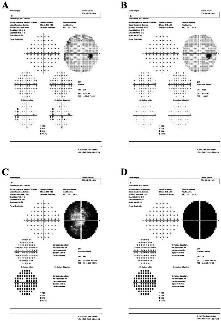
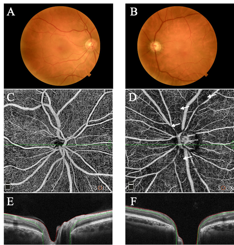
We present a case of a 61-year-old woman with an atypical non-arteritic anterior ischemic optic neuropathy (NA-AION) as a unique manifestation of COVID-19. Furthermore, the patient worsened after Pfizer-BioNTech COVID-19 vaccine administration. Our findings suggest that NA-AION could result from microangiopathic/thrombotic events that may occur during SARS-CoV-2 infection and/or vaccination against COVID-19. This report sheds light on possible ophthalmologic complications of COVID-19.

Keywords: optic nerve‌; optical coherence tomography angiography‌; vaccine‌; ‌SARS-CoV-2.


Conflict of interest statement

Declaration of conflicting interestsThe author(s) declared no potential conflicts of interest with respect to the research, authorship, and/or publication of this article.

Figures

Figure 1.
Figure 1.
Humphrey visual field (VF) 24–2 testing. Panel A: VF performed on March 31st 2021, showed a moderate reduction in peripheral retinal sensitivity in the right eye. Panel B: VF showed an almost complete recovery of retinal sensitivity in peripheral fields of the right eye. Panel C: VF performed on March 31st 2021, showed marked concentrical reduction of retinal sensitivity in the left eye. Panel D: total loss of visual field in the left eye.
Figure 2.
Figure 2.
Panel A: the retinography of the right eye shows a normal optic nerve. Panel B: the retinography of the left eye shows a normal optic nerve. Panels C-D: optical coherence tomography angiography (OCTA) of the papillary and peri-papillary area in 6 × 6 SOLIX (Optovue) device. The right eye shows a normal vascularization of the superficial and deep vascular layer (Panel C). A diffuse reduction in the left eye is evident mainly in the deep vascular layer (Panel D). White arrows indicate the ischemic area. Panels E-F: normal OCT appearance of the optic nerve head in the right eye and pathologic cupping in the left eye.

References

    1. Hayreh SS. Ischemic optic neuropathy. Prog Retin Eye Res 2009; 28: 34–62. 2008/12/10. DOI: 10.1016/j.preteyeres.2008.11.002. - DOI - PubMed
    1. Rho J, Dryden SC, McGuffey CD, et al.. A Case of Non-Arteritic Anterior Ischemic Optic Neuropathy with COVID-19. Cureus 2020; 12: e11950. 2021/01/12. DOI: 10.7759/cureus.11950. - DOI - PMC - PubMed
    1. Moschetta L, Fasolino G, Kuijpers RW. Non-arteritic anterior ischaemic optic neuropathy sequential to SARS-CoV-2 virus pneumonia: preventable by endothelial protection? BMJ case reports 2021; 14: e240542. DOI: 10.1136/bcr-2020-240542. - DOI - PMC - PubMed
    1. Savastano A, Crincoli E, Savastano MC, et al.. Peripapillary Retinal Vascular Involvement in Early Post-COVID-19 Patients. J Clin Med 2020; 9. 2020/09/12. DOI: 10.3390/jcm9092895. - DOI - PMC - PubMed
    1. Cines DB, Bussel JB. SARS-CoV-2 Vaccine-Induced Immune Thrombotic Thrombocytopenia. The New England journal of medicine 2021; 384: 2254–2256. 2021/04/16. DOI: 10.1056/NEJMe2106315. - DOI - PMC - PubMed

LinkOut - more resources