. 2023 Jan 4;23(1):273-288.
doi: 10.1021/acs.cgd.2c00992. Epub 2022 Dec 15.

Seeking Rules Governing Mixed Molecular Crystallization

Affiliations

Seeking Rules Governing Mixed Molecular Crystallization

Norbert M Villeneuve et al. Cryst Growth Des. .

Abstract

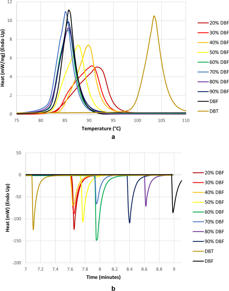
Mixed crystals result when components of the structure are randomly replaced by analogues in ratios that can be varied continuously over certain ranges. Mixed crystals are useful because their properties can be adjusted by increments, simply by altering the ratio of components. Unfortunately, no clear rules exist to predict when two compounds are similar enough to form mixed crystals containing substantial amounts of both. To gain further understanding, we have used single-crystal X-ray diffraction, computational methods, and other tools to study mixed crystallizations within a selected set of structurally related compounds. This work has allowed us to begin to clarify the rules governing the phenomenon by showing that mixed crystals can have compositions and properties that vary continuously over wide ranges, even when the individual components do not normally crystallize in the same way. Moreover, close agreement of the results of our experiments and computational modeling demonstrates that reliable predictions about mixed crystallization can be made, despite the complexity of the phenomenon.


Conflict of interest statement

The authors declare no competing financial interest.

Figures

Figure 1
Figure 1
(a) Representation of the structure of monoclinic P21/n crystals of DBT, as viewed along the b-axis. (b) View of molecules linked along the b-axis by C–H···π interactions (broken lines). (c) Optical micrograph showing an area of approximately 1 × 1 cm2 containing needles formed by DBT. In the structural images, selected molecules are shown in a space-filling representation, and atoms of carbon appear in gray, hydrogen in white, and sulfur in yellow.
Figure 2
Figure 2
(a) Representation of the structure of orthorhombic Pnma crystals of DBF, as viewed along the c-axis. (b) Molecules linked in the ac-plane by C–H···π interactions (broken lines). (c) Optical micrograph showing an area of approximately 1 × 1 cm2 containing thin plates formed by DBF. In the structural images, selected molecules are shown in a space-filling representation, and atoms of carbon appear in gray, hydrogen in white, and oxygen in red.
Figure 3
Figure 3
Hirshfeld surfaces (top images) and the corresponding two-dimensional fingerprint plots (bottom images) for molecules in monoclinic P21/n crystals of DBT and orthorhombic Pnma crystals of DBF, FLU, and CBZ. The Hirshfeld surfaces are colored according to the local value of de (distance from the surface to the nearest atomic nucleus in another molecule), and the colors range from cool (blue) to hot (red) as de decreases. The fingerprint plots show the frequency of finding points on the surface with particular values of de and di (distances to the nearest external and internal atomic nuclei). The colors at each point range from cool (blue) to hot (red) as the frequency increases.
Figure 4
Figure 4
Plots showing energies and densities in the low-energy regions of the predicted polymorphic landscapes of (a) DBT, (b) DBF, (c) FLU, and (d) CBZ. Each plotted point represents a predicted crystal structure, and the color and shape of the marker identify the crystal system according to the legend provided below the plots. On each plot, two points of special interest are enclosed in black circles or squares (P21/n or Pnma structures, respectively) and labeled as “observed” or “alternate.” These structures are highlighted because they match experimentally determined forms (“observed”) or because they are unreported but isostructural with respect to the known crystal structure of one or more of the other three compounds (“alternate”).
Figure 5
Figure 5
Optical micrograph showing a mixed Pnma crystal grown from a solution in MeOH containing an initial DBT/DBF ratio of 2:3, with an overlaid compositional map of a rectangular part of the crystal obtained by Raman microscopy. Local composition was determined by measuring the relative intensities of characteristic Raman bands (υ701 for DBT and υ730 for DBF). The scale ranges from black to white as the local value of χDBT increases from 0.33 to 0.50.
Figure 6
Figure 6
Overview of the method for generating and optimizing mixed-crystal supercells, as illustrated by partially replacing DBF with DBT in the normal Pnma packing of DBF.
Figure 7
Figure 7
(a) Plots showing the relationship between χDBT and the lattice energies of mixed crystals of DBF and DBT, as calculated for structures in which DBF and DBT have been swapped in their normal Pnma and P21/n packing arrangements (green and red data points, respectively). Structures marked as “observed” (open black diamonds and squares) correspond to predicted structures that match the experimentally observed DBF and DBT structures taken from the CSD and used as the initial packing arrangements to construct mixed-crystal models. The energy of the predicted DBF structure that is isostructural to P21/n crystals of DBT is shown as a solid black square. Red crosses correspond to P21/n mixed-crystal configurations that maintain the same molecular packing as in P21/n crystals of pure DBT after minimization of the lattice energy. Open red diamonds denote P21/n mixed-crystal configurations that undergo structural rearrangement during energy minimization. (b) Plots of the free energy of simulated mixed crystals of DBT and DBF as a function of χDBT at 298 K, with dashed lines corresponding to the composition-weighted energy of the pure unmixed components.
Figure 8
Figure 8
Plots of the formula unit volume of Pnma mixed crystals of DBT and DBF as a function of χDBT. Experimental data points (×) were obtained by single-crystal X-ray diffraction at 100 K (Table 4). Computational results (green dots) exhibit a similar volume increase in simulated Pnma mixed crystals. The plot also includes reported values of formula unit volume (+) for P21/n crystals of pure DBT at 100 K and for Pnma crystals of pure DBF at 169 K.,
Figure 9
Figure 9
Analyses of Pnma mixed crystals of DBT and DBF by differential scanning calorimetry, with related data for P21/n crystals of pure DBT and Pnma crystals of pure DBF added for comparison. (a) Melting endotherms as a function of composition. (b) Recrystallization exotherms as a function of composition. The colors of the scans identify the compositions according to the legends. In (a), heat flow is plotted as a function of temperature, which was increased at a rate of 10 °C/min. Data in (b) are shown as a function of time to avoid distortions caused by self-heating of samples during crystallization. At the start of the experiment (t = 0), the temperature was 30 °C. After a hold of 1 min, the temperature was raised to 110 °C at a rate of 20 °C/min, then cooled back to 30 °C at the same rate.
Figure 10
Figure 10
Sublimed crystals of pure DBT imaged by optical microscopy under polarized light. The thin plates correspond to the metastable Pnma polymorph and the needles to the previously reported P21/n form.

References

    1. Schoen H. M.; Grove C. S. Jr.; Palermo J. A. The Early History of Crystallization. J. Chem. Ed. 1956, 33, 373–375. 10.1021/ed033p373. - DOI
    1. Urwin S. J.; Levilain G.; Marziano I.; Merritt J. M.; Houson I.; Ter Horst J. H. A Structured Approach To Cope with Impurities during Industrial Crystallization Development. Org. Process Res. Dev. 2020, 24, 1443–1456. 10.1021/acs.oprd.0c00166. - DOI - PMC - PubMed
    1. Iuzzolino L. Survey of Crystallographic Data and Thermodynamic Stabilities of Pharmaceutical Solvates: A Step toward Predicting the Formation of Drug Solvent Adducts. Cryst. Growth Des. 2021, 21, 4362–4371. 10.1021/acs.cgd.1c00265. - DOI
    1. Werner J. E.; Swift J. A. Organic Solvates in the Cambridge Structural Database. CrystEngComm 2021, 23, 1555–1565. 10.1039/D0CE01749C. - DOI
    1. Boothroyd S.; Kerridge A.; Broo A.; Buttar D.; Anwar J. Why Do Some Molecules Form Hydrates or Solvates?. Cryst. Growth Des. 2018, 18, 1903–1908. 10.1021/acs.cgd.8b00160. - DOI