. 2022 Dec 31:2022:3027514.
doi: 10.1155/2022/3027514. eCollection 2022.

6-Gingerol Alleviates Ferroptosis and Inflammation of Diabetic Cardiomyopathy via the Nrf2/HO-1 Pathway

Affiliations

6-Gingerol Alleviates Ferroptosis and Inflammation of Diabetic Cardiomyopathy via the Nrf2/HO-1 Pathway

Shenglin Wu et al. Oxid Med Cell Longev. .

Abstract

Background: Diabetes mellitus (DM) can induce cardiomyocyte injury and lead to diabetic cardiomyopathy (DCM) which presently has no specific treatments and consequently increase risk of mortality.

Objective: To characterize the therapeutic effect of 6-gingerol (6-G) on DCM and identify its potential mechanism.

Methods: In vivo streptozotocin- (STZ-) induced DM model was established by using a high-fat diet and STZ, followed by low-dose (25 mg/kg) and high-dose (75 mg/kg) 6-G intervention. For an in vitro DCM model, H9c2 rat cardiomyoblast cells were stimulated with high glucose (glucose = 33 mM) and palmitic acid (100 μM) and then treated with 6-G (100 μM). Histological and echocardiographic analyses were used to assess the effect of 6-G on cardiac structure and function in DCM. Western blotting, ELISA, and real-time qPCR were used to assess the expression of ferroptosis, inflammation, and the Nrf2/HO-1 pathway-related proteins and RNAs. Protein expression of collagen I and collagen III was assessed by immunohistochemistry, and kits were used to assay SOD, MDA, and iron levels.

Results: The results showed that 6-G decreased cardiac injury in both mouse and cell models of DCM. The cardiomyocyte hypertrophy and interstitial fibrosis were attenuated by 6-G treatment in vivo and resulted in an improved heart function. 6-G inhibited the expression of ferroptosis-related protein FACL4 and the content of iron and enhanced the expression of anti-ferroptosis-related protein GPX4. In addition, 6-G also diminished the secretion of inflammatory cytokines, including IL-1β, IL-6, and TNF-α. 6-G treatment activated the Nrf2/HO-1 pathway, enhanced antioxidative stress capacity proved by increased activity of SOD, and decreased MDA production. Compared with in vivo, 6-G treatment of H9c2 cells treated with high glucose and palmitic acid could produce a similar effect.

Conclusion: These findings suggest that 6-G could protect against DCM by the mechanism of ferroptosis inhibition and inflammation reduction via enhancing the Nrf2/HO-1 pathway.


Conflict of interest statement

The authors have declared that no competing interest exists.

Figures

Figure 1
Figure 1
Representative echocardiographic pictures of each group.
Figure 2
Figure 2
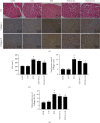
6-Gingerol prevents STZ-induced mouse heart remodeling and fibrosis. (a) Hematoxylin and eosin (HE) staining showing the morphological changes of myocardial tissue (n = 6). Immunohistochemistry shows the expression of collagen I/III (n = 6). (b) Cross-sectional area (CSA) of cardiomyocytes (n = 6). (c, d) The positive area of collagens I and III (n = 6). All values are expressed as mean ± SEM. #p < 0.05, compared with the CON or 6-G group; p < 0.05, compared with the STZ group.
Figure 3
Figure 3
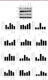
6-Gingerol attenuates STZ-induced ferroptosis and inflammation in vivo. (a) Prussian blue staining showing the level of iron ion. (b) Representative blots of FACL4 and GPX4 in the mouse heart (n = 6). (c, d) Histograms showing the fold change of FACL4 and GPX4. (e, f) The relative mRNA expression of FACL4 and GPX4 (n = 4); IL-1β (g), IL-6 (h), and TNF-α (i) activities in mouse serum (n = 6). Relative mRNA expression of IL-1β (j), IL-6 (k), and TNF-α (l) (n = 4). Heart tissue levels of SOD (m) and MDA (n) in mice. All data for proteins and mRNA was normalized to GAPDH before relative quantitative analysis. All values are expressed as mean ± SEM. #p < 0.05, compared with the CON or 6-G group; p < 0.05, compared with the STZ group.
Figure 4
Figure 4
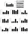
6-Gingerol inhibits ferroptosis and inflammation in vitro. (a) Cell proliferation of H9c2 in different concentrations of 6-G was determined by a CCK-8 cell proliferation assay. (b) Representative blots of FACL4 and GPX4 in H9c2 cells (n = 4). (c, d) Histograms showing the fold change of FACL4 and GPX4. Relative mRNA expression of FACL4 (e), GPX4 (f), IL-1β (g), IL-6 (h), and TNF-α (i) (n = 4). All data for protein and mRNA levels were normalized to GAPDH before quantitative analysis. All values are expressed as mean ± SEM. #p < 0.05, compared with the CON or 6-G group; p < 0.05, compared with the HG group.
Figure 5
Figure 5
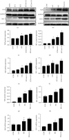
6-G upregulates the expression of Nrf2 and HO-1 in DCM both in vivo and in vitro. (a) Representative blots for Nrf2 and HO-1 in the mouse heart (n = 6). (b) Representative blots of Nrf2 and HO-1 in H9c2 cells (n = 4). (c, d) Histograms showing the fold change of Nrf2 and HO-1 in mouse hearts. (e, f) Relative mRNA expression of Nrf2 and HO-1 in mouse hearts (n = 6). (g, h) Histograms showing the fold change of Nrf2 and HO-1 in H9c2 cells. (i, j) Relative mRNA expression of Nrf2 and HO-1 in H9c2 cells (n = 4). All data for proteins and mRNA was normalized to GAPDH before relative quantitative analysis. All values are expressed as mean ± SEM. #p < 0.05, compared with the CON or 6-G group; p < 0.05, compared with the HG or STZ group.

References

    1. Wild S., Roglic G., Green A., Sicree R., King H. Global prevalence of diabetes. Diabetes Care . 2004;27(5):1047–1053. doi: 10.2337/diacare.27.5.1047. - DOI - PubMed
    1. Grundy S. M., Benjamin I. J., Burke G. L., et al. Diabetes and cardiovascular disease. Circulation . 1999;100(10):1134–1146. doi: 10.1161/01.CIR.100.10.1134. - DOI - PubMed
    1. Yap J., Tay W. T., Teng T. K., et al. Association of diabetes mellitus on cardiac remodeling, quality of life, and clinical outcomes in heart failure with reduced and preserved ejection fraction. Journal of the American Heart Association . 2019;8(17, article e013114) doi: 10.1161/JAHA.119.013114. - DOI - PMC - PubMed
    1. Tate M., Grieve D. J., Ritchie R. H. Are Targeted Therapies for Diabetic Cardiomyopathy on the Horizon? Clinical Science. Clinical Science . 2017;131:897–915. - PubMed
    1. Brunvand L., Heier M., Brunborg C., et al. Advanced glycation end products in children with type 1 diabetes and early reduced diastolic heart function. BMC Cardiovascular Disorders . 2017;17(1):p. 133. doi: 10.1186/s12872-017-0551-0. - DOI - PMC - PubMed