Review
. 2023 Feb 7;56(3):297-307.
doi: 10.1021/acs.accounts.2c00696. Epub 2023 Jan 10.

Membranized Coacervate Microdroplets: from Versatile Protocell Models to Cytomimetic Materials

Affiliations
Review

Membranized Coacervate Microdroplets: from Versatile Protocell Models to Cytomimetic Materials

Ning Gao et al. Acc Chem Res. .

Abstract

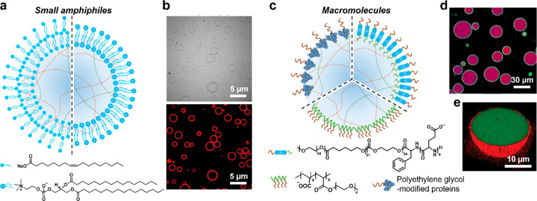
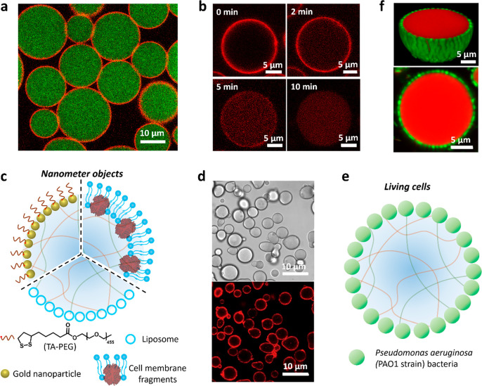
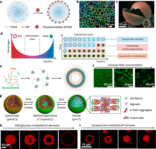
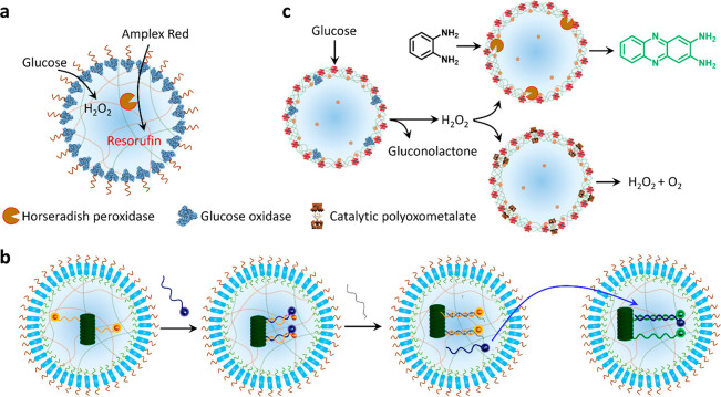
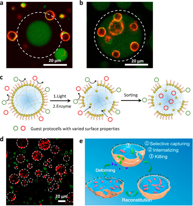
Although complex coacervate microdroplets derived from associative phase separation of counter-charged electrolytes have emerged as a broad platform for the bottom-up construction of membraneless, molecularly crowded protocells, the absence of an enclosing membrane limits the construction of more sophisticated artificial cells and their use as functional cytomimetic materials. To address this problem, we and others have recently developed chemical-based strategies for the membranization of preformed coacervate microdroplets. In this Account, we review our recent work on diverse coacervate systems using a range of membrane building blocks and assembly processes. First, we briefly introduce the unusual nature of the coacervate/water interface, emphasizing the ultralow interfacial tension and broad interfacial width as physiochemical properties that require special attention in the judicious design of membranized coacervate microdroplets. Second, we classify membrane assembly into two different approaches: (i) interfacial self-assembly by using diverse surface-active building blocks such as molecular amphiphiles (fatty acids, phospholipids, block copolymers, protein-polymer conjugates) or nano- and microscale objects (liposomes, nanoparticle surfactants, cell fragments, living cells) with appropriate wettability; and (ii) coacervate droplet-to-vesicle reconfiguration by employing auxiliary surface reconstruction agents or triggering endogenous transitions (self-membranization) under nonstoichiometric (charge mismatched) conditions. We then discuss the key cytomimetic behaviors of membranized coacervate-based model protocells. Customizable permeability is achieved by synergistic effects operating between the molecularly crowded coacervate interior and surrounding membrane. In contrast, metabolic-like endogenous reactivity, diffusive chemical signaling, and collective chemical operations occur specifically in protocell networks comprising diverse populations of membranized coacervate microdroplets. In each case, these cytomimetic behaviors can give rise to functional microscale materials capable of promising cell-like applications. For example, immobilizing spatially segregated enzyme-loaded phospholipid-coated coacervate protocells in concentrically tubular hydrogels delivers prototissue-like bulk materials that generate nitric oxide in vitro, enabling platelet deactivation and inhibition of blood clot formation. Alternatively, therapeutic protocells with in vivo vasoactivity, high hemocompatibility, and increased blood circulation times are constructed by spontaneous assembly of hemoglobin-containing cell-membrane fragments on the surface of enzyme-loaded coacervate microdroplets. Higher-order properties such as artificial endocytosis are achieved by using nanoparticle-caged coacervate protocell hosts that selectively and actively capture guest nano- and microscale objects by responses to exogenous stimuli or via endogenous enzyme-mediated reactions. Finally, we discuss the current limitations in the design and programming of membranized coacervate microdroplets, which may help to guide future directions in this emerging research area. Taken together, we hope that this Account will inspire new advances in membranized coacervate microdroplets and promote their application in the development of integrated protocell models and functional cytomimetic materials.


Conflict of interest statement

The authors declare no competing financial interest.

Figures

Figure 1
Figure 1
Liquid interfaces for membrane assembly. Scheme showing key properties of water/oil and coacervate/supernatant interfaces. The interfacial width is the distance between two locations where the densities of the coacervate and supernatant are 90% of their own bulk densities.
Figure 2
Figure 2
Membranized coacervate microdroplets based on molecular/macromolecular interfacial self-assembly. (a) Scheme showing coacervate microdroplets coated with a multilayer fatty acid (left side) or a phospholipid bilayer (right side). (b) Bright-field (top) and fluorescence (bottom) images of phospholipid (DPPC, Dil stain, red fluorescence) bilayer-bounded DEAE-dextran/DNA coacervate microdroplets. Reproduced with permission from ref (27). Copyright 2021 American Chemical Society. (c) Scheme showing coacervate microdroplets coated with macromolecules (terpolymer, top right), PEG-modified proteins (top left), or comb polyelectrolytes (bottom). (d) Confocal laser scanning microscopy (CLSM) image showing terpolymer-bounded coacervate microdroplets (purple). Reproduced with permission from ref (30). Copyright 2017 American Chemical Society. (e) 3D CLSM image showing PEG-modified protein-coated coacervate microdroplet: red fluorescence, RITC-labeled protein; green fluorescence, FITC-tagged coacervate. Reproduced with permission from ref (32). Copyright 2019 Wiley-VCH.
Figure 3
Figure 3
Membranized coacervate microdroplets based on nano- and microscale interfacial self-assembly. (a) CLSM image of caged coacervate droplets; red fluorescence membrane (jammed Au/RITC-labeled TA-PEG nanoparticles); green fluorescence interior (FITC-labeled CM-dextran coacervate). The Au nanoparticles are initially coated with tannic acid to prevent aggregation. (b) Time series of CLSM images showing changes in membrane and interior red fluorescence for a Au/RITC-TA-PEG nanoparticle-caged coacervate droplet after light illumination for different time intervals. Membrane disassembly and translocation of the decapped Au nanoparticles into the coacervate interior occurs within 10 min. Reproduced with permission from ref (1). Copyright 2022 American Chemical Society. (c) Scheme showing coating of a coacervate microdroplet with nanometer-sized objects; TA-PEG/gold nanoparticles (top left), liposomes (bottom), or cell membrane fragments (top right)., (d) Bright-field (top) and fluorescence (bottom) images of Dil-stained erythrocyte membrane-fragment-encapsulated DEAE-dextran/DNA coacervate microdroplets. Reproduced with permission from ref (2). Copyright 2020 Springer Nature. (e) Scheme showing continuous shell of living P. aeruginosa cells surrounding a coacervate microdroplet. (f) 2D CLSM (bottom) and 3D reconstruction (top) images of bacteria-coated PDDA/ATP coacervate microdroplets (P. aeruginosa, green fluorescence; coacervate, red fluorescence). Reproduced with permission from ref (39). Copyright 2022 Springer Nature.
Figure 4
Figure 4
Coacervate droplet-to-vesicle reconfiguration. (a) Scheme showing POM-mediated coacervate droplet-to-vesicle transition via surface complexation and osmotic pressure-induced swelling of PDDA/ATP coacervate droplets., (b,c) Corresponding dark-field microscopy and (c) SEM images of the coacervate vesicles showing the continuous POM/PDDA membrane. Reproduced with permission from ref (47). Copyright 2020 Springer Nature. (d) Scheme showing counter-directional chemical (SDS and POM) gradients and corresponding morphological transitions of PDDA/ATP coacervate droplets within the intersection zone of the different membrane-forming agents. Coacervate vesicles with various structures are produced as a function of relative position and time due to local changes in the SDS/POM ratio. The initial population of identical membrane-free coacervate droplets is transformed into a segregated community of spatially and functionally differentiated protocells. Reproduced with permission from ref (3). Copyright 2019 Springer Nature. (e) Scheme showing coacervate droplet-to-vesicle transition by inducing charge mismatching in coacervate constituents (RNA and nucleoprotein) under substoichiometric (disproportionate) conditions. (f) CLSM images showing the structure of nucleoprotein/RNA condensates at different RNA concentrations. Homogeneous coacervate droplets prepared from stoichiometric mixtures transform into membrane-bounded coacervate vesicles under charge-mismatched conditions. Reproduced with permission from ref (43). Copyright 2020 National Academy of Sciences. (g) Scheme showing enzyme-mediated transitions between isotropic coacervate microdroplets, multicompartmentalized coacervate droplets and coacervate vesicles with a single water-filled lumen and thin outer membrane. The coacervate microdroplets are prepared using CSF and negatively charged alginate. (h,i) CLSM images showing GOx-mediated alginate/CSF coacervate droplet-to-vesicle transition (h) and reverse transition in the presence of urease (i). Assembly and disassembly of the membrane is associated with an increase or decrease in the osmotic pressure gradient, which in turn gives rise to expansion or contraction of the water-filled lumen, respectively. Reproduced with permission from ref (4). Copyright 2022 Wiley-VCH.
Figure 5
Figure 5
Microscale reactivity and chemical signaling. (a) Scheme showing membrane-to-interior chemical signaling in membranized coacervate droplets by integration of a PEG-modified GOx membrane and HRP-containing coacervate interior. (b) Scheme showing DNA signaling in populations of terpolymer (turquoise) membrane-enclosed semipermeable coacervate droplets containing a DNA-decorated nanoscaffold (dark green). A complementary ssDNA input strand (blue) acts as a fluorescence reporter that can be displaced by a fuel strand (gray) (left side). Efflux of the reporter strand is used as a communication signal (blue arrow) for activating a second population of the protocells (right side). Reproduced with permission from ref (46). Copyright 2020 American Chemical Society. (c) Scheme showing chemical communication pathways in a community of coacervate vesicle-based protocells consisting of GOx/POM, HRP/POM, or RuPOM catalytic membranes. Hydrogen peroxide is generated by the GOx/POM population and employed as a diffusive signaling molecule for competing transformations in the HRP/POM (peroxidase activity) and RuPOM (catalase-like activity) populations. Reproduced with permission from ref (47). Copyright 2020 Springer Nature.
Figure 6
Figure 6
Prototissue construction and therapeutic protocells. (a) Scheme showing three-layer prototissue-like vessel harboring phospholipid-enveloped PDDA/DNA coacervate vesicles (CVs) decorated with GOx in the outer hydrogel layer, HRP in the middle hydrogel layer and catalase (CAT) in the inner hydrogel layer. (b) Varying the spatial sequence of the enzyme-CV modules in the presence of identical exterior inputs (Glu and Hu) gives rise to different outputs in the internal lumen. Reproduced with permission from ref (28). Copyright 2022 Springer Nature (c) Scheme showing the design of membranized coacervate microdroplet-based therapeutic protocells that perform GOx/hemoglobin-mediated generation of NO in the presence of glucose and hydroxyurea (Hu) for blood vessel vasodilation. GOx and hemoglobin are spatially positioned in the coacervate interior and membrane, respectively. Reproduced with permission from ref (2). Copyright 2020 Springer Nature.
Figure 7
Figure 7
Artificial phagocytosis and protocell sorting. (a,b) CLSM images of red/green fluorescence overlay images showing a single FITC-labeled GOx-containing Au/TA-AE-PEG6k-caged coacervate droplet surrounded by RITC-labeled PCVs and recorded before (a) and 60 min after (b) addition of glucose. Unjamming of the membrane by enzyme-mediated cleavage of polymer TA-AE-PEG6k results in PCV transfer across the membrane. The focal plane is aligned with the PCVs not the caged coacervate droplet. White dash circles delineate the boundary of the caged coacervate droplet. (c) Scheme showing triggered uptake and sorting of guest protocells in Au/PEG nanoparticle-caged PDDA/CM-dextran coacervate protocells via the light or enzyme-mediated disassembly of the membrane. (d) CLSM images showing the selective capture of RITC-labeled PCVs (red fluorescence) within the interior of the nanoparticle-caged protocells (white dashed circles), while FITC-labeled PEG-modified colloidosomes (green) remain in the external environment. Reproduced with permission from ref (1). Copyright 2022 American Chemical Society. (e) Scheme showing artificial phagocytosis of E. coli bacterial cells within membranized coacervate microdroplets. The membrane is generated by the interfacial assembly of yeast membrane fragments and is locally disrupted by mechanical agitation, leading to aperture formation and capture of the bacteria, which subsequently die in the presence of cationized amylose. Addition of yeast membrane fragments results in resealing of the apertures. Reproduced with permission from ref (35). Copyright 2021 American Chemical Society.

References

    1. Gao N.; Xu C.; Yin Z.; Li M.; Mann S. Triggerable protocell capture in nanoparticle-caged coacervate microdroplets. J. Am. Chem. Soc. 2022, 144, 3855–3862. 10.1021/jacs.1c11414. - DOI - PMC - PubMed
    1. Liu S.; Zhang Y.; Li M.; Xiong L.; Zhang Z.; Yang X.; He X.; Wang K.; Liu J.; Mann S. Enzyme-mediated nitric oxide production in vasoactive erythrocyte membrane-enclosed coacervate protocells. Nat. Chem. 2020, 12, 1165–1173. 10.1038/s41557-020-00585-y. - DOI - PubMed
    1. Tian L.; Li M.; Patil A. J.; Drinkwater B. W.; Mann S. Artificial morphogen-mediated differentiation in synthetic protocells. Nat. Commun. 2019, 10, 3321.10.1038/s41467-019-11316-4. - DOI - PMC - PubMed
    1. Yin Z.; Tian L.; Patil A. J.; Li M.; Mann S. Spontaneous membranization in a silk-based coacervate protocell model. Angew. Chem. Int. Ed. 2022, 61, e20220230210.1002/anie.202202302. - DOI - PMC - PubMed
    1. Abbas M.; Lipiński W. P.; Wang J.; Spruijt E. Peptide-based coacervates as biomimetic protocells. Chem. Soc. Rev. 2021, 50, 3690–3705. 10.1039/D0CS00307G. - DOI - PubMed

Publication types