. 2023 Feb 16;91(2):e0033322.
doi: 10.1128/iai.00333-22. Epub 2023 Jan 10.

Aspergillus fumigatus Drives Tissue Damage via Iterative Assaults upon Mucosal Integrity and Immune Homeostasis

Affiliations

Aspergillus fumigatus Drives Tissue Damage via Iterative Assaults upon Mucosal Integrity and Immune Homeostasis

Uju Joy Okaa et al. Infect Immun. .

Abstract

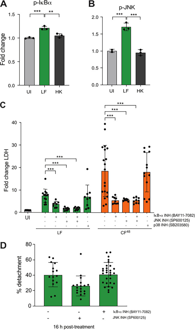
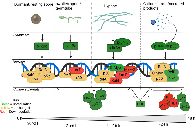
The human lung is constantly exposed to Aspergillus fumigatus spores, the most prevalent worldwide cause of fungal respiratory disease. Pulmonary tissue damage is a unifying feature of Aspergillus-related diseases; however, the mechanistic basis of damage is not understood. In the lungs of susceptible hosts, A. fumigatus undergoes an obligatory morphological switch involving spore germination and hyphal growth. We modeled A. fumigatus infection in cultured A549 human pneumocytes, capturing the phosphoactivation status of five host signaling pathways, nuclear translocation and DNA binding of eight host transcription factors, and expression of nine host response proteins over six time points encompassing exposures to live fungus and the secretome thereof. The resulting data set, comprised of more than 1,000 data points, reveals that pneumocytes mount differential responses to A. fumigatus spores, hyphae, and soluble secreted products via the NF-κB, JNK, and JNK + p38 pathways, respectively. Importantly, via selective degradation of host proinflammatory (IL-6 and IL-8) cytokines and growth factors (FGF-2), fungal secreted products reorchestrate the host response to fungal challenge as well as driving multiparameter epithelial damage, culminating in cytolysis. Dysregulation of NF-κB signaling, involving sequential stimulation of canonical and noncanonical signaling, was identified as a significant feature of host damage both in vitro and in a mouse model of invasive aspergillosis. Our data demonstrate that composite tissue damage results from iterative (repeated) exposures to different fungal morphotypes and secreted products and suggest that modulation of host responses to fungal challenge might represent a unified strategy for therapeutic control of pathologically distinct types of Aspergillus-related disease.

Keywords: Aspergillus fumigatus; fungal infection; lung infection; virulence.


Conflict of interest statement

The authors declare no conflict of interest.

Figures

FIG 1
FIG 1
Temporal quantitative analysis of epithelial decay following challenge with live A. fumigatus spores (LF) or CF. (A) Visualization of concanavalin A-FITC-labeled A549 monolayers incubated with the A. fumigatus tdTomatoATCC4664 strain (MOI = 1) and a 5-fold diluted culture filtrate thereof (CF48). (B) Percentage of detachment of A549 cells following infection with 106 A. fumigatus CEA10 spores at indicated time points. (C) Percentage of detachment of A549 cells following 16 h challenge with a 5-fold diluted filtrate from CEA10 fungal cultures (inoculum of 106 spores/mL) grown for 16 h (CF16) or 48 h (CF48). (D) Percentage of detachment of A549 cells following challenge with a 5-fold diluted filtrate from CEA10 fungal cultures at indicated time points. (E) Fold change LDH release (relative to PBS challenge) at indicated time points with 106 A. fumigatus CEA10 spores. (F) Fold change LDH release (relative to PBS challenge) at 24 h postexposure to CF16. (G) Fold change LDH release (relative to PBS challenge) at indicated time points with a 5-fold diluted filtrate from CEA10 fungal cultures. (H) Fold change decrease in TEER between A549 monolayers incubated for 24 and 0 h with A. fumigatus CEA10 spores and a 5-fold diluted filtrate from CEA10 fungal cultures. Data represent 3 biological replicates with 1 to 5 technical replicates. Error bars show ± SEM. Data were analyzed by nonparametric one-way ANOVA (Kruskal-Wallis test) with Dunn’s multiple comparisons. Significance was calculated relative to challenge with vehicle control (PBS) unless otherwise shown by brackets. ****, P ≤ 0.0001; ***, P ≤ 0.001; **, P ≤ 0.01; and *, P ≤ 0.05.
FIG 2
FIG 2
Differential cytokine secretion by A549 cells in response to live A. fumigatus spores (LF) and CF48. (A to I) Secreted cytokines GM-CSF (A), IL-8 (B), adiponectin (C), CD40L (D), IL-6 (E), FGF-2 (F), MIP-3a (G), G-CSF (H), and monocyte chemoattractant protein-1 (MCP-1) (I) were quantified in cell culture supernatant following exposure to A. fumigatus spores (1 × 10⁷ spores/mL) (MOI = 10) or 5-fold diluted CF48 for 24 h. (J to L) Cytokines IL-6 (J), IL-8 (K), and FGF-2 (L) were quantified in A549-free culture supernatants collected after exposure of epithelial monolayers to A. fumigatus for 24 h (CF24) followed by incubation with CF48 or with culture filtrates recovered from a 24 h infection of AECs with CF48 (CDN). Data represent 3 biological replicates with 1 to 5 technical replicates. Error bars show ± SEM. Data were analyzed by nonparametric one-way ANOVA (Kruskal-Wallis test) with Dunn’s multiple comparisons. Significance was calculated relative to challenge with vehicle control (PBS) and between each treatment as shown. ****, P ≤ 0.0001; ***, P ≤ 0.001; **, P ≤ 0.01; and *, P ≤ 0.05.
FIG 3
FIG 3
Different fungal morphotypes induce differential and dynamic host responses in A549 cells. (A to C) Fold change (relative to uninfected control [UI]) phosphorylation of NF-κB (p-IkBα [A]) and MAPK (p-JNK [B]and p-p38 [C]) signaling following exposure to A. fumigatus spores (1 × 10⁷ spores/mL) (MOI = 10) for indicated time points or 5-fold diluted CF48 for 4 h. (D to E) Fold change (relative to uninfected control, UI) in DNA binding activity of noncanonical NF-κB transcription factors (p52 [D] and RelB [E]) following exposure to A. fumigatus spores (107 spores/mL) for indicated time points or 5-fold diluted CF48 (inoculum of 106 spores/mL) for 4 h. Data represent 3 biological replicates with 1 to 5 technical replicates. Error bars show ± SEM. Data were analyzed by nonparametric one-way ANOVA (Kruskal-Wallis test) with Dunn’s multiple comparisons. Significance was calculated relative to challenge with vehicle control (PBS) as shown. ****, P ≤ 0.0001; ***, P ≤ 0.001; **, P ≤ 0.01; and *, P ≤ 0.05.
FIG 4
FIG 4
Signaling activation is dependent on fungal viability and plays a role in epithelial damage. (A and B) Fold change (relative to uninfected control [UI]) phosphorylation of NF-κB (p-IkBα [A]) and MAPK (p-JNK [B]) signaling following 4 h exposure to live fungus (LF) and heat-killed (HK) morphotypes (107 spores/mL) of A. fumigatus pregrown to hyphae for 12 h (MOI = 10). (C) Fold change LDH release (relative to PBS challenge) following chemical inhibition of NF-κB (IkBα inhibitor BAY11-7082) and MAPK (JNK inhibitor SP600125 and p38 inhibitor SB203580) signaling pathways and exposure to A. fumigatus spores (106 spores/mL) MOI = 1- or 5-fold diluted CF48 for 24 h. (D) Fold change detachment (relative to PBS challenge) following chemical inhibition of NF-κB (IkBα inhibitor BAY11-7082) and MAPK (JNK inhibitor SP600125) signaling pathways and exposure to A. fumigatus spores (106 spores/mL) MOI = 1 for 16 h. Data represent 2 or 3 biological replicates with 1 to 5 technical replicates. Error bars show ± SEM. Data were analyzed by nonparametric one-way ANOVA (Kruskal-Wallis test) with Dunn’s multiple comparisons. Significance was calculated relative to challenge with vehicle control (PBS) and between each treatment as shown. ****, P ≤ 0.0001; ***, P ≤ 0.001; **, P ≤ 0.01; and *, P ≤ 0.05.
FIG 5
FIG 5
A. fumigatus ΔpacC, ΔgliP, and ΔprtT mutants and respective CF48 fail to induce AEC detachment and lysis. (A) Percentage of detachment of A549 cells following infection with 106 A. fumigatus ΔpacC, ΔgliP, and ΔprtT mutants and respective parental isolate (PI) and CF48 thereof for 16 h. (B) Fold change LDH release (relative to PBS challenge) following infection with 106 A. fumigatus ΔpacC, ΔgliP, and ΔprtT mutants and respective parental isolate (PI) and CF48 thereof for 24 h. Data represent 3 biological replicates with 1 to 5 technical replicates. Error bars show ± SEM. Data were analyzed by nonparametric one-way ANOVA (Kruskal-Wallis test) with Dunn’s multiple comparisons. Significance was calculated relative to the parental isolate (PI) as shown. ****, P ≤ 0.0001; ***, P ≤ 0.001; **, P ≤ 0.01; and *, P ≤ 0.05.
FIG 6
FIG 6
A. fumigatus ΔpacC, ΔgliP, and ΔprtT mutants and respective CF48 fail to activate AEC host signaling proteins and transcription factors. (A to C) Fold change (relative to PBS challenge) phosphorylation of NF-κB (p-IkBα [A]) and MAPK (p-JNK [B] and p-p38 [C]) signaling following exposure to A. fumigatus ΔpacC, ΔgliP and ΔprtT mutants and respective parental isolate (PI) (1 × 10⁷ spores/mL) for 8 h or respective 5-fold diluted CF48 for 4 h. (D to E) Fold change (relative to PBS challenge) in DNA binding activity of noncanonical NF-κB transcription factors (p52 [D] and RelB [E]) following exposure to A. fumigatus ΔpacC, ΔgliP and ΔprtT mutants and respective parental isolate (PI) (1 × 10⁷ spores/mL) for 8 h or respective 5-fold diluted CF48 for 4 h. Data represent 3 biological replicates with 1 to 5 technical replicates. Error bars show ± SEM. Data were analyzed by nonparametric one-way ANOVA (Kruskal-Wallis test) with Dunn’s multiple comparisons. Significance was calculated relative to the parental isolate (PI) as shown. ****, P ≤ 0.0001, ***, P ≤ 0.001; **, P ≤ 0.01; and *, P ≤ 0.05.
FIG 7
FIG 7
Immunohistochemistry to assess RelB and p65 immunoreactivity in vivo indicates subvertion of the NF-κB signaling axis following A. fumigatus infection of leukopenic mice. (A) Hematoxylin and eosin (H&E) staining, RelB/p65 immunochemistry, and GMS/green light staining of histological sections from leukopenic mice infected with A. fumigatus dTomatoATCC46645 for 24 and 48 h. (B to E) Quantification of p65 (B and C) and RelB (D and E) staining from scoring infected lesions and uninfected tissue in histological sections from leukopenic mice. Using the ImageJ software, nuclei were classified into 4 bins as follows: negative (intense blue), weak (light blue), moderate (light brown), and strong intensity (dark brown). Values of each category were expressed as percent relative to the total number of nuclei evaluated and reported as having positive (moderate + strong) or negative (negative + weak) immunohistochemical phenotypes. ****, P ≤ 0.0001; ***, P ≤ 0.001; **, P ≤ 0.01; and *, P ≤ 0.05.
FIG 8
FIG 8
Kinetics of intracellular signaling events in alveolar type II epithelial cells in response to A. fumigatus infection. A. fumigatus conidia interact with epithelial cells activating the canonical (p50/p65) NF-κB pathway and JNK/c-Fos or c-Myc signaling specifically during hyphal growth leading to IL-8 and GM-CSF synthesis. As the fungus matures, secreted products (in the culture filtrate) interact with epithelial cells and activate 3 MAPK pathways (JNK, p38, and ERK1/2) and the noncanonical (RelB and p52) NF-κB transcription factors. This results in targeted modulation of secreted products biased toward degradation of proinflammatory mediators. Finally, cell damage measured by LDH release (cellular damage) increases as the fungal hyphae mature and secrete more soluble factors. Green and red colors indicate activation and suppression, respectively. Bold lettering indicates strong responses.

References

    1. Brown GD, Denning DW, Gow NAR, Levitz SM, Netea MG, White TC. 2012. Hidden killers: human fungal infections. Sci Transl Med 4:165rv13. 10.1126/scitranslmed.3004404. - DOI - PubMed
    1. Thompson GR, III, Young JH. 2021. Aspergillus infections. N Engl J Med 385:1496–1509. 10.1056/NEJMra2027424. - DOI - PubMed
    1. Bertuzzi M, Schrettl M, Alcazar-Fuoli L, Cairns TC, Muñoz A, Walker LA, Herbst S, Safari M, Cheverton AM, Chen D, Liu H, Saijo S, Fedorova ND, Armstrong-James D, Munro CA, Read ND, Filler SG, Espeso EA, Nierman WC, Haas H, Bignell EM. 2014. The pH-responsive PacC transcription factor of Aspergillus fumigatus governs epithelial entry and tissue invasion during pulmonary aspergillosis. PLoS Pathog 10:e1004413. 10.1371/journal.ppat.1004413. - DOI - PMC - PubMed
    1. Naglik JR, Moyes DL, Wächtler B, Hube B. 2011. Candida albicans interactions with epithelial cells and mucosal immunity. Microbes Infect 13:963–976. 10.1016/j.micinf.2011.06.009. - DOI - PMC - PubMed
    1. Ruan J, Qi Z, Shen L, Jiang Y, Xu Y, Lan L, Luo L, Yin Z. 2015. Crosstalk between JNK and NF-kappaB signaling pathways via HSP27 phosphorylation in HepG2 cells. Biochem Biophys Res Commun 456:122–128. 10.1016/j.bbrc.2014.11.045. - DOI - PubMed

Publication types