. 2023 Feb 14;11(1):e0394322.
doi: 10.1128/spectrum.03943-22. Epub 2023 Jan 10.

Epitranscriptomic N6-Methyladenosine Profile of SARS-CoV-2-Infected Human Lung Epithelial Cells

Affiliations

Epitranscriptomic N6-Methyladenosine Profile of SARS-CoV-2-Infected Human Lung Epithelial Cells

Stacia Phillips et al. Microbiol Spectr. .

Abstract

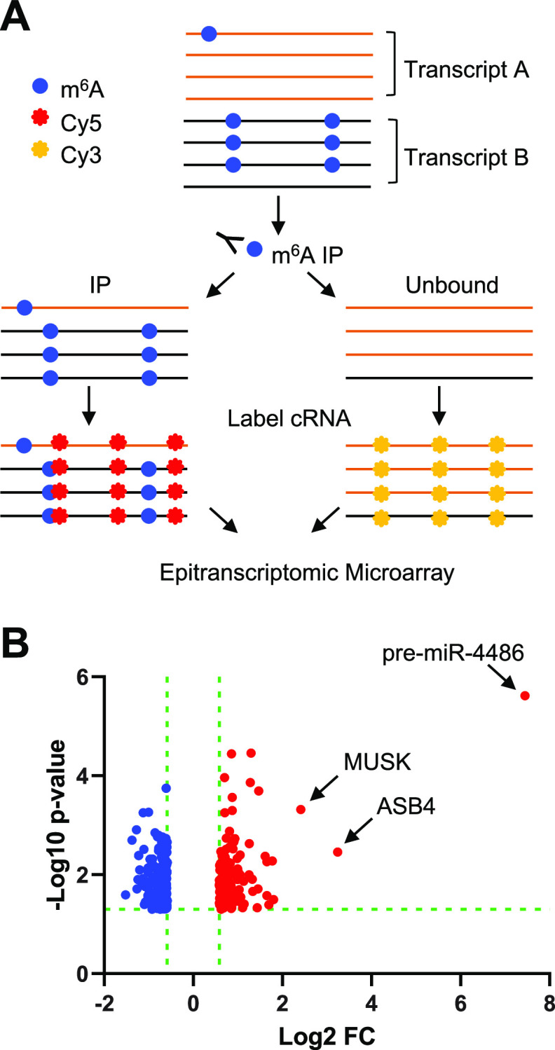
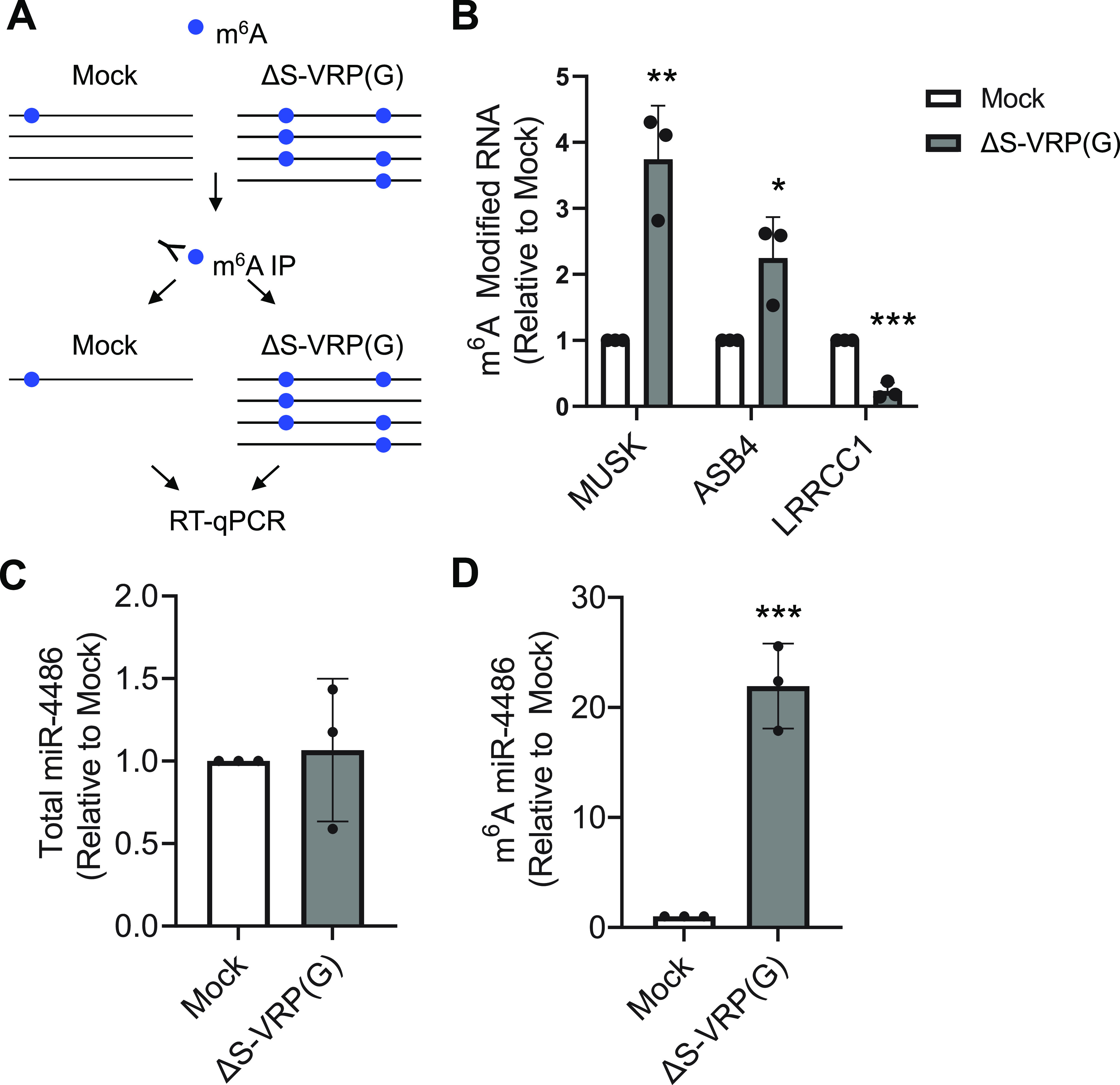
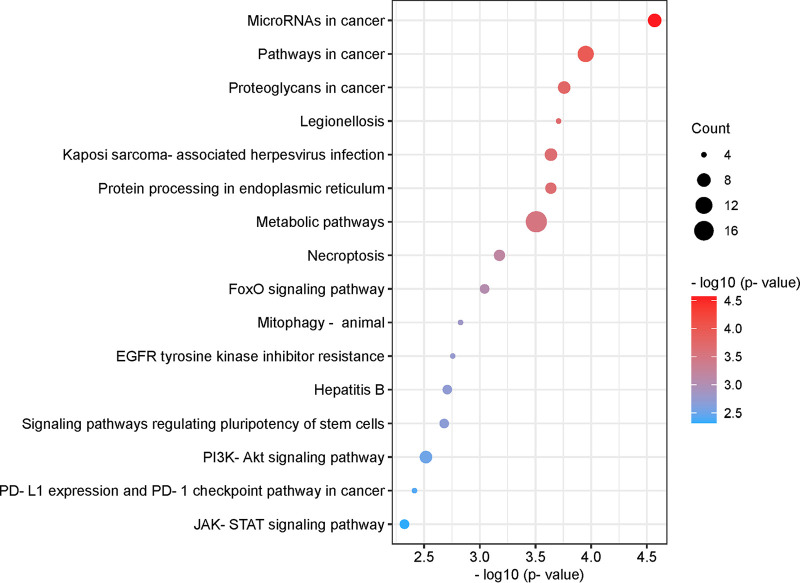
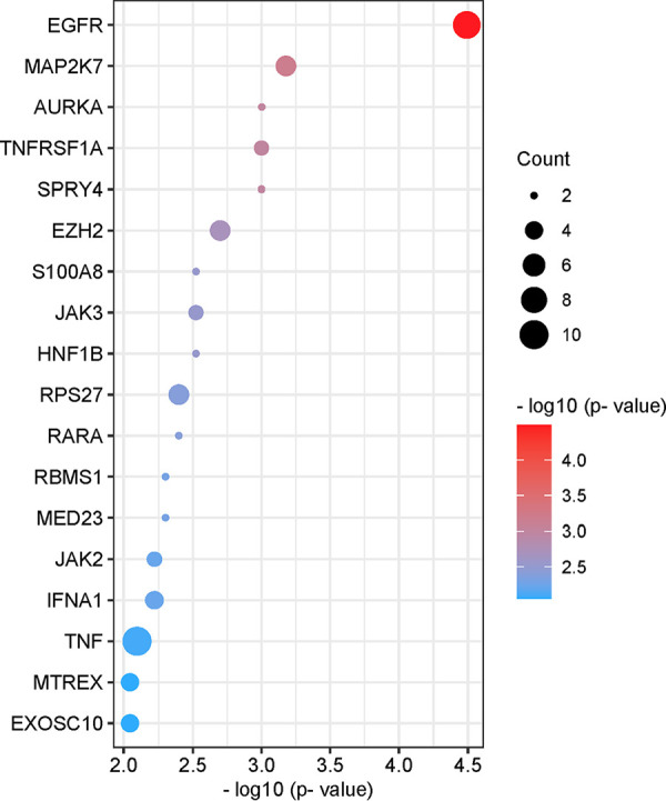
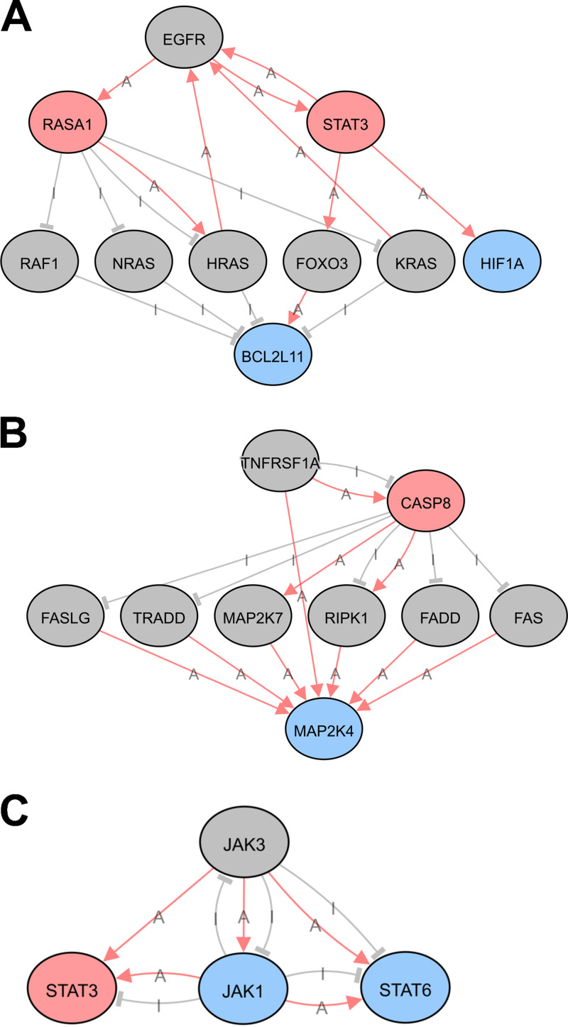
N6-methyladenosine (m6A) is a dynamic posttranscriptional RNA modification that plays an important role in determining transcript fate. The functional consequence of m6A deposition is dictated by a group of host proteins that specifically recognize and bind the m6A modification, leading to changes in RNA stability, transport, splicing, or translation. The cellular m6A methylome undergoes changes during certain pathogenic conditions such as viral infections. However, how m6A modification of host cell transcripts and noncoding RNAs change during severe acute respiratory syndrome coronavirus (SARS-CoV-2) infection has not been reported. Here, we define the epitranscriptomic m6A profile of SARS-CoV-2-infected human lung epithelial cells compared to uninfected controls. We identified mRNA and long and small noncoding RNA species that are differentially m6A modified in response to SARS-CoV-2 infection. The most significantly differentially methylated transcript was the precursor of microRNA-4486 (miRNA-4486), which showed significant increases in abundance and percentage of methylated transcripts in infected cells. Pathway analyses revealed that differentially methylated transcripts were significantly associated with several cancer-related pathways, protein processing in the endoplasmic reticulum, cell death, and proliferation. Upstream regulators predicted to be associated with the proteins encoded by differentially methylated mRNAs include several proteins involved in the type-I interferon response, inflammation, and cytokine signaling. IMPORTANCE Posttranscriptional modification of viral and cellular RNA by N6-methyladenosine (m6A) plays an important role in regulating the replication of many viruses and the cellular immune response to infection. We therefore sought to define the epitranscriptomic m6A profile of human lung epithelial cells infected with SARS-CoV-2. Our analyses demonstrate the differential methylation of both coding and noncoding cellular RNAs in SARS-CoV-2-infected cells compared to uninfected controls. Pathway analyses revealed that several of these RNAs may be involved in the cellular response to infection, such as type-I interferon. Our study implicates m6A modification of infected-cell RNA as a mechanism of posttranscriptional gene regulation during SARS-CoV-2 infection.

Keywords: N6-methyladenosine; SARS-CoV-2; epitranscriptomics; infection; lung epithelial cells; microarray.


Conflict of interest statement

The authors declare no conflict of interest.

Figures

FIG 1
FIG 1
SARS-CoV-2 infection of A549-hACE2 cells. A549-hACE2 cells were infected with SARS-CoV-2 (strain USA-WA1/2020) at an MOI of 1 for 24 h. (A) Immunofluorescent staining was performed to visualize infected cells by the presence of SARS-CoV-2 nucleocapsid (green). Nuclei of cells are stained with DAPI (blue). (B) SARS-CoV-2 spike RNA in infected cells (n = 3, biological triplicate) was quantified by RT-qPCR. ND, not detected.
FIG 2
FIG 2
Epitranscriptomic m6A microarray of SARS-CoV-2-infected A549-hACE2 cells. (A) Schematic overview of the method. Total cellular RNA from each sample (SARS-CoV-2-infected and mock-infected controls, biological triplicate, n = 3 each group) was used for immunoprecipitation using an m6A-specific antibody. Methylated and unmethylated RNA fractions were fluorescently labeled (Cy3 or Cy5) prior to array hybridization (refer to Materials and Methods for details). (B) Volcano plot of transcripts containing higher (red) and lower (blue) levels of m6A modification in infected cells compared to mock-infected control cells. The miRNA precursor (pre-mir-4486) with the most significant m6A change is labeled.
FIG 3
FIG 3
Validation of differential methylation for selected transcripts in A549-hACE2 cells. (A) Schematic of m6A RIP. A549-hACE2 cells were mock infected or infected with ΔS-VRP(G) for 24 h. Total RNA was used for m6A-RIP. (B) Total RNA from cells infected with ΔS-VRP(G) was used for m6A-RIP. The fold enrichment of m6A-modfied RNA in the IP fraction of infected cell RNA versus mock-infected controls is indicated. (C) Total cellular RNA from mock-infected or ΔS-VRP(G)-infected cells was used for RT-qPCR analysis of total levels of mature miR-4486. (D) Total cellular RNA from mock-infected or ΔS-VRP(G)-infected cells was used for m6A RNA-IP to determine the levels of m6A-modified mature miR-4486. (C and D) Statistical significance was determined by t test compared to mock control. *, P < 0.05; **, P < 0.005; ***, P < 0.0005.
FIG 4
FIG 4
Pathway analysis of differentially methylated mRNAs. List of pathways associated with differentially methylated mRNAs with the highest combined significance (pORA and pAcc ≤0.005). Significance is indicated on the x axis and by sphere color. The size of the sphere corresponds to the number of differentially methylated mRNAs associated with each pathway (count). Bubble plot was created using https://www.bioinformatics.com.cn/en, a free online platform for data analysis and visualization.
FIG 5
FIG 5
Upstream regulators of differentially methylated mRNAs. List of upstream regulators associated with differentially methylated mRNAs with the highest significance (P ≤ 0.01). Significance is indicated on the x axis and by sphere color. The size of the sphere corresponds to the number of differentially methylated mRNAs associated with each pathway (count). Bubble plot was created using https://www.bioinformatics.com.cn/en, a free online platform for data analysis and visualization.
FIG 6
FIG 6
Predicted upstream regulator networks. Network map of selected upstream regulators of differentially methylated mRNA transcripts. Upstream regulators are EGFR (A), TNFRSF1A (B), and JAK3 (C). Colors represent the change in m6A modification of indicated transcripts in response to SARS-CoV-2 infection in human lung epithelial cells. Gray circles: no change; pink circles: hypermethylated; blue circles: hypomethylated. Lines indicate known functional interactions between pathway nodes. Pink arrows, activation (A); gray bars, inhibition (I). Images were obtained using iPathwayGuide from AdvaitaBio.

References

    1. Liu J, Yue Y, Han D, Wang X, Fu Y, Zhang L, Jia G, Yu M, Lu Z, Deng X, Dai Q, Chen W, He C. 2014. A METTL3-METTL14 complex mediates mammalian nuclear RNA N6-adenosine methylation. Nat Chem Biol 10:93–95. doi:10.1038/nchembio.1432. - DOI - PMC - PubMed
    1. Warda AS, Kretschmer J, Hackert P, Lenz C, Urlaub H, Hobartner C, Sloan KE, Bohnsack MT. 2017. Human METTL16 is a N(6)-methyladenosine (m(6)A) methyltransferase that targets pre-mRNAs and various non-coding RNAs. EMBO Rep 18:2004–2014. doi:10.15252/embr.201744940. - DOI - PMC - PubMed
    1. Jia G, Fu Y, Zhao X, Dai Q, Zheng G, Yang Y, Yi C, Lindahl T, Pan T, Yang YG, He C. 2011. N6-methyladenosine in nuclear RNA is a major substrate of the obesity-associated FTO. Nat Chem Biol 7:885–887. doi:10.1038/nchembio.687. - DOI - PMC - PubMed
    1. Zheng G, Dahl JA, Niu Y, Fedorcsak P, Huang CM, Li CJ, Vagbo CB, Shi Y, Wang WL, Song SH, Lu Z, Bosmans RP, Dai Q, Hao YJ, Yang X, Zhao WM, Tong WM, Wang XJ, Bogdan F, Furu K, Fu Y, Jia G, Zhao X, Liu J, Krokan HE, Klungland A, Yang YG, He C. 2013. ALKBH5 is a mammalian RNA demethylase that impacts RNA metabolism and mouse fertility. Mol Cell 49:18–29. doi:10.1016/j.molcel.2012.10.015. - DOI - PMC - PubMed
    1. Patil DP, Pickering BF, Jaffrey SR. 2018. Reading m(6)A in the transcriptome: m(6)A-binding proteins. Trends Cell Biol 28:113–127. doi:10.1016/j.tcb.2017.10.001. - DOI - PMC - PubMed

Publication types