. 2023 Apr;307(1):e221210.
doi: 10.1148/radiol.221210. Epub 2023 Jan 10.

Surveillance Imaging after Primary Diagnosis of Ductal Carcinoma in Situ

Affiliations

Surveillance Imaging after Primary Diagnosis of Ductal Carcinoma in Situ

Danalyn Byng et al. Radiology. 2023 Apr.

Abstract

Background Guidelines recommend annual surveillance imaging after diagnosis of ductal carcinoma in situ (DCIS). Guideline adherence has not been characterized in a contemporary cohort. Purpose To identify uptake and determinants of surveillance imaging in women who underwent treatment for DCIS. Materials and Methods A stratified random sample of women who underwent breast-conserving surgery for primary DCIS between 2008 and 2014 was retrospectively selected from 1330 facilities in the United States. Imaging examinations were recorded from date of diagnosis until first distant recurrence, death, loss to follow-up, or end of study (November 2018). Imaging after treatment was categorized into 10 12-month periods starting 6 months after diagnosis. Primary outcome was per-period receipt of asymptomatic surveillance imaging (mammography, MRI, or US). Secondary outcome was diagnosis of ipsilateral invasive breast cancer. Multivariable logistic regression with repeated measures and generalized estimating equations was used to model receipt of imaging. Rates of diagnosis with ipsilateral invasive breast cancer were compared between women who did and those who did not undergo imaging in the 6-18-month period after diagnosis using inverse probability-weighted Kaplan-Meier estimators. Results A total of 12 559 women (median age, 60 years; IQR, 52-69 years) were evaluated. Uptake of surveillance imaging was 75% in the first period and decreased over time (P < .001). Across the first 5 years after treatment, 52% of women participated in consistent annual surveillance. Surveillance was lower in Black (adjusted odds ratio [OR], 0.80; 95% CI: 0.74, 0.88; P < .001) and Hispanic (OR, 0.82; 95% CI: 0.72, 0.94; P = .004) women than in White women. Women who underwent surveillance in the first period had a higher 6-year rate of diagnosis of invasive cancer (1.6%; 95% CI: 1.3, 1.9) than those who did not (1.1%; 95% CI: 0.7, 1.4; difference: 0.5%; 95% CI: 0.1, 1.0; P = .03). Conclusion Half of women did not consistently adhere to imaging surveillance guidelines across the first 5 years after treatment, with racial disparities in adherence rates. © RSNA, 2023 Supplemental material is available for this article. See also the editorial by Rahbar and Dontchos in this issue.


Conflict of interest statement

Disclosures of conflicts of interest: D.B. Employed by Vara after manuscript submission. S.M.T. No relevant relationships. C.N.R. No relevant relationships. T.L. No relevant relationships. A.M. No relevant relationships. A.B.F. No relevant relationships. E.S.F. On the DataSafety Monitoring Board for the ISPY-2 trial. A.H.P. Royalties from UpToDate for coauthoring a chapter on breast cancer survivorship. A.M.T. No relevant relationships. V.P.R. No relevant relationships. W.H.v.H. Grants from the Royal Cancer Society/Koningin Wilhelmina Fonds; member of the OECI Accreditation and Designation Board; Chair of the OECI Working Group Health Economics. L.J.G. Grants from the Department of Defense and the National Institutes of Health; consulting fees from Hologic and Medscape Reference. T.H. No relevant relationships. E.S.H. On the DataSafety Monitoring Board of Merckand AstraZeneca; on the NIH NCI Breast Cancer Steering Committee. M.D.R. No relevant relationships.

Figures

None
Graphical abstract
Images in a 68-year-old woman with a history of benign excisional biopsy
performed during breast cancer screening. (A) Diagnostic mammogram (synthetic
mammography, craniocaudal view) shows new calcifications and an adjacent mass
(arrow). At subsequent biopsy, an estrogen receptor–positive,
progesterone receptor–positive, intermediate-grade ductal carcinoma in
situ is identified. At this time, the patient underwent lumpectomy and radiation
therapy. (B) Surveillance mammogram (synthetic mammography, craniocaudal view)
obtained 3 years after A shows a new spiculated mass (arrow). Subsequent biopsy
revealed an estrogen receptor–positive, progesterone
receptor–positive, human epidermal growth factor receptor
2–negative, intermediate-grade invasive ductal carcinoma.
Figure 1:
Images in a 68-year-old woman with a history of benign excisional biopsy performed during breast cancer screening. (A) Diagnostic mammogram (synthetic mammography, craniocaudal view) shows new calcifications and an adjacent mass (arrow). At subsequent biopsy, an estrogen receptor–positive, progesterone receptor–positive, intermediate-grade ductal carcinoma in situ is identified. At this time, the patient underwent lumpectomy and radiation therapy. (B) Surveillance mammogram (synthetic mammography, craniocaudal view) obtained 3 years after A shows a new spiculated mass (arrow). Subsequent biopsy revealed an estrogen receptor–positive, progesterone receptor–positive, human epidermal growth factor receptor 2–negative, intermediate-grade invasive ductal carcinoma.
Curation of analytic cohort. DCIS = ductal carcinoma in situ, NCDB =
National Cancer Database. * Women were censored from a surveillance
period if they died or had a new breast cancer diagnosis before the end of
the period.
Figure 2:
Curation of analytic cohort. DCIS = ductal carcinoma in situ, NCDB = National Cancer Database. * Women were censored from a surveillance period if they died or had a new breast cancer diagnosis before the end of the period.
Surveillance imaging. (A) Surveillance imaging by modality. For each
period, the number of women with available follow-up is shown below the bar
chart. (B) Screening group classification based on surveillance imaging
uptake in the first five surveillance periods. Consistent screeners were
women who underwent at least one surveillance imaging study (with any
modality) during each available period before an event; nonscreeners were
women who did not undergo any screening before an event; all other women
were labeled as inconsistent screeners.
Figure 3:
Surveillance imaging. (A) Surveillance imaging by modality. For each period, the number of women with available follow-up is shown below the bar chart. (B) Screening group classification based on surveillance imaging uptake in the first five surveillance periods. Consistent screeners were women who underwent at least one surveillance imaging study (with any modality) during each available period before an event; nonscreeners were women who did not undergo any screening before an event; all other women were labeled as inconsistent screeners.
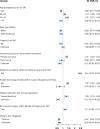
Predictors of surveillance screening over time. The relationship
between longitudinal uptake of asymptomatic surveillance imaging and
patient, tumor, and treatment characteristics in the study cohort (n =
12 559) was modeled using repeated measures multivariable logistic
regression with generalized estimating equations. See Figure S3 for full
model result. OR = odds ratio.
Figure 4:
Predictors of surveillance screening over time. The relationship between longitudinal uptake of asymptomatic surveillance imaging and patient, tumor, and treatment characteristics in the study cohort (n = 12 559) was modeled using repeated measures multivariable logistic regression with generalized estimating equations. See Figure S3 for full model result. OR = odds ratio.
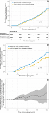
Cumulative rate of ipsilateral invasive breast cancer diagnosis in
women who did and those who did not undergo early surveillance imaging. (A)
The Kaplan-Meier curve indicates the unweighted cumulative rate of
ipsilateral invasive breast cancer diagnosis in women who did and those who
who did not undergo surveillance imaging within 12 months of surgery. P
value is based on log-rank test. (B) The Kaplan-Meier curve indicates the
inverse probability of treatment-weighted cumulative rate of ipsilateral
invasive breast cancer diagnosis in women who did and those who did not
undergo surveillance imaging within 12 months of surgery. P value is based
on weighted log-rank test. (C) Absolute difference in the rate of
ipsilateral invasive breast cancer diagnosis between women who did and those
who did not undergo surveillance imaging within 12 months of surgery. Shaded
region indicates 95% CI.
Figure 5:
Cumulative rate of ipsilateral invasive breast cancer diagnosis in women who did and those who did not undergo early surveillance imaging. (A) The Kaplan-Meier curve indicates the unweighted cumulative rate of ipsilateral invasive breast cancer diagnosis in women who did and those who who did not undergo surveillance imaging within 12 months of surgery. P value is based on log-rank test. (B) The Kaplan-Meier curve indicates the inverse probability of treatment-weighted cumulative rate of ipsilateral invasive breast cancer diagnosis in women who did and those who did not undergo surveillance imaging within 12 months of surgery. P value is based on weighted log-rank test. (C) Absolute difference in the rate of ipsilateral invasive breast cancer diagnosis between women who did and those who did not undergo surveillance imaging within 12 months of surgery. Shaded region indicates 95% CI.

Comment in

References

    1. Erbas B , Provenzano E , Armes J , Gertig D . The natural history of ductal carcinoma in situ of the breast: a review . Breast Cancer Res Treat 2006. ; 97 ( 2 ): 135 – 144 . - PubMed
    1. Jones JL . Overdiagnosis and overtreatment of breast cancer: progression of ductal carcinoma in situ: the pathological perspective . Breast Cancer Res 2006. ; 8 ( 2 ): 204 . - PMC - PubMed
    1. Worni M , Akushevich I , Greenup R , et al. . Trends in treatment patterns and outcomes for ductal carcinoma in situ . J Natl Cancer Inst 2015. ; 107 ( 12 ): djv263 . - PMC - PubMed
    1. Mannu GS , Wang Z , Broggio J , et al. . Invasive breast cancer and breast cancer mortality after ductal carcinoma in situ in women attending for breast screening in England, 1988-2014: population based observational cohort study . BMJ 2020. ; 369 : m1570 . - PMC - PubMed
    1. Van Zee KJ , Subhedar P , Olcese C , Patil S , Morrow M . Relationship Between Margin Width and Recurrence of Ductal Carcinoma In Situ: Analysis of 2996 Women Treated With Breast-conserving Surgery for 30 Years . Ann Surg 2015. ; 262 ( 4 ): 623 – 631 . - PMC - PubMed