. 2023 May 2;119(4):982-997.
doi: 10.1093/cvr/cvad005.

Dietary essential amino acids for the treatment of heart failure with reduced ejection fraction

Affiliations

Dietary essential amino acids for the treatment of heart failure with reduced ejection fraction

Maurizio Ragni et al. Cardiovasc Res. .

Abstract

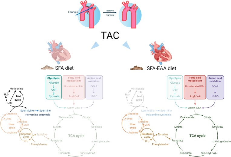
Aims: Heart failure with reduced ejection fraction (HFrEF) is a leading cause of mortality worldwide, requiring novel therapeutic and lifestyle interventions. Metabolic alterations and energy production deficit are hallmarks and thereby promising therapeutic targets for this complex clinical syndrome. We aim to study the molecular mechanisms and effects on cardiac function in rodents with HFrEF of a designer diet in which free essential amino acids-in specifically designed percentages-substituted for protein.

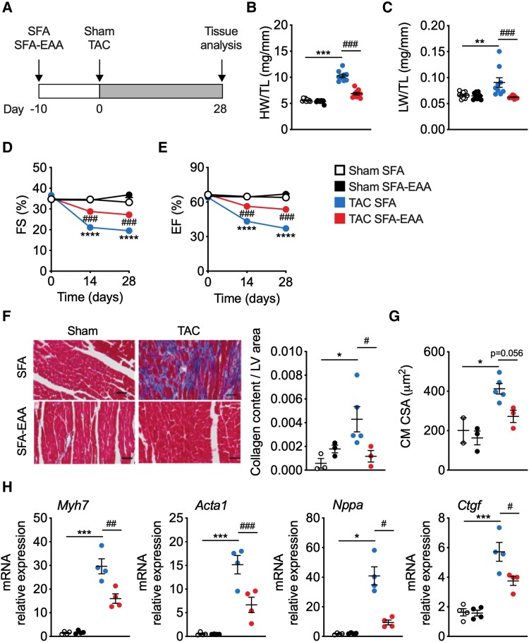
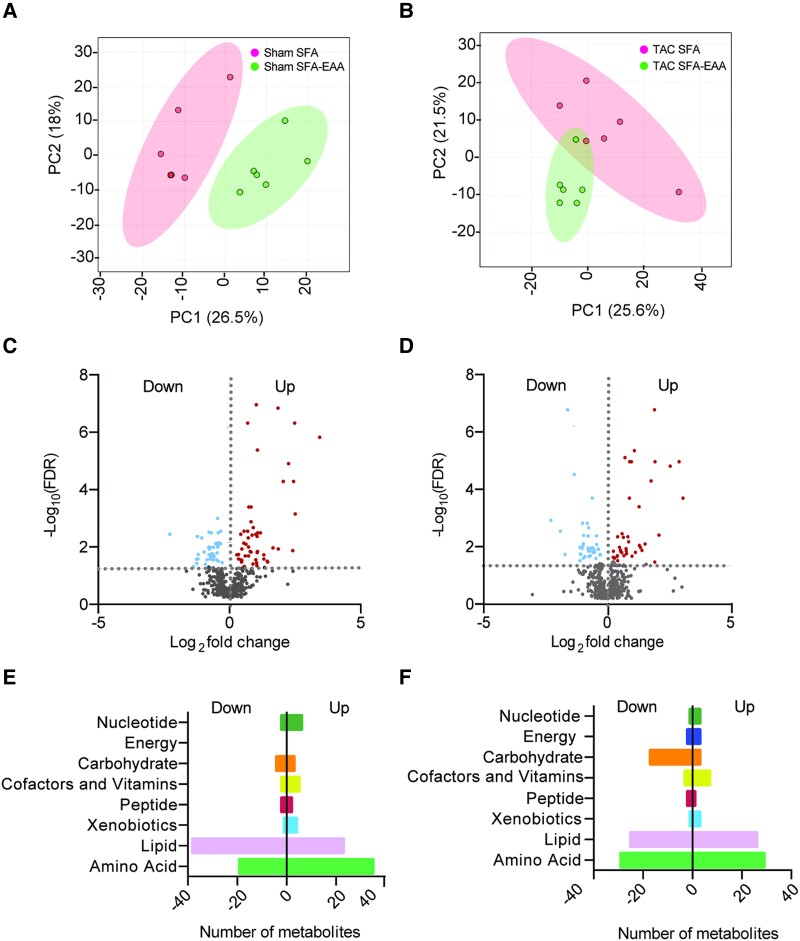
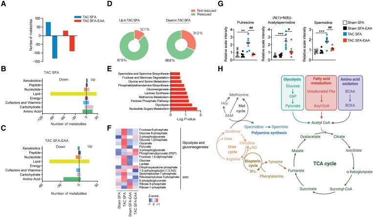
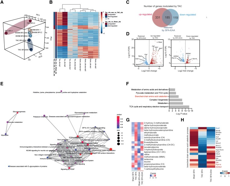
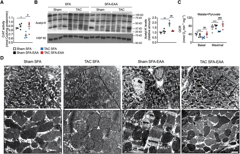
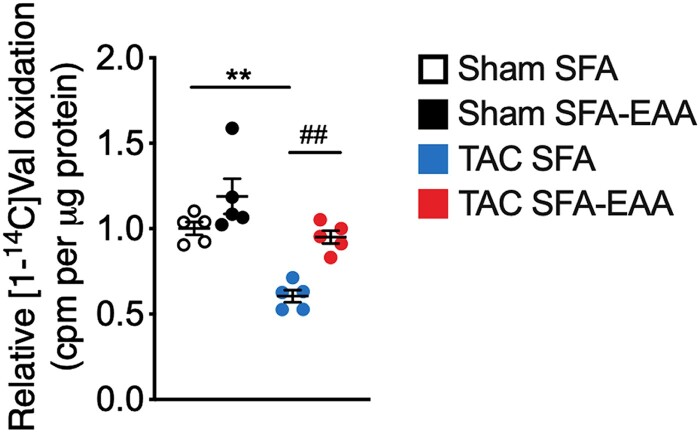
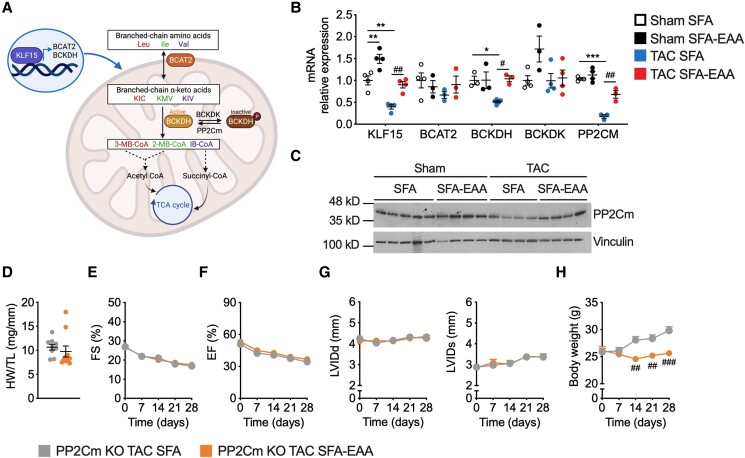
Methods and results: Wild-type mice were subjected to transverse aortic constriction (TAC) to induce left ventricle (LV) pressure overload or sham surgery. Whole-body glucose homeostasis was studied with glucose tolerance test, while myocardial dysfunction and fibrosis were measured with echocardiogram and histological analysis. Mitochondrial bioenergetics and morphology were investigated with oxygen consumption rate measurement and electron microscopy evaluation. Circulating and cardiac non-targeted metabolite profiles were analyzed by ultrahigh performance liquid chromatography-tandem mass spectroscopy, while RNA-sequencing was used to identify signalling pathways mainly affected. The amino acid-substituted diet shows remarkable preventive and therapeutic effects. This dietary approach corrects the whole-body glucose metabolism and restores the unbalanced metabolic substrate usage-by improving mitochondrial fuel oxidation-in the failing heart. In particular, biochemical, molecular, and genetic approaches suggest that renormalization of branched-chain amino acid oxidation in cardiac tissue, which is suppressed in HFrEF, plays a relevant role. Beyond the changes of systemic metabolism, cell-autonomous processes may explain at least in part the diet's cardioprotective impact.

Conclusion: Collectively, these results suggest that manipulation of dietary amino acids, and especially essential amino acids, is a potential adjuvant therapeutic strategy to treat systolic dysfunction and HFrEF in humans.

Keywords: Amino acids; Heart failure; Mitochondrial function; Nutrition; Transcriptomic reprogramming.


Conflict of interest statement

Conflict of interest: None declared.

Figures

Graphical abstract
Graphical abstract
Figure 1
Figure 1
SFA-EAA diet prevents systolic dysfunction caused by pressure overload. (A) Schematic overview of SFA and SFA-EAA feeding protocol to male C57BL/6N mice exposed to sham or transverse aortic constriction (TAC) surgery. Mice were fed with the two diets for 10 days before surgery and for additional 4 weeks after surgery, when cardiac parameters were assessed (if not otherwise indicated). (B and C) Heart weight (B) and lung weight (C) normalized to tibia length (HW/TL) (LW/TL) (n = 9 mice per group). (D) Percentage fractional shortening (FS %) and (E) relative ejection fraction (EF %) in sham- and TAC-operated mice fed with SFA or SFA-EAA diet were quantified by echocardiography (n = 10 mice per group). (F) Left, representative cross-sections of LV from mice fed with SFA or SFA-EAA diet, subjected to sham or TAC surgery, and stained with Azan’s trichrome collagen staining. Scale bars 50 µm. Right, quantification of the fibrotic area showed as the ratio between collagen content vs. area of LV (n = 3 mice per group). (G) Quantification of cardiomyocyte cross-sectional area (CSA) using wheat germ agglutinin (WGA) staining. (H) Gene expression analysis (qRT-PCR) of hypertrophy and fibrosis markers in LV tissue (n = 4 animals per group). All data are presented as mean ± SEM. *P < 0.05, **P < 0.01, and ***P < 0.001 vs. sham-operated mice fed with SFA diet; #P < 0.05, ##P < 0.01, and ###P < 0.001 vs. TAC-operated mice fed with SFA diet; comparison was performed by two-way ANOVA followed by post hoc Tukey’s test.
Figure 2
Figure 2
Metabolic reprogramming mediated by the SFA-EAA diet is associated with extensive changes in cardiac metabolite levels related to energy metabolism in sham- and TAC-operated mice. A and B: Principal component analysis (PCA) of metabolome data. Cardiac tissue (LV) was obtained from sham- (A) and TAC-operated (B) mice fed with SFA or SFA-EAA diet as in Figure 1A (n = 6 mice per group). (C and D) Volcano plot showing upregulated (red dots), downregulated (light-blue dots), and unchanged (grey dots) metabolites in LV of sham- (C) and TAC-operated (D) mice fed with SFA-EAA vs. SFA diet (n = 6 mice per group; P < 0.05). E and F: Biochemical classification of metabolites in LV of sham- (E) and TAC-operated (F) mice, shown as the number of metabolites for each class significantly modulated by SFA-EAA vs. SFA diet (n = 6 mice per group). Statistical analysis was performed with two-way ANOVA and an estimate of the false discovery rate (FDR) for multiple comparisons.
Figure 3
Figure 3
SFA-EAA diet preserves cardiac metabolism upon pressure overload. (A) Bars represent the number of metabolites significantly upregulated or downregulated (P ≤ 0.05) by TAC in LV of mice fed with SFA and SFA-EAA diet as in Figure 1A (n = 6 mice per group). (B and C) Biochemical classification of metabolites in LV of TAC-operated mice, shown as the number of metabolites for each class significantly modulated by SFA (B) and SFA-EAA diet (C) (n = 6 mice per group). (D) Percentage of metabolites in LV of SFA-fed mice, upregulated (left) or downregulated (right) by the pressure overload and that were rescued or not by the SFA-EAA diet (n = 6 mice per group). (E) Pathway-enrichment analysis of metabolites significantly (P ≤ 0.05) increased upon TAC in the SFA group (n = 6 mice per group). (F) Heatmap of cardiac metabolites showing the levels (red = high, blue = low) of differentially regulated intermediates involved in glycolytic, gluconeogenesis, and pentose phosphate pathway in sham- and TAC-operated mice fed with SFA and SFA-EAA diet (n = 6 mice per group). (G) Polyamine metabolite levels in LV of sham- and TAC-operated mice fed with SFA and SFA-EAA diet. The relative scale intensity was determined by rescaling each metabolite (n = 6) to set the median equal to 1.0. All data are presented as mean ± SEM. **P < 0.01 and ***P < 0.001 vs. sham-operated mice fed with SFA diet; #P < 0.05, ##P < 0.01, and ###P < 0.001 vs. TAC-operated mice fed with SFA diet. Statistical analysis was performed with two-way ANOVA followed by Tukey’s post hoc test (panel G) and Fisher's Exact Test (E). (H) Schematic diagram showing the main metabolic pathways affected by the SFA-EAA diet in cardiac tissue of TAC-operated mice. Diagram was created with BioRender.com.
Figure 4
Figure 4
SFA-EAA diet preserves cardiac transcriptomic profile upon pressure overload. (A) Three-dimension principal component analysis (3D PCA) of RNA-seq data in LV samples obtained from sham- and TAC-operated mice fed with SFA or SFA-EAA diet as reported in Figure 1A. (B) Heatmaps of cardiac transcripts showing the levels (red = high, blue = low) of differentially regulated genes following sham and TAC surgery in mice on the different dietary regimens. Statistical criteria is false discovery rate (FDR < 0.05) (n = 2 mice per group). The upregulated (dark red), downregulated (dark blue), or not changed (grey) gene groups are also reported in TAC-operated mice fed with SFA-EAA vs. SFA diet or in TAC- vs. sham-operated mice fed with SFA diet (the two left columns). (C) Venn diagram showing upregulated (red circle) and downregulated (blue circle) genes in LV following pressure overload, and the genes entirely restored by the SFA-EAA diet (overlap). (D) Volcano plot showing cardiac genes upregulated (red in the left panel) and downregulated (red in the right panel) by TAC surgery and their restoration by the SFA-EAA diet (blue in the left and right panels, respectively) (n = 2 mice per group; FDR < 0.05). (E) Pathway analysis of cardiac genes significantly modulated by the SFA-EAA diet in TAC-operated mice. The P-values indicate the enrichment level of the pathway term; the colour intensity and size indicate the significativity level and the number of proteins included in a single pathway, respectively. (F) Joint pathway analysis of transcriptomics and metabolomics data (n = 6 mice per group). (G) Heatmap of the levels of BCAA catabolites in sham- and TAC-operated mice fed with SFA or SFA-EAA diet (n = 6 mice per group). (H) Heatmap of the expression profile of BCAA oxidation genes in mice fed with SFA or SFA-EAA diet and subjected to sham or TAC surgery. Differential expression analysis was performed using the GLM approach in edgeR using a false discovery rate (FDR), while pathway analysis was made with Integrated Molecular Pathway Level Analysis (IMPaLA) using a P-value from Fisher's Exact Test.
Figure 5
Figure 5
SFA-EAA diet restores TAC-induced mitochondrial dysfunction and structural damage. (A) Carnitine acetyltransferase (CrAT) activity in LV obtained from sham- and TAC-operated mice fed with SFA or SFA-EAA diet as reported in Figure 1A. CrAT activity is expressed as nmol of free coenzyme A (CoA) released per minute and normalized to mg of total proteins (n = 4 mice per group). (B) Immunoblot analysis and quantification of lysine acetylation (acetyl-K) of total mitochondrial proteins in LV. One experiment representative of three reproducible ones is shown. (C) Oxygen consumption rates (OCRs) in the presence of malate/pyruvate in mitochondria obtained from LV (n = 4 mice per group). Basal: ADP-stimulated respiration (state 3); Maximal: maximal respiration (i.e. the FCCP-induced uncoupled state) (n = 5 mice per group). (D) Transmission electron microscopy images showing the mitochondrial morphology in LV. A magnified view of the regions outlined by the lined boxes is reported below. Globular, swollen mitochondria are predominant in TAC-operated mice fed with the SFA diet, with signs of constriction suggesting early stage (arrow) and late-stage (arrowhead) fission events. Scale bar (shown only in TAC SFA-EAA lower pannel), 1.8 µm (upper pannels) and 0.3 µm (lower pannels) (n = 2 mice per group). All data are presented as mean ± SEM. *P < 0.05, **P < 0.01, and ***P < 0.001 vs. sham-operated mice fed with SFA diet; #P < 0.05, ##P < 0.01, and ###P < 0.001 vs. TAC-operated mice fed with SFA diet. Statistical analysis was performed using two-way ANOVA followed by the Tukey’s post hoc test.
Figure 6
Figure 6
Valine oxidation rate in LV obtained from sham- and TAC-operated mice fed with SFA or SFA-EAA diet as reported in Figure 1A, normalized to tissue mass (n = 3 mice per group). **P < 0.01 vs. sham-operated mice fed with SFA diet; ##P < 0.01, vs. TAC-operated mice fed with SFA diet using two-way ANOVA followed by the Tukey’s post hoc test.
Figure 7
Figure 7
Cardiac BCAA catabolism is required for the beneficial effects of the SFA-EAA diet in HFrEF. (A) KLF15 promotes the expression of enzymes crucial to the BCAA oxidation (e.g. BCAT2, BCKDH, and PP2Cm), producing acetyl-CoA and succinyl-CoA intermediates of the TCA cycle. KLF15 and BCAA degradation enzymes are downregulated in failing hearts, increasing cardiotoxic branched-chain ketoacids, mTOR activity, and cell growth. Ile, isoleucine; Leu, leucine; Val, valine; BCAT2, mitochondrial BCAA aminotransferase; BCKDH, branched-chain α-ketoacid dehydrogenase complex; BCKDK, branched-chain ketoacid dehydrogenase kinase; PP2Cm, protein phosphatase 2Cm; KIC, ketoisocaproic acid; KMV, α-keto-β-methylvaleric acid; KIV, α-ketoisovaleric acid; 3-MB-CoA, 3-methylcrotonyl-CoA; 2-MB-CoA, 2-methyl-3-hydroxy-butyryl-CoA; IB-CoA, 3-hydroxy-isobutyryl-CoA; TCA, tricarboxylic acid cycle. The BCKDH complex is inactive when phosphorylated by BCKDK and active when dephosphorylated by PP2Cm. (B) Relative mRNA levels of BCAA catabolic enzyme genes in LV obtained from sham- and TAC-operated mice fed with SFA or SFA-EAA diet as reported in Figure 1A (n = 3 mice per group). (C) Western blot analysis of PP2Cm in LV of mice fed with SFA or SFA-EAA diet and subjected to sham or TAC surgery. One immunoblot experiment representative of three reproducible ones (n = 4–5 mice per group). (D) Heart weight normalized to tibia length (HW/TL) in TAC-operated PP2Cm-null mice fed with SFA and SFA-EAA diet as in Figure 1A (n = 10 mice per group). (E) Percentage fractional shortening (FS %) and (F) relative ejection fraction (EF %) in TAC-operated PP2Cm-null mice fed with SFA or SFA-EAA diet were quantified by echocardiography (n = 10 mice per group). G: Left ventricular internal diameter end diastole (LVIDd) (left panel), left ventricular internal diameter end systole (LVIDs) (right panel) and body weight measured once a week (H) in TAC-operated PP2Cm-null mice fed with SFA and SFA-EAA diet (n = 10 mice per group). All data are presented as mean ± SEM. *P < 0.05, **P < 0.01, and ***P < 0.001 vs. sham-operated mice fed with SFA diet; #P < 0.05, ##P < 0.01, and ###P < 0.001 vs. TAC-operated mice fed with SFA diet. Statistical analysis was performed with two-way ANOVA followed by Tukey’s post hoc test (panel B) and unpaired Student’s t-test (panel D–H). The cartoon illustration in panel A was created with BioRender.com.

References

    1. Benjamin EJ, Muntner P, Alonso A, Bittencourt MS, Callaway CW, Carson AP, Chamberlain AM, Chang AR, Cheng S, Das SR, Delling FN, Djousse L, Elkind MSV, Ferguson JF, Fornage M, Jordan LC, Khan SS, Kissela BM, Knutson KL, Kwan TW, Lackland DT, Lewis TT, Lichtman JH, Longenecker CT, Loop MS, Lutsey PL, Martin SS, Matsushita K, Moran AE, Mussolino ME, O’Flaherty M, Pandey A, Perak AM, Rosamond WD, Roth GA, Sampson UKA, Satou GM, Schroeder EB, Shah SH, Spartano NL, Stokes A, Tirschwell DL, Tsao CW, Turakhia MP, VanWagner LB, Wilkins JT, Wong SS, Virani SS. Heart disease and stroke statistics-2019 update: a report from the American heart association. Circulation 2019;139:e56–e528. - PubMed
    1. Savarese G, Lund LH. Global public health burden of heart failure. Card Fail Rev 2017;3:7–11. - PMC - PubMed
    1. Rossignol P, Hernandez AF, Solomon SD, Zannad F. Heart failure drug treatment. Lancet. 2019;393:1034–1044. - PubMed
    1. Gheorghiade M, Larson CJ, Shah SJ, Greene SJ, Cleland JGF, Colucci WS, Dunnmon P, Epstein SE, Kim RJ, Parsey RV, Stockbridge N, Carr J, Dinh W, Krahn T, Kramer F, Wahlander K, Deckelbaum LI, Crandall D, Okada S, Senni M, Sikora S, Sabbah HN, Butler J. Developing new treatments for heart failure: Focus on the Heart. Circ Hear Fail 2016;9:e002727. - PubMed
    1. Zhou B, Tian R. Mitochondrial dysfunction in pathophysiology of heart failure. J Clin Invest. 2018;128:3716–3726. - PMC - PubMed

Publication types

Substances