

The comprehensive comparison of imaging sign from CT angiography and noncontrast CT for predicting intracranial hemorrhage expansion: A comparative study
- PMID: 36626412
- PMCID: PMC9750542
- DOI: 10.1097/MD.0000000000031914
The comprehensive comparison of imaging sign from CT angiography and noncontrast CT for predicting intracranial hemorrhage expansion: A comparative study
Abstract
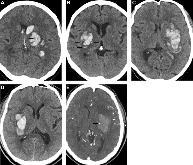
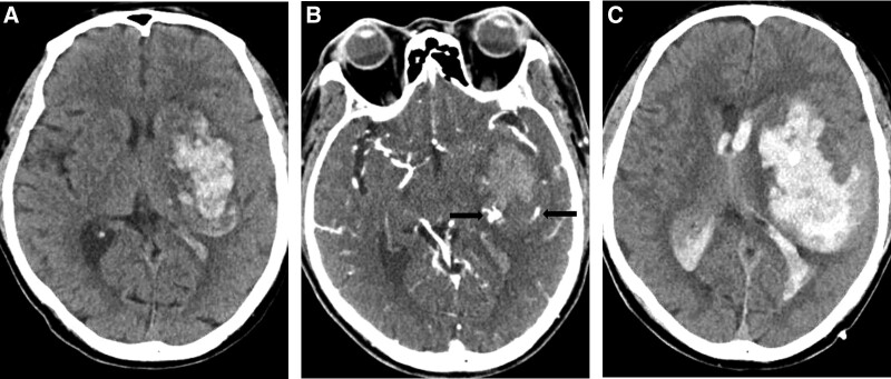
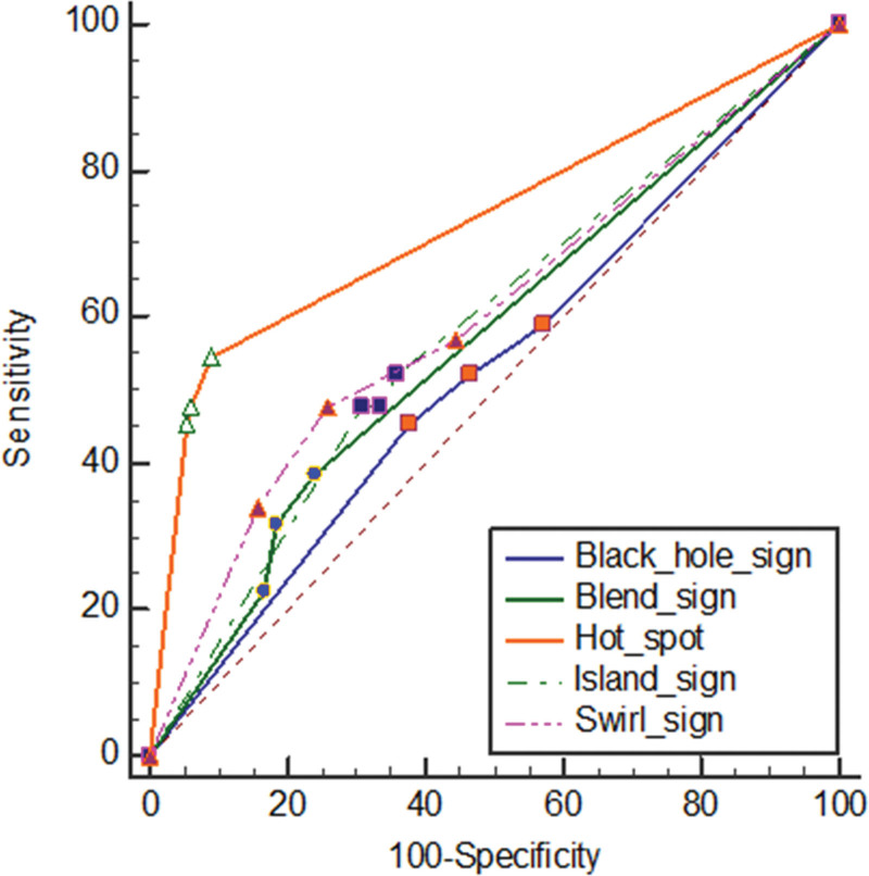
Expansion of intracranial hemorrhage (ICH) is an important predictor of poor clinical outcomes. Various imaging markers on non-contrast computed tomography (NCCT) or computed tomographic angiography (CTA) have been reported as predictors of ICH expansion. We aimed to compare the associations between various CT imaging markers and ICH expansion. Patients with spontaneous ICH who underwent initial NCCT, CTA, and subsequent NCCT between January 2016 and December 2019 were retrospectively identified. ICH expansion was defined as a volume increase of > 33% or > 6 mL. We analyzed the presence of imaging markers such as the black hole sign, blend sign, island sign, or swirl sign on initial NCCT or spot sign on CTA. An alternative free-response receiver operating characteristic curve analysis was performed using a 4-point scoring system based on the consensus of the reviewers. The predictive value of each marker was assessed using univariate and multivariate logistic regression analyses. A total of 250 patients, including 60 (24.0%) with ICH expansion, qualified for the analysis. Among the patients with spontaneous ICH, 118 (47.2%) presented with a black hole sign, 52 (20.8%) with a blend sign, 93 (37.2%) with an island sign, 79 (31.6%) with a swirl sign, and 56 (22.4%) with a spot sign. In univariate logistic regression, the initial ICH volume (P = .038), initial intraventricular hemorrhage (IVH) presence (P < .001), swirl sign (P < .001), and spot sign (P < .001) were associated with ICH expansion. Multivariate analysis confirmed that the presence of initial IVH (odds ratio, 4.111; P = .002) and spot sign (odds ratio, 109.5; P < .001) were independent predictors of ICH expansion. Initial ICH volume, IVH, swirl sign, and spot sign are associated with ICH expansion. The presence of spot signs and IVH were independent predictors of ICH expansion.
Copyright © 2022 the Author(s). Published by Wolters Kluwer Health, Inc.
Conflict of interest statement
The authors have no conflicts of interest to disclose.
Figures
Similar articles
-
Diagnostic value of swirl sign on noncontrast computed tomography and spot sign on computed tomographic angiography to predict intracranial hemorrhage expansion.Clin Neurol Neurosurg. 2019 Jul;182:130-135. doi: 10.1016/j.clineuro.2019.05.013. Epub 2019 May 14. Clin Neurol Neurosurg. 2019. PMID: 31121472
-
Triage of 5 Noncontrast Computed Tomography Markers and Spot Sign for Outcome Prediction After Intracerebral Hemorrhage.Stroke. 2018 Oct;49(10):2317-2322. doi: 10.1161/STROKEAHA.118.021625. Stroke. 2018. PMID: 30355120
-
Non-contrast computed tomography features predict intraventricular hemorrhage growth.Eur Radiol. 2023 Nov;33(11):7807-7817. doi: 10.1007/s00330-023-09707-9. Epub 2023 May 22. Eur Radiol. 2023. PMID: 37212845 Free PMC article.
-
Computed Tomography Imaging Predictors of Intracerebral Hemorrhage Expansion.Curr Neurol Neurosci Rep. 2021 Mar 12;21(5):22. doi: 10.1007/s11910-021-01108-z. Curr Neurol Neurosci Rep. 2021. PMID: 33710468 Review.
-
Standards for Detecting, Interpreting, and Reporting Noncontrast Computed Tomographic Markers of Intracerebral Hemorrhage Expansion.Ann Neurol. 2019 Oct;86(4):480-492. doi: 10.1002/ana.25563. Epub 2019 Aug 24. Ann Neurol. 2019. PMID: 31364773 Review.
Cited by
-
Research advances in predicting the expansion of hypertensive intracerebral hemorrhage based on CT images: an overview.PeerJ. 2024 Jun 7;12:e17556. doi: 10.7717/peerj.17556. eCollection 2024. PeerJ. 2024. PMID: 38860211 Free PMC article. Review.
-
The role of imaging in predicting 3-month prognosis of primary intracerebral hemorrhage: a single-center, prospective observational study in tertiary care hospital.Quant Imaging Med Surg. 2025 Jun 6;15(6):5674-5688. doi: 10.21037/qims-24-1299. Epub 2025 Jun 3. Quant Imaging Med Surg. 2025. PMID: 40606359 Free PMC article.
References
-
- Brott T, Broderick J, Kothari R, et al. . Early hemorrhage growth in patients with intracerebral hemorrhage. Stroke. 1997;28:1–5. - PubMed
-
- Li Q, Shen YQ, Xie XF, et al. . Expansion-prone hematoma: defining a population at high risk of hematoma growth and poor outcome. Neurocrit Care. 2019;30:601–8. - PubMed
-
- Davis SM, Broderick J, Hennerici M, et al. . Hematoma growth is a determinant of mortality and poor outcome after intracerebral hemorrhage. Neurology. 2006;66:1175–81. - PubMed
-
- Delcourt C, Huang Y, Arima H, et al. . Hematoma growth and outcomes in intracerebral hemorrhage: the INTERACT1 study. Neurology. 2012;79:314–9. - PubMed
-
- He GN, Guo HZ, Han X, et al. . Comparison of CT black hole sign and other CT features in predicting hematoma expansion in patients with ICH. J Neurol. 2018;265:1883–90. - PubMed
MeSH terms
LinkOut - more resources
Full Text Sources
