. 2023 Feb 20;6(2):591-602.
doi: 10.1021/acsabm.2c00892. Epub 2023 Jan 10.

Engineered Nonviral Protein Cages Modified for MR Imaging

Affiliations

Engineered Nonviral Protein Cages Modified for MR Imaging

Megan A Kaster et al. ACS Appl Bio Mater. .

Abstract

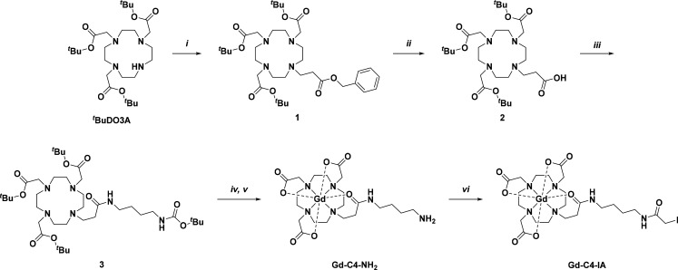
Diagnostic medical imaging utilizes magnetic resonance (MR) to provide anatomical, functional, and molecular information in a single scan. Nanoparticles are often labeled with Gd(III) complexes to amplify the MR signal of contrast agents (CAs) with large payloads and high proton relaxation efficiencies (relaxivity, r1). This study examined the MR performance of two structurally unique cages, AaLS-13 and OP, labeled with Gd(III). The cages have characteristics relevant for the development of theranostic platforms, including (i) well-defined structure, symmetry, and size; (ii) the amenability to extensive engineering; (iii) the adjustable loading of therapeutically relevant cargo molecules; (iv) high physical stability; and (v) facile manufacturing by microbial fermentation. The resulting conjugates showed significantly enhanced proton relaxivity (r1 = 11-18 mM-1 s-1 at 1.4 T) compared to the Gd(III) complex alone (r1 = 4 mM-1 s-1). Serum phantom images revealed 107% and 57% contrast enhancements for Gd(III)-labeled AaLS-13 and OP cages, respectively. Moreover, proton nuclear magnetic relaxation dispersion (1H NMRD) profiles showed maximum relaxivity values of 50 mM-1 s-1. Best-fit analyses of the 1H NMRD profiles attributed the high relaxivity of the Gd(III)-labeled cages to the slow molecular tumbling of the conjugates and restricted local motion of the conjugated Gd(III) complex.

Keywords: NMRD; gadolinium; magnetic resonance imaging; magnetism; nonviral protein cages.


Conflict of interest statement

The authors declare no competing financial interest.

Figures

Figure 1
Figure 1
Engineered AaLS-13 and OP protein cages. Surface representation of (a) AaLS-13 (PDB 5MQ7) and (b) OP (PDB 6FDB) cages. AaLS-13 assembles into 38 nm spherical cages, possessing a negatively supercharged interior, from 72 pentameric subunits. OP forms ∼13 nm positively supercharged cubic cages from eight trimeric capsomers.
Figure 2
Figure 2
Synthetic scheme for Gd-C4-IA. (i) tBuDO3A (1 equiv), benzyl acrylate (2 equiv), DIEA (5.9 equiv), MeCN, N2 (g), RT, 47%; (ii) 1 (1 equiv), Pd/C (catalyst), MeOH, H2 (g), RT, 27%; (iii) 2 (1 equiv), tBu (4-aminobutyl)carbamate (1.5 equiv), NHS (3 equiv), DIEA (5 equiv), DIC (5 equiv), DMF, N2 (g), RT, quantitative yield; (iv) 3 (1 equiv), TFA, CH2Cl2, N2 (g), RT, crude; (v) 4 (1 equiv), GdCl3·6H2O (1.3 equiv), H2O, N2 (g), RT, 34% over 2 steps; (vi) Gd-C4–NH2 (1 equiv), iodoacetic anhydride (3 equiv), K2CO3 (3 equiv), DMF, N2 (g), 0 °C, 20%.
Figure 3
Figure 3
Thiol-mediated functionalization of AaLS-13 and OP and transmission electron microscopy (TEM) images of AaLS-13 and OP-3intC. (a) Transparent surface of a pentamer used to construct an AaLS-13 cage (left) and a trimer used to construct OP (right). Monomers are shown as gray ribbons. Targeted cysteine residue on AaLS-13 (Cys127) and positions on OP targeted for cysteine mutations (Ser38, Arg66, and Arg103) are highlighted as yellow spheres. (b) Representations of Gd(III) labeling the cysteine reactive sites. TEM images of (c) AaLS-13 and (d) OP-3intC cages, unmodified (left) or labeled with Gd-complexes (right). Scale bar is equal to 50 nm.
Figure 4
Figure 4
1H relaxivity profiles of Gd-C4-IA and Gd(III)-labeled protein cages. (a) Gd-C4-IA and Gd-AaLS-13 in sodium phosphate (pH 8.0) buffer at 25 °C and 37 °C. (b) Gd-C4-IA, Gd-OP-3intC, Gd-OP-2intC, Gd-OP-1intC, and Gd-OP-1extC in Tris (pH 7.6) buffer at 25 °C and 37 °C. Solid and dotted lines are the best fit profiles at 25 and 37 °C, respectively, obtained with the parameters reported in Table 3 and Tables S4–S6.
Figure 5
Figure 5
T1-weighted MR solution phantom images of (a) Gd-AaLS-13 and (b) Gd-OP-3intC at 3, 7, and 9.4 T. (a) Control sample of 10% FBS in 50 mM sodium phosphate (pH 8.0), 200 mM NaCl, 5 mM EDTA. The Gd-AaLS-13 sample was prepared at 67 μM with respect to monomer, and Gd(III) concentration was measured by ICP-MS. (b) Control sample of 10% FBS in 25 mM Tris-HCl (pH 7.6), 200 mM NaCl, 5 mM EDTA. The Gd-OP-3intC sample was prepared at 20 μM with respect to monomer, and Gd(III) concentration was measured by ICP-MS. 1Values at 3 T were measured using a dual gradient echo method with two different flip angles. 2Values at 7 and 9.4 T were obtained using a saturation recovery method.

References

    1. Currie S.; Hoggard N.; Craven I. J.; Hadjivassiliou M.; Wilkinson I. D. Understanding MRI: basic MR physics for physicians. Postgrad Med. J. 2013, 89 (1050), 209–223. 10.1136/postgradmedj-2012-131342. - DOI - PubMed
    1. Jaffer F. A.; Weissleder R. Molecular imaging in the clinical arena. JAMA 2005, 293 (7), 855–862. 10.1001/jama.293.7.855. - DOI - PubMed
    1. Wahsner J.; Gale E. M.; Rodriguez-Rodriguez A.; Caravan P. Chemistry of MRI Contrast Agents: Current Challenges and New Frontiers. Chem. Rev. 2019, 119 (2), 957–1057. 10.1021/acs.chemrev.8b00363. - DOI - PMC - PubMed
    1. Caravan P.; Ellison J. J.; McMurry T. J.; Lauffer R. B. Gadolinium(III) Chelates as MRI Contrast Agents: Structure, Dynamics, and Applications. Chem. Rev. 1999, 99 (9), 2293–2352. 10.1021/cr980440x. - DOI - PubMed
    1. Caravan P. Strategies for increasing the sensitivity of gadolinium based MRI contrast agents. Chem. Soc. Rev. 2006, 35 (6), 512–523. 10.1039/b510982p. - DOI - PubMed

Publication types