. 2023 Jan 10;14(1):79.
doi: 10.1038/s41467-022-35608-4.

Decoding of the ubiquitin code for clearance of colliding ribosomes by the RQT complex

Affiliations

Decoding of the ubiquitin code for clearance of colliding ribosomes by the RQT complex

Yoshitaka Matsuo et al. Nat Commun. .

Abstract

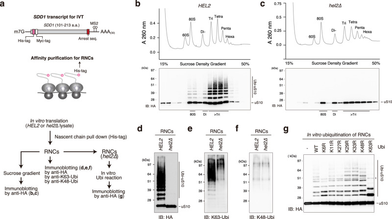
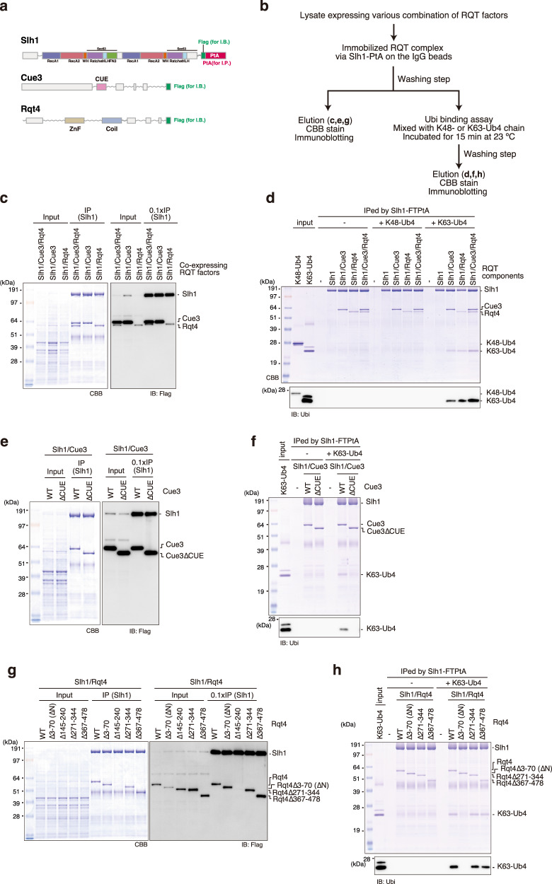
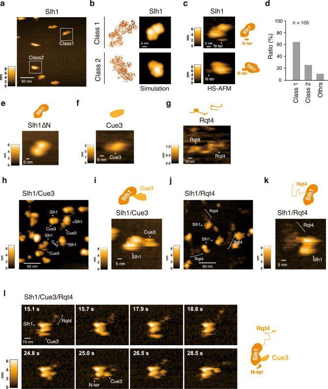
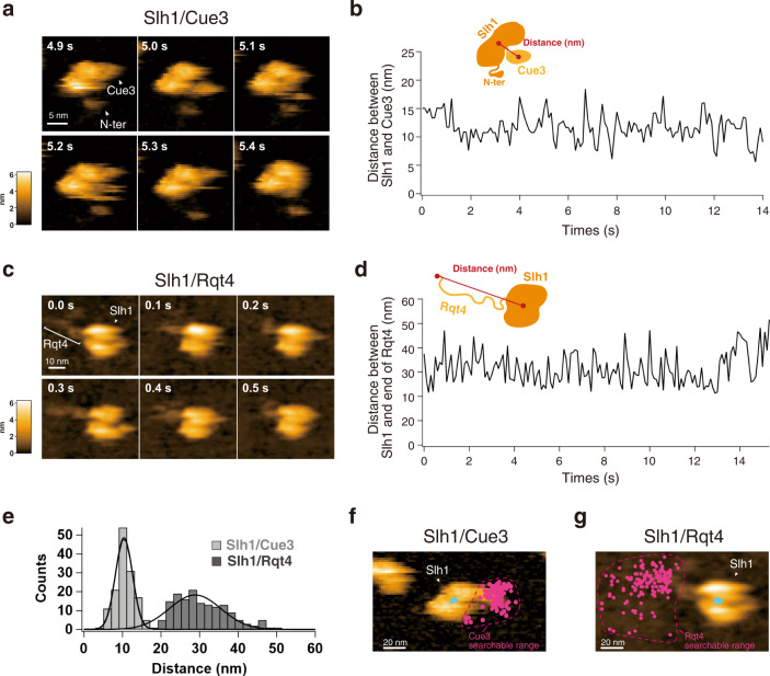
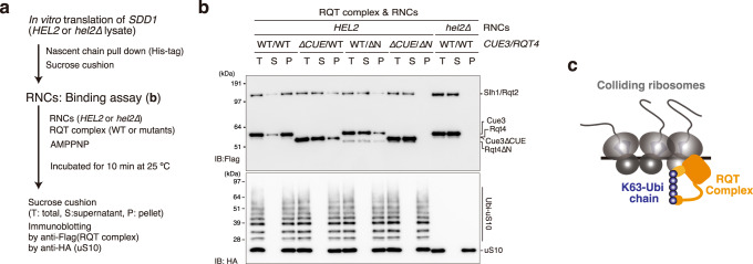
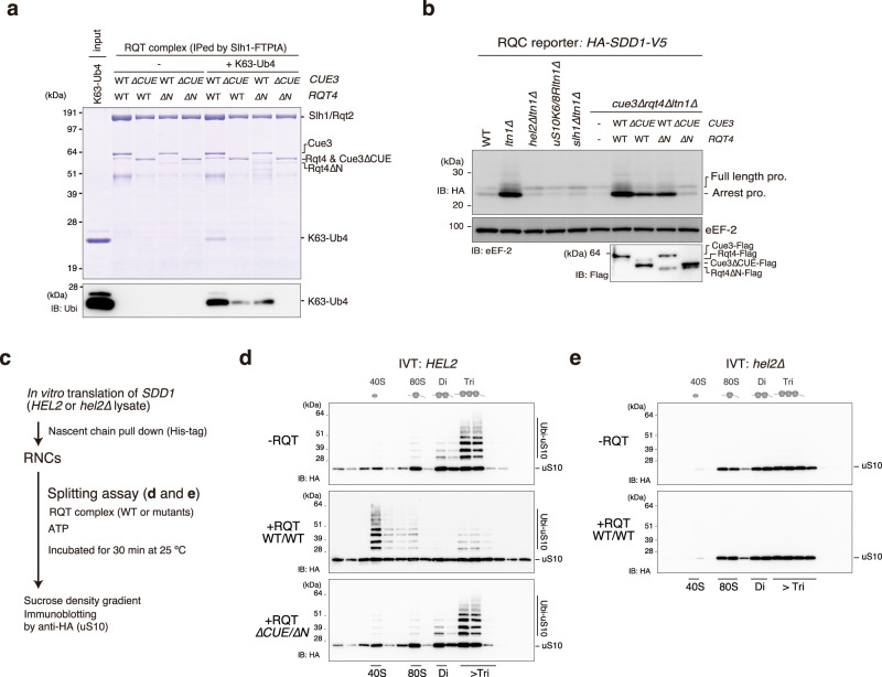
The collision sensor Hel2 specifically recognizes colliding ribosomes and ubiquitinates the ribosomal protein uS10, leading to noncanonical subunit dissociation by the ribosome-associated quality control trigger (RQT) complex. Although uS10 ubiquitination is essential for rescuing stalled ribosomes, its function and recognition steps are not fully understood. Here, we show that the RQT complex components Cue3 and Rqt4 interact with the K63-linked ubiquitin chain and accelerate the recruitment of the RQT complex to the ubiquitinated colliding ribosome. The CUE domain of Cue3 and the N-terminal domain of Rqt4 bind independently to the K63-linked ubiquitin chain. Their deletion abolishes ribosomal dissociation mediated by the RQT complex. High-speed atomic force microscopy (HS-AFM) reveals that the intrinsically disordered regions of Rqt4 enable the expansion of the searchable area for interaction with the ubiquitin chain. These findings provide mechanistic insight into the decoding of the ubiquitin code for clearance of colliding ribosomes by the RQT complex.


Conflict of interest statement

The authors declare no competing interests.

Figures

Fig. 1
Fig. 1. In vitro reconstitution of the ubiquitinated colliding ribosomes.
a Schematic drawing of the SDD1 model mRNA used in the in vitro translation (IVT) assay (Top). Schematic of the in vitro experiments (bottom). b, c The purified RNCs in the IVT reaction using HEL2-containing or hel2-knockout (hel2∆) IVT extract were separated by sucrose density gradient centrifugation and detected by UV absorbance at a wavelength of 260 nm. HA-tagged uS10 in each fraction was detected by immunoblotting using an anti-HA antibody. Immunoblotting of purified RNCs using anti-HA (d), K63-linkage (e), and K48-linkage-specific anti-ubiquitin antibodies (f). g In vitro ubiquitination assay. Purified RNCs derived from the hel2-knockout (hel2∆) IVT reaction mixed with Hel2 (E3), Uba1 (E1), Ubc4 (E2), ATP, and ubiquitin or the indicated ubiquitin mutants. After the reaction, HA-tagged uS10 was detected by immunoblotting using an anti-HA antibody. All experiments were performed at least twice with highly reproducible results.
Fig. 2
Fig. 2. The RQT complex interacts with the K63-linked ubiquitin chain via two accessory proteins.
a Domain structure of each component of the RQT complex. b Schematic of the experiments. c, e, g Purification of the complex containing the indicated RQT factors. Different combinations of the indicated RQT factors were co-expressed in yeast cells. The complex was affinity-purified via Slh1-Flag-TEV-protein A using IgG magnetic beads. Copurified RQT factors were separated by 8% Nu-PAGE and detected by Coomassie brilliant blue (CBB) staining or immunoblotting using an anti-Flag antibody. d, f, h Pull-down assay of the RQT complex with the K63- or K48-linked tetraubiquitin chain. The complex containing the indicated RQT factors was immobilized on IgG magnetic beads and mixed with the K63- or K48-linked tetraubiquitin chain. After binding and washing steps (as indicated in 2b), the proteins in the final elution were separated by 10% Nu-PAGE and detected by CBB staining or immunoblotting using an anti-ubiquitin antibody. All experiments were performed at least twice with highly reproducible results.
Fig. 3
Fig. 3. The dynamics of the RQT complex.
a HS-AFM image of Slh1. Two major particles were indicated as Class1 and Class2. b The pseudo-AFM images of Slh1 belonging to Class1 and Class2 particles, which were simulated using predicted Slh1 structure lacking N-terminal region by Alphafold2. c The HS-AFM images and schematized molecular features of Slh1. d Classification of Slh1 particles. All particles used for the classification were presented in the supplementary Fig. 4. e HS-AFM images of Slh1 lacking N-terminal region (Slh1∆N). f HS-AFM image of Cue3. g HS-AFM image of Rqt4. h, i HS-AFM images of Slh1/Cue3 complex. j, k HS-AFM images of Slh1/Rqt4 complex. l The time-lapse HS-AFM images of the RQT complex. All experiments were performed at least twice with highly reproducible results.
Fig. 4
Fig. 4. The movable range of the accessory proteins of RQT complex.
a The time-lapse HS-AFM images of the Slh1/Cue3 complex. b Time course of the distance between Slh1 and Cue3 as described in the schematic diagram. The distance was calculated for each frame per 0.1 s. c The time-lapse HS-AFM images of the Slh1/Rqt4 complex. d Time course of the distance between center of Slh1 and the most distant point of Rqt4 from center of Slh1 as described in the schematic diagram. The distance was calculated for each frame per 0.1 s. e The histogram of the Slh1-Cue3 distance (b) and the Slh1-end of Rqt4 distance (d). f The 2D plot of the center positions of Cue3 (magenta) and Slh1 (cyan) of all frames on the Slh1/Cue3 HS-AFM image at 0 s frame. (g) The 2D plot of the most distant positions of Rqt4 (magenta) and the center position of Slh1 (cyan) of all frames on the Slh1/Rqt4 HS-AFM image at 0 s frame. All experiments were performed at least twice with highly reproducible results.
Fig. 5
Fig. 5. The recruitment of RQT complex into the colliding ribosome in the ubiquitin decoding-dependent manner.
a Schematic of the experiments. b Binding assay between the colliding ribosome and the RQT complex. The ubiquitinated or nonubiquitinated colliding ribosomes were prepared by HEL2-containing or hel2-knockout (hel2∆) IVT reaction and mixed with the indicated RQT complex in the presence of AMPPMP as described in (a). After the binding reaction, free and bound RQT complexes were separated using a sucrose cushion. The RQT factors and uS10 in each fraction were detected by immunoblotting using anti-Flag and anti-HA antibodies, respectively. c Model of the recognition of colliding ribosomes by the RQT complex. All experiments were performed at least twice with highly reproducible results.
Fig. 6
Fig. 6. Decoding of K63-linked polyubiquitination is essential for the disassembly of the colliding ribosome by the RQT complex.
a Pull-down assay of the trimer RQT complex with the K63-linked tetraubiquitin chain. The RQT complex was immobilized on IgG magnetic beads and mixed with the K63-linked tetraubiquitin chain. After binding and washing steps, the proteins in the final elution were separated by 10% Nu-PAGE and detected by CBB staining or immunoblotting using an anti-ubiquitin antibody. b Immunoblotting of the HA-Sdd1-V5 reporter peptide expressed in the deletion strains for RQT- and RQC-related genes. c Schematic of the in vitro splitting assay. In vitro splitting assay using ubiquitinated (HEL2) (d) or nonubiquitinated (hel2∆) colliding ribosomes (e). The ubiquitinated or nonubiquitinated colliding ribosomes were prepared by HEL2-containing or hel2-knockout (hel2∆) IVT reaction and mixed with the indicated RQT complex in the presence of ATP as described in 6c. After the splitting reaction, the ribosomes were separated by sucrose density gradient centrifugation. HA-tagged uS10 in each fraction was detected by immunoblotting using an anti-HA antibody. All experiments were performed at least twice with highly reproducible results.

References

    1. Yan LL, Simms CL, McLoughlin F, Vierstra RD, Zaher HS. Oxidation and alkylation stresses activate ribosome-quality control. Nat. Commun. 2019;10:5611. doi: 10.1038/s41467-019-13579-3. - DOI - PMC - PubMed
    1. Han P, et al. Genome-wide survey of ribosome collision. Cell Rep. 2020;31:107610. doi: 10.1016/j.celrep.2020.107610. - DOI - PMC - PubMed
    1. Arpat AB, et al. Transcriptome-wide sites of collided ribosomes reveal principles of translational pausing. Genome Res. 2020;30:985–999. doi: 10.1101/gr.257741.119. - DOI - PMC - PubMed
    1. Kim KQ, Zaher HS. Canary in a coal mine: collided ribosomes as sensors of cellular conditions. Trends Biochem Sci. 2022;47:82–97. doi: 10.1016/j.tibs.2021.09.001. - DOI - PMC - PubMed
    1. Yip MCJ, Shao S. Detecting and rescuing stalled ribosomes. Trends Biochem Sci. 2021;46:731–743. doi: 10.1016/j.tibs.2021.03.008. - DOI - PMC - PubMed

Publication types

MeSH terms