. 2023 Jan 10;15(1):2.
doi: 10.1186/s13073-022-01145-4.

Single-cell transcriptomics reveals a mechanosensitive injury signaling pathway in early diabetic nephropathy

Affiliations

Single-cell transcriptomics reveals a mechanosensitive injury signaling pathway in early diabetic nephropathy

Shuya Liu et al. Genome Med. .

Abstract

Background: Diabetic nephropathy (DN) is the leading cause of end-stage renal disease, and histopathologic glomerular lesions are among the earliest structural alterations of DN. However, the signaling pathways that initiate these glomerular alterations are incompletely understood.

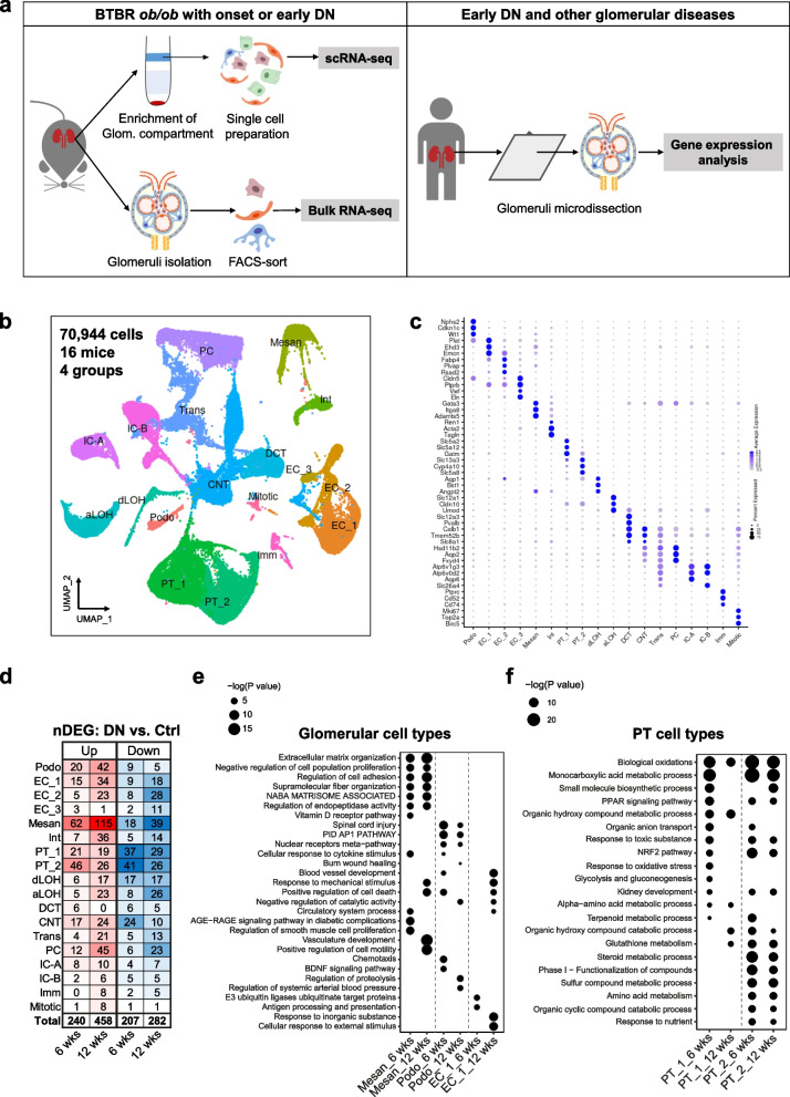
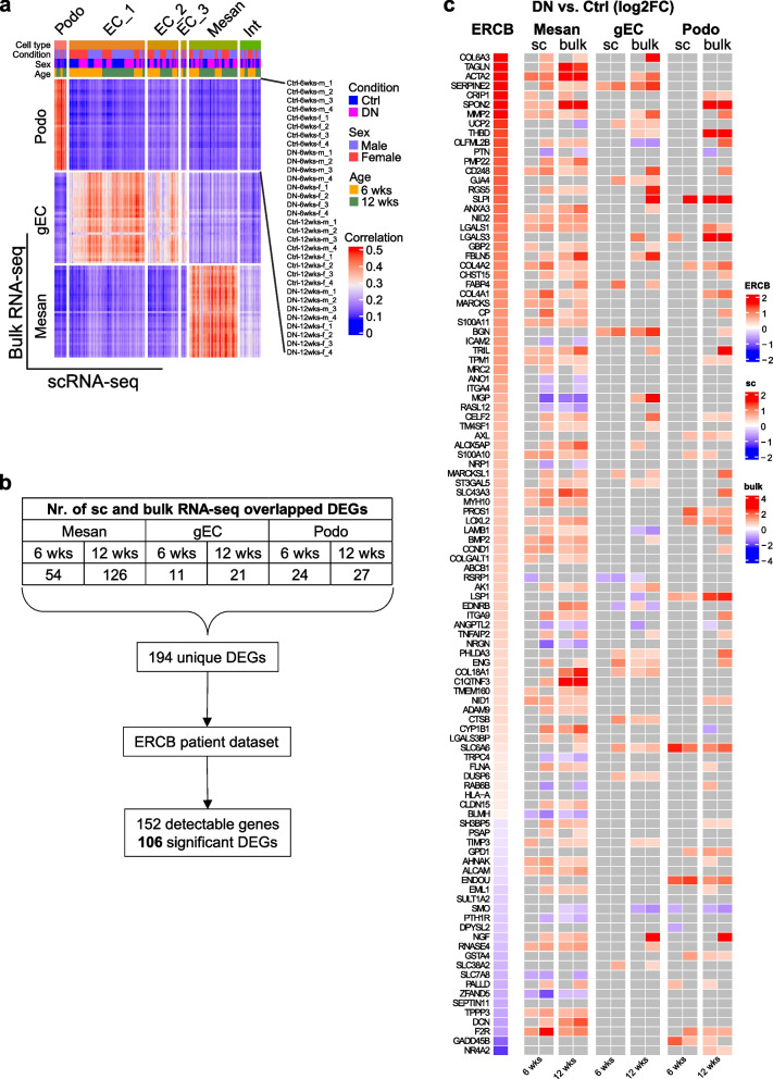
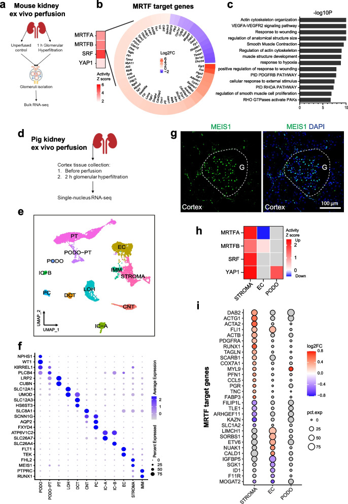
Methods: To delineate the cellular and molecular basis for DN initiation, we performed single-cell and bulk RNA sequencing of renal cells from type 2 diabetes mice (BTBR ob/ob) at the early stage of DN.

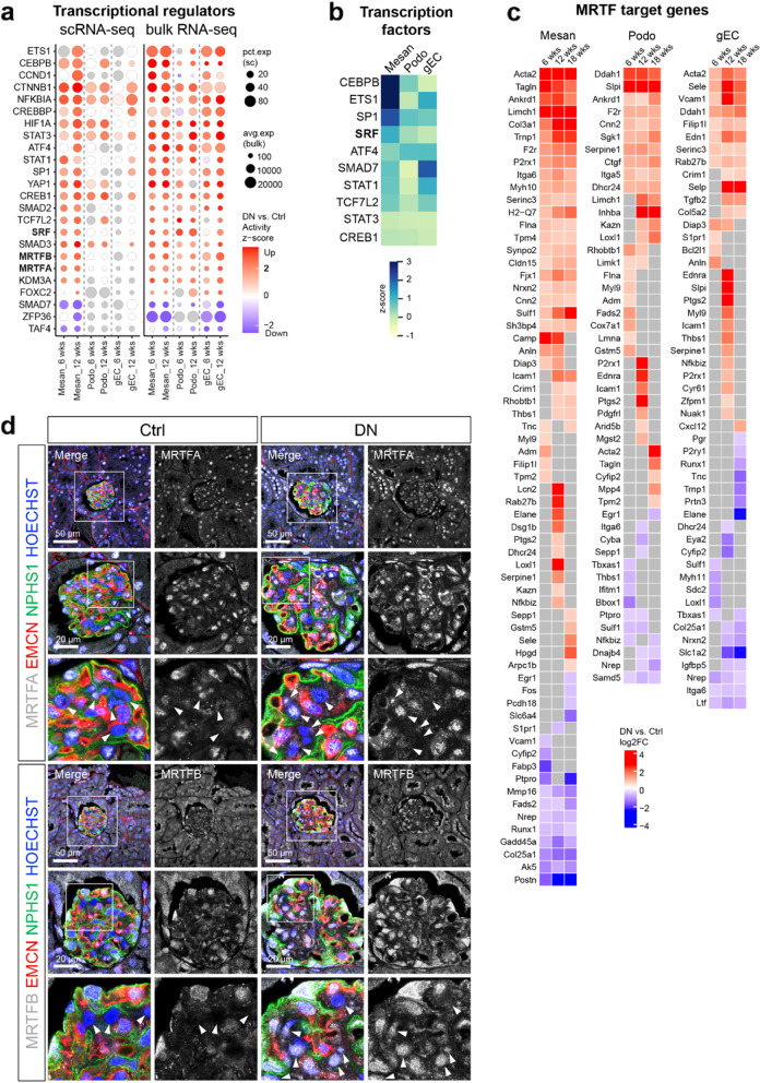
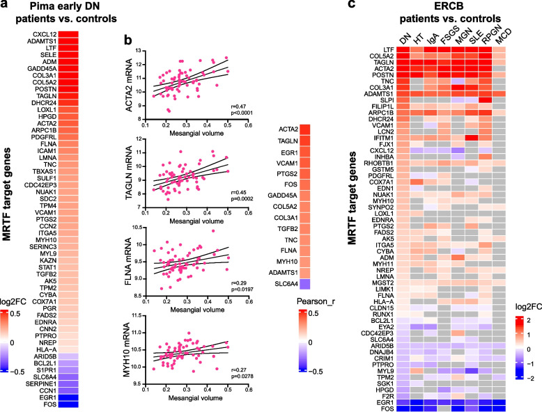
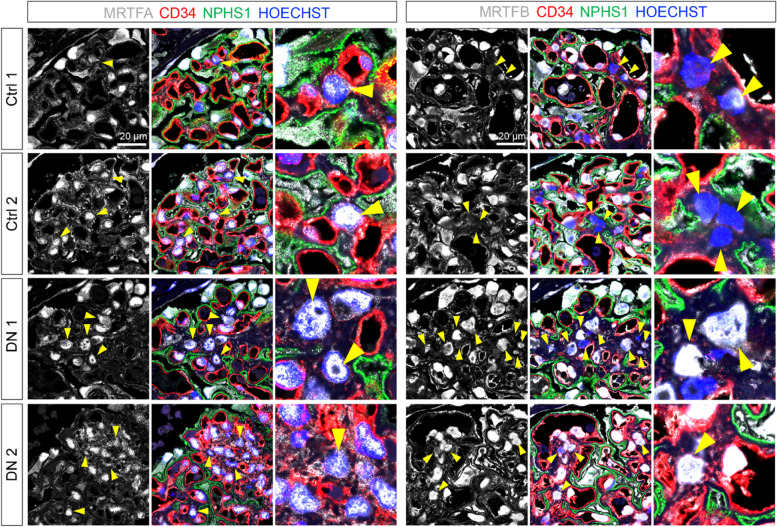
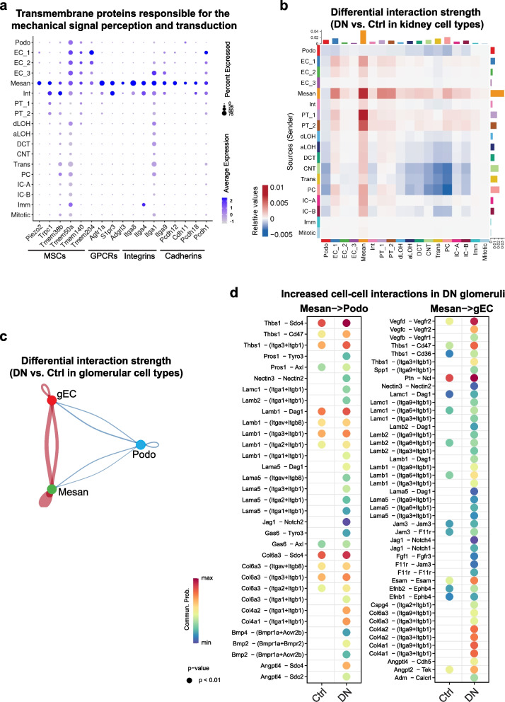
Results: Analysis of differentially expressed genes revealed glucose-independent responses in glomerular cell types. The gene regulatory network upstream of glomerular cell programs suggested the activation of mechanosensitive transcriptional pathway MRTF-SRF predominantly taking place in mesangial cells. Importantly, activation of MRTF-SRF transcriptional pathway was also identified in DN glomeruli in independent patient cohort datasets. Furthermore, ex vivo kidney perfusion suggested that the regulation of MRTF-SRF is a common mechanism in response to glomerular hyperfiltration.

Conclusions: Overall, our study presents a comprehensive single-cell transcriptomic landscape of early DN, highlighting mechanosensitive signaling pathways as novel targets of diabetic glomerulopathy.


Conflict of interest statement

DD is an employee of Boehringer Ingelheim Pharma GmbH & Co. KG. T.B. Huber reports having consultancy agreements with AstraZeneca, Bayer, Boehringer-Ingelheim, DaVita, Fresenius Medical Care, Novartis, and Retrophin; receiving research funding from Amicus Therapeutics, Fresenius Medical Care; and being on the editorial board of Kidney International and the advisory board of Nature Review Nephrology. The other authors declare that they have no competing interests.

Figures

Fig. 1
Fig. 1
scRNA-seq of BTBR ob/ob mouse kidneys with early DN. a Experimental scheme. b UMAP plot of annotated cell types. c Dot plot of defining marker genes for each cell type. d Total number of significant DEGs (nDEGs) in each cell type. Top enriched pathways in glomerular e and PT cell types f at 6 and 12 weeks. UMAP, uniform manifold approximation and projection; Ctrl, control; DN, diabetic nephropathy; Podo, podocyte; EC, endothelial cell; Mesan, mesangial cell; Int, interstitial cell; PT, proximal tubule; dLOH, descending limb of loop of Henle; aLOH, ascending limb of loop of Henle; DCT, distal convoluted tubule; CNT, connecting tubule; PC, collecting duct principal cell; IC-A, A-type collecting duct intercalated cell; IC-B, B-type collecting duct intercalated cell; Trans, transition cell; Imm, immune cell; Mitotic, mitotic cell
Fig. 2
Fig. 2
Shared features of human and experimental DN. a Heatmap displaying the Pearson correlation coefficient between scRNA-seq and bulk RNA-seq data. b Overview of significant glomerular DEGs detected in both single-cell and bulk RNA-seq datasets, as well as in the ERCB patient dataset. cHeatmap showing the significant regulation of DEGs identified in microdissected glomeruli and in glomerular cells from DN patients and mice, respectively. Nonsignificant genes are shown in grey. ERCB, European Renal cDNA Bank
Fig. 3
Fig. 3
Mechanosensitive transcriptional regulators are activated in DN glomeruli. a Dot plot displaying the commonly changed transcriptional regulations in both single-cell and bulk RNA-seq data estimated by IPA. b Heatmap showing the activity z scores of transcription factors estimated by SCENIC. c Heatmap showing the log2FC (DN vs. Ctrl) values of MRTF transcriptional target genes in mice glomerular cells. Nonsignificant genes are shown in gray (a, c). d Immunofluorescence staining of the mouse kidney paraffin sections showing the expression and localization of MRTFA and MRTFB in control and DN glomeruli. Glomerular endothelial cells were labeled by EMCN (endomucin), podocytes were labeled by NPHS1 (Nephrin), nuclei were counterstained with HOECHST. Arrows indicate mesangial cells, which were negative for EMCN and NPHS1
Fig. 4
Fig. 4
MRTF transcriptional target genes are upregulated in DN patients with type 2 diabetes. a Heatmap showing the log2FC (DN vs. Ctrl) values of MRTF transcriptional target genes in microdissected glomeruli from Pima early DN patients. b Pearson correlations between MRTF target gene expression levels and mesangial volumes in Pima early DN patients. c Heatmap showing the log2FC (DN vs. Ctrl) values of MRTF transcriptional target genes in microdissected glomeruli from ERCB patient datasets. Nonsignificant genes are shown in gray. DN, diabetic nephropathy; HT, hypertensive nephropathy; IgA, IgA nephropathy; FSGS, focal segmental glomerulosclerosis; MGN, membranous nephropathy; SLE, lupus nephritis; RPGN, ANCA-associated glomerulonephritis; MCD, minimal change disease
Fig. 5
Fig. 5
Immunofluorescence staining confirms the activation of MRTFB in mesangial cells during early DN. Immunofluorescence staining of the human kidney samples (ctrl: n = 2, DN: n = 5). Representative images showing the expression and localization of MRTFA and MRTFB in control and DN glomeruli. Glomerular endothelial cells were labeled by CD34, podocytes were labeled by NPHS1 (Nephrin), and nuclei were counterstained with HOECHST. Arrows indicate mesangial cells, which were negative for CD34 and NPHS1
Fig. 6
Fig. 6
Mesangial cells exhibit dominant signaling networks in DN glomeruli. a Dot plot showing mesangial marker genes encoding transmembrane proteins responsible for the perception and transduction of mechanical signals. MSCs, mechanosensitive ion channels; GPCRs, G protein-coupled receptors. b Change of cell–cell interactions in all pairs of kidney cell types in DN vs. Ctrl mice. c Change of cell–cell interactions in all pairs of glomerular cell types in DN vs. Ctrl mice. d Increased cell–cell interactions for mesangial cell signaling to podocytes and glomerular endothelial cell in DN mice
Fig. 7
Fig. 7
Kidney ex vivo perfusion activates mechanosensitive signaling pathways. a Experimental scheme of mouse kidney ex vivo perfusion. b Heatmap showing the log2FC values of MRTF transcriptional target genes in perfused versus control glomeruli. c Top enriched pathways of MRTF transcriptional target genes. d Experimental scheme of pig kidney ex vivo perfusion. e UMAP plot of annotated cell types from snRNA-seq of perfused and control pig kidney tissue. g Immunofluorescence staining against MEIS1 in the pig kidney cortex. Nuclei were counterstained with DAPI. G, glomerulus. Dashed lines indicate the area of glomerulus. Scale bar: 100 μm. f Dot plot displaying defining marker genes for each cell type. STROMA, stromal cells. h Heatmap showing z scores of mechanosensitive transcriptional regulators estimated by IPA. i Dot plot displaying MRTF transcriptional target genes significantly changed in perfused kidney tissue. Nonsignificant regulation or genes are shown in gray g, h

Comment in

References

    1. Thomas MC, Brownlee M, Susztak K, Sharma K, Jandeleit-Dahm KA, Zoungas S, Rossing P, Groop PH, Cooper ME. Diabetic kidney disease. Nat Rev Dis Primers. 2015;1:15018. doi: 10.1038/nrdp.2015.18. - DOI - PMC - PubMed
    1. Anders HJ, Huber TB, Isermann B, Schiffer M. CKD in diabetes: diabetic kidney disease versus nondiabetic kidney disease. Nat Rev Nephrol. 2018;14:361–377. doi: 10.1038/s41581-018-0001-y. - DOI - PubMed
    1. Wilson PC, Wu H, Kirita Y, Uchimura K, Ledru N, Rennke HG, Welling PA, Waikar SS, Humphreys BD. The single-cell transcriptomic landscape of early human diabetic nephropathy. Proc Natl Acad Sci U S A. 2019;116:19619–19625. doi: 10.1073/pnas.1908706116. - DOI - PMC - PubMed
    1. Wilson PC, Muto Y, Wu H, Karihaloo A, Waikar SS, Humphreys BD. Multimodal single cell sequencing implicates chromatin accessibility and genetic background in diabetic kidney disease progression. Nat Commun. 2022;13:5253. doi: 10.1038/s41467-022-32972-z. - DOI - PMC - PubMed
    1. Stefansson VTN, Nair V, Melsom T, Looker HC, Mariani LH, Fermin D, Eichinger F, Menon R, Subramanian L, Ladd P, et al. Molecular programs associated with glomerular hyperfiltration in early diabetic kidney disease. Kidney Int. 2022;102(6):1345–1358. doi: 10.1016/j.kint.2022.07.033. - DOI - PMC - PubMed

Publication types