

Alpinetin inhibits neuroinflammation and neuronal apoptosis via targeting the JAK2/STAT3 signaling pathway in spinal cord injury
- PMID: 36627822
- PMCID: PMC10018110
- DOI: 10.1111/cns.14085
Alpinetin inhibits neuroinflammation and neuronal apoptosis via targeting the JAK2/STAT3 signaling pathway in spinal cord injury
Abstract
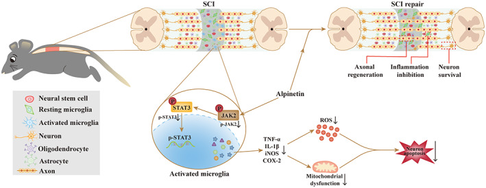
Background: A growing body of research shows that drug monomers from traditional Chinese herbal medicines have antineuroinflammatory and neuroprotective effects that can significantly improve the recovery of motor function after spinal cord injury (SCI). Here, we explore the role and molecular mechanisms of Alpinetin on activating microglia-mediated neuroinflammation and neuronal apoptosis after SCI.
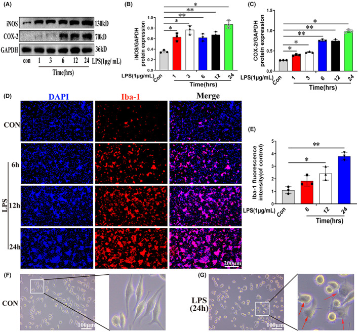
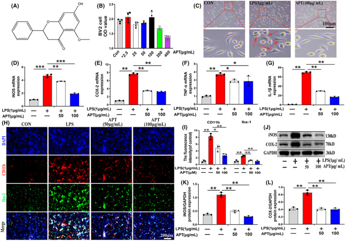
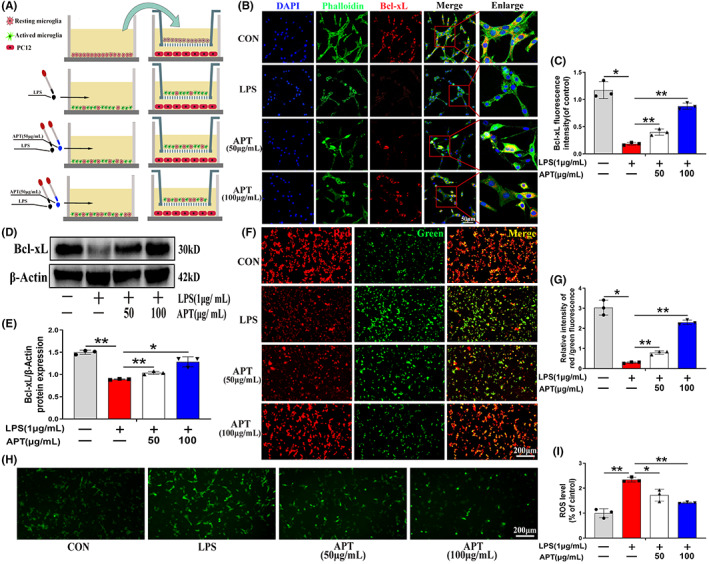
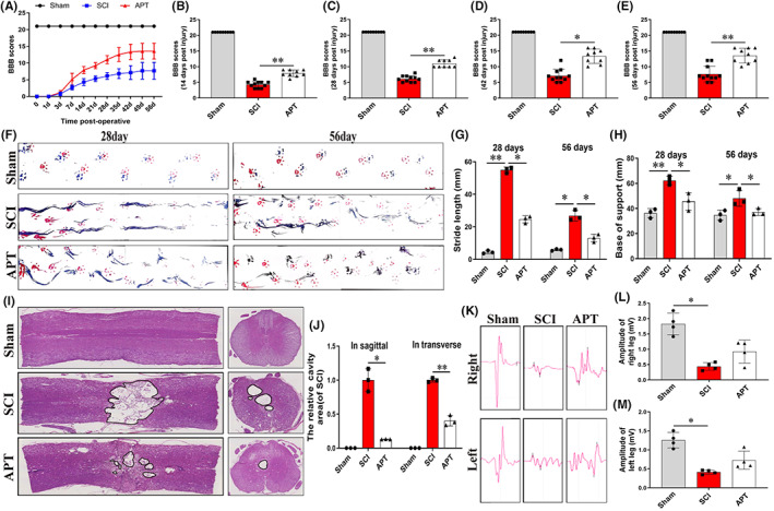
Methods: Stimulation of microglia with lipopolysaccharide (LPS) to simulate neuroinflammation models in vitro, the effect of Alpinetin on the release of pro-inflammatory mediators in LPS-induced microglia and its mechanism were detected. In addition, a co-culture system of microglia and neuronal cells was constructed to assess the effect of Alpinetin on activating microglia-mediated neuronal apoptosis. Finally, rat spinal cord injury models were used to study the effects on inflammation, neuronal apoptosis, axonal regeneration, and motor function recovery in Alpinetin.
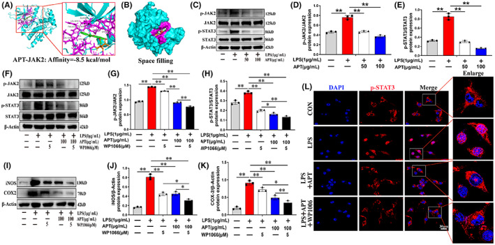
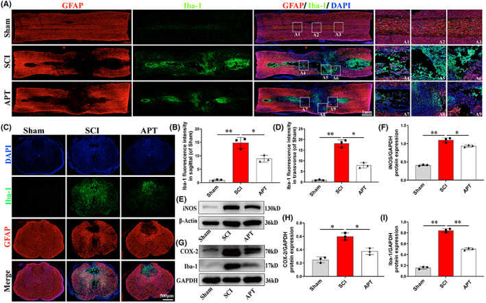
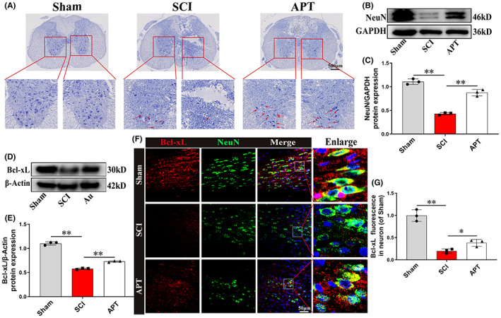
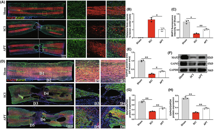
Results: Alpinetin inhibits microglia-mediated neuroinflammation and activity of the JAK2/STAT3 pathway. Alpinetin can also reverse activated microglia-mediated reactive oxygen species (ROS) production and decrease of mitochondrial membrane potential (MMP) in PC12 neuronal cells. In addition, in vivo Alpinetin significantly inhibits the inflammatory response and neuronal apoptosis, improves axonal regeneration, and recovery of motor function.
Conclusion: Alpinetin can be used to treat neurodegenerative diseases and is a novel drug candidate for the treatment of microglia-mediated neuroinflammation.
Keywords: Alpinetin; JAK2/STAT3 pathway; neurinflammation; neuronal apoptosis; spinal cord injury.
© 2023 The Authors. CNS Neuroscience & Therapeutics published by John Wiley & Sons Ltd.
Conflict of interest statement
The authors confirm that they have no conflict of interest.
Figures
Similar articles
-
Photobiomodulation inhibits the activation of neurotoxic microglia and astrocytes by inhibiting Lcn2/JAK2-STAT3 crosstalk after spinal cord injury in male rats.J Neuroinflammation. 2021 Nov 5;18(1):256. doi: 10.1186/s12974-021-02312-x. J Neuroinflammation. 2021. PMID: 34740378 Free PMC article.
-
Electroacupuncture stimulation inhibited astrogliosis and microglia polarisation to alleviate spinal cord injury via Janus kinase 2/signal transducer and activator of transcription 3 signalling pathway.Folia Histochem Cytobiol. 2025;63(1):28-40. doi: 10.5603/fhc.104273. Folia Histochem Cytobiol. 2025. PMID: 40421823
-
[Linarin inhibits microglia activation-mediated neuroinflammation and neuronal apoptosis in mouse spinal cord injury by inhibiting the TLR4/NF-κB pathway].Nan Fang Yi Ke Da Xue Xue Bao. 2024 Aug 20;44(8):1589-1598. doi: 10.12122/j.issn.1673-4254.2024.08.18. Nan Fang Yi Ke Da Xue Xue Bao. 2024. PMID: 39276055 Free PMC article. Chinese.
-
JAK2/STAT3 as a new potential target to manage neurodegenerative diseases: An interactive review.Eur J Pharmacol. 2024 May 5;970:176490. doi: 10.1016/j.ejphar.2024.176490. Epub 2024 Mar 15. Eur J Pharmacol. 2024. PMID: 38492876 Review.
-
TGF-β signaling pathway in spinal cord injury: Mechanisms and therapeutic potential.J Neurosci Res. 2024 Jan;102(1):e25255. doi: 10.1002/jnr.25255. Epub 2023 Oct 10. J Neurosci Res. 2024. PMID: 37814990 Review.
Cited by
-
Caffeic acid phenethyl ester inhibits neuro-inflammation and oxidative stress following spinal cord injury by mitigating mitochondrial dysfunction via the SIRT1/PGC1α/DRP1 signaling pathway.J Transl Med. 2024 Mar 25;22(1):304. doi: 10.1186/s12967-024-05089-8. J Transl Med. 2024. PMID: 38528569 Free PMC article.
-
Epidermal Neural Crest Stem Cell Conditioned Medium Enhances Spinal Cord Injury Recovery via PI3K/AKT-Mediated Neuronal Apoptosis Suppression.Neurochem Res. 2024 Oct;49(10):2854-2870. doi: 10.1007/s11064-024-04207-8. Epub 2024 Jul 18. Neurochem Res. 2024. PMID: 39023805 Free PMC article.
-
Ganoderma lucidum polysaccharide attenuates retinal ischemia-reperfusion injury by regulating microglial M1/M2 polarization, suppressing neuroinflammation and inhibiting JAK2/STAT3 pathway.Biochem Biophys Rep. 2025 Jan 29;41:101926. doi: 10.1016/j.bbrep.2025.101926. eCollection 2025 Mar. Biochem Biophys Rep. 2025. PMID: 39944465 Free PMC article.
-
HSP70 protects PC12 cells against TBHP-induced apoptosis and oxidative stress by activating the Nrf2/HO-1 signaling pathway.In Vitro Cell Dev Biol Anim. 2024 Sep;60(8):868-878. doi: 10.1007/s11626-024-00924-0. Epub 2024 May 28. In Vitro Cell Dev Biol Anim. 2024. PMID: 38807023
-
Micro electrical fields induced MSC-sEVs attenuate neuronal cell apoptosis by activating autophagy via lncRNA MALAT1/miR-22-3p/SIRT1/AMPK axis in spinal cord injury.J Nanobiotechnology. 2023 Nov 27;21(1):451. doi: 10.1186/s12951-023-02217-2. J Nanobiotechnology. 2023. PMID: 38012570 Free PMC article.
References
-
- Zipser CM, Cragg JJ, Guest JD, et al. Cell‐based and stem‐cell‐based treatments for spinal cord injury: evidence from clinical trials. Lancet Neurol. 2022;21:659‐670. - PubMed
-
- Han M, Yang H, Lu X, et al. Three‐dimensional‐cultured MSC‐derived exosome‐hydrogel hybrid microneedle Array patch for spinal cord repair. Nano Lett. 2022;22:6391‐6401. - PubMed
-
- Van Broeckhoven J, Sommer D, Dooley D, Hendrix S, Franssen A. Macrophage phagocytosis after spinal cord injury: when friends become foes. Brain. 2021;144:2933‐2945. - PubMed
Publication types
MeSH terms
Substances
LinkOut - more resources
Full Text Sources
Medical
Miscellaneous
