. 2022 Nov 14;7(2):bvac173.
doi: 10.1210/jendso/bvac173. eCollection 2022 Dec 15.

Four-Compartment Diffusion Model of Cortisol Disposition: Comparison With 3 Alternative Models in Current Clinical Use

Affiliations

Four-Compartment Diffusion Model of Cortisol Disposition: Comparison With 3 Alternative Models in Current Clinical Use

Richard I Dorin et al. J Endocr Soc. .

Abstract

Context: Estimated rates of cortisol elimination and appearance vary according to the model used to obtain them. Generalizability of current models of cortisol disposition in healthy humans is limited.

Objective: Development and validation of a realistic, mechanistic model of cortisol disposition that accounts for the major factors influencing plasma cortisol concentrations in vivo (Model 4), and comparison to previously described models of cortisol disposition in current clinical use (Models 1-3).

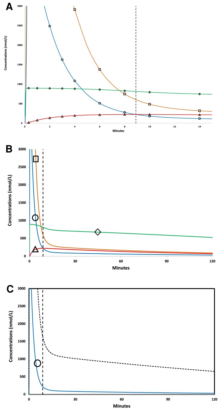
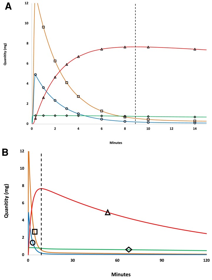
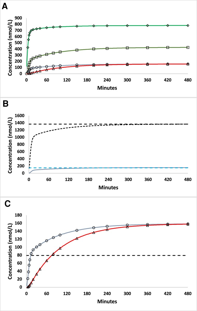
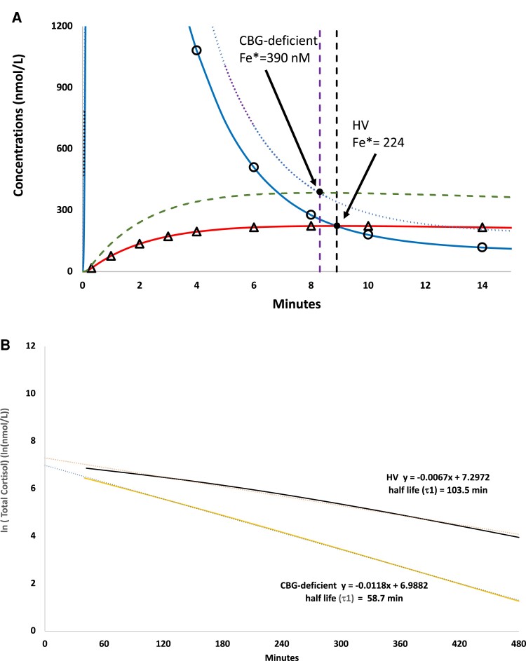
Methods: The 4 models were independently applied to cortisol concentration data obtained for the hydrocortisone bolus experiment (20 mg) in 2 clinical groups: healthy volunteers (HVs, n = 6) and corticosteroid binding globulin (CBG)-deficient (n = 2). Model 4 used Fick's first law of diffusion to model free cortisol flux between vascular and extravascular compartments. Pharmacokinetic parameter solutions for Models 1-4 were optimized by numerical methods, and model-specific parameter solutions were compared by repeated measures analysis of variance. Models and respective parameter solutions were compared by mathematical and simulation analyses, and an assessment tool was used to compare performance characteristics of the four models evaluated herein.

Results: Cortisol half-lives differed significantly between models (all P < .001) with significant model-group interaction (P = .02). In comparative analysis, Model 4 solutions yielded significantly reduced free cortisol half-life, improved fit to experimental data (both P < .01), and superior model performance.

Conclusion: The proposed 4-compartment diffusion model (Model 4) is consistent with relevant experimental observations and met the greatest number of empiric validation criteria. Cortisol half-life solutions obtained using Model 4 were generalizable between HV and CBG-deficient groups and bolus and continuous modes of hydrocortisone infusion.

Keywords: albumin; compartmental modeling; computer-assisted; corticosteroid binding globulin (CBG); cortisol; hydrocortisone; metabolic clearance rate; numerical analysis; septic shock.


Figures

Figure 1.
Figure 1.
Model 4 simulation showing time-varying cortisol concentrations for 20 mg hydrocortisone bolus. (A) Cortisol concentrations as a function of time (0-15 minutes) following IV hydrocortisone bolus (20 mg infused over 20 seconds) in all 4 compartments, including vascular (plasma) free cortisol (XF, circle symbol, blue), vascular (plasma) albumin-bound cortisol (XFA, square symbol, orange), vascular (plasma) CBG-bound cortisol (XFC, diamond symbol, green), and extravascular (free) cortisol (XFe, red curve, triangle symbol, red). In this example a unique intersection point, where extravascular and plasma free cortisol concentrations are momentarily equal (XF = XFe) occurred at T* = 8.9 minutes, indicated by dashed vertical line, and at free cortisol concentration of 223 nmol/L (XF = XFe = 223 nmol/L). (B) The same data with axes expanded in cortisol concentration and time (0-120 minutes), symbols as in Panel A. (C) Model 4 predicted concentrations for 2 more commonly measured analytes: plasma free cortisol (XF, solid line, blue) and plasma total cortisol (XTotF, dotted line, black), which is the sum of free, albumin-bound, and CBG-bound cortisol concentrations in the vascular (plasma) compartment.
Figure 2.
Figure 2.
Model 4 simulation showing time-varying cortisol mass for 20 mg hydrocortisone bolus. (A) Cortisol mass in all 4 compartments as a function of time, symbols and colors are as in Fig. 1. (A) Cortisol mass by compartment for 0 to 15 minutes. (B) The same data with axes expanded to 120 minutes.
Figure 3.
Figure 3.
Model 4 simulation for continuous hydrocortisone infusion (8 mg/hour). (A) Model 4 predicted cortisol concentrations as a function of time for continuous infusion of hydrocortisone (8 mg/h) for all 4 compartments, symbols and line format as in Fig. 1. (B) Predicted concentration of vascular (plasma) total (XTotF, black line) and free (XF, blue line) cortisol. Steady-state cortisol concentrations (95% of asymptote) are shown by dashed lines. (C) Comparison of predicted plasma free (XF, blue line) and extravascular (XFe, red line) cortisol concentrations as a function of time. The dashed horizontal line indicates the free cortisol concentration at 50% of the asymptotic, steady-state value.
Figure 4.
Figure 4.
Simulation of computer-generated cortisol concentrations for 20 mg hydrocortisone bolus obtained using Model 4 for CBG-deficient vs HV groups. (A) Model-predicted plasma free (XF) and extravascular (XFe) cortisol concentrations for HV and CBG-deficient groups. For the CBG-deficient simulation, XF and XFe are indicated as dotted (purple) and dashed (green) lines, respectively. For HV, XF and XFe are indicated by circles (blue line) and triangles (orange line), respectively. Vertical lines indicate times to intersection point postbolus (T*) for HV (intersection (Fe* at 224 nmol/L, black) and CBG-deficient (intersection Fe* at 390 nmol/L, purple). (B) Model-predicted, log-transformed total cortisol concentrations (XTotF) for HV and CBG-deficient groups and Model 1 half-life solutions (τ1) for the log-transformed cortisol concentrations (terminal half-life shown in bold).

References

    1. Bikle DD. The free hormone hypothesis: when, why, and how to measure the free hormone levels to assess vitamin D, thyroid, sex hormone, and cortisol status. JBMR Plus. 2021;5(1):e10418. - PMC - PubMed
    1. Keenan DM, Roelfsema F, Veldhuis JD. Endogenous ACTH concentration-dependent drive of pulsatile cortisol secretion in the human. Am J Physiol Endocrinol Metab. 2004;287(4):E652–E661. - PubMed
    1. Mendel CM. The free hormone hypothesis: a physiologically based mathematical model. Endocr Rev. 1989;10(3):232–274. - PubMed
    1. Burt MG, Mangelsdorf BL, Rogers A, et al. . Free and total plasma cortisol measured by immunoassay and mass spectrometry following ACTH(1)(-)(2)(4) stimulation in the assessment of pituitary patients. J Clin Endocrinol Metab. 2013;98(5):1883–1890. - PubMed
    1. Genere N, Kaur RJ, Athimulam S, et al. . Interpretation of abnormal dexamethasone suppression test is enhanced with use of synchronous free cortisol assessment. J Clin Endocrinol Metab. 2022;107(3):e1221–e1230. - PMC - PubMed

LinkOut - more resources