Review
. 2023 Jan;13(1):e1170.
doi: 10.1002/ctm2.1170.

Neutrophil, neutrophil extracellular traps and endothelial cell dysfunction in sepsis

Affiliations
Review

Neutrophil, neutrophil extracellular traps and endothelial cell dysfunction in sepsis

Hao Zhang et al. Clin Transl Med. 2023 Jan.

Abstract

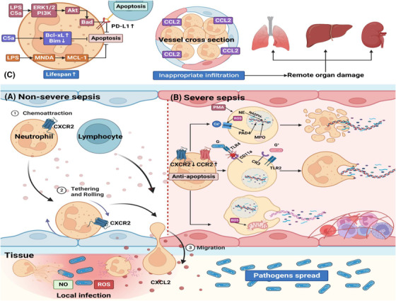
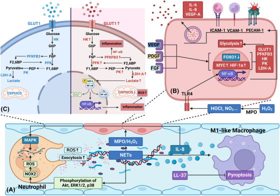
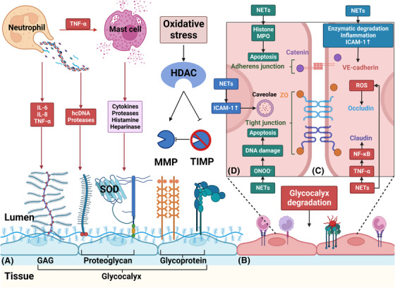
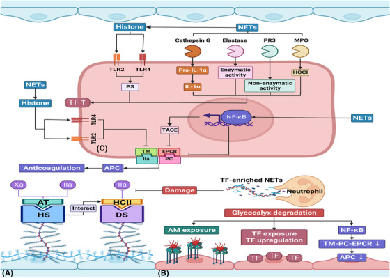
Sepsis is a persistent systemic inflammatory condition involving multiple organ failures resulting from a dysregulated immune response to infection, and one of the hallmarks of sepsis is endothelial dysfunction. During its progression, neutrophils are the first line of innate immune defence against infection. Aside from traditional mechanisms, such as phagocytosis or the release of inflammatory cytokines, reactive oxygen species and other antibacterial substances, activated neutrophils also release web-like structures composed of tangled decondensed DNA, histone, myeloperoxidase and other granules called neutrophil extracellular traps (NETs), which can efficiently ensnare bacteria in the circulation. In contrast, excessive neutrophil activation and NET release may induce endothelial cells to shift toward a pro-inflammatory and pro-coagulant phenotype. Furthermore, neutrophils and NETs can degrade glycocalyx on the endothelial cell surface and increase endothelium permeability. Consequently, the endothelial barrier collapses, contributing to impaired microcirculatory blood flow, tissue hypoperfusion and life-threatening organ failure in the late phase of sepsis.

Keywords: endothelial cell dysfunction; neutrophil; neutrophil extracellular traps; sepsis.


Conflict of interest statement

The authors declare that they have no conflict of interest

Figures

FIGURE 1
FIGURE 1
Neutrophil, NET formation and sepsis. (A) During non‐severe sepsis, neutrophils that express CXCR2 are recruited from blood to the infection site responding to CXCL2. Neutrophils migrate to the location of infection and kill pathogens through the release of antibacterial substances, such as ROS, NO and NETs. Other immune cells such as lymphocytes can also migrate to the infection site to prevent pathogens spread. (B) However, during severe sepsis, neutrophils show increased lifespan and impaired function due to the down‐regulation of CXCR2 and up‐regulation of CCR2. On the one hand, impaired migration results in pathogens' spread. On the other hand, many neutrophils are confined to vessels and release NETs, resulting in vascular inflammation, endothelial damage, and thrombosis. NET formation can be classified into three types. The first type is suicidal NETosis since NETs are released via cell lysis. The second type allows NET release and conventional live neutrophil functions, such as phagocytosis, to coexist. The third type is mtDNA NETosis. Viable neutrophils release mtDNA to form NETs, and this process does not depend on cell death but is dependent on ROS. (C) LPS and C5a in the peripheral circulation can induce neutrophils’ resistance to apoptosis via three signalling pathways. Moreover, neutrophils induce lymphocyte apoptosis via PD‐L1 up‐regulation. Additionally, CCR2, which is absent in neutrophils under normal conditions, is upregulated in neutrophils through TLR activation. CCR2 drives inappropriate infiltration of neutrophils into remote organs which produce CCL2 and further elicit tissue damage in remote organs such as lungs, liver, and kidneys. C5a, complement component 5a; CCL2, CC ligand 2; CCR2, CC receptor 2; CR3, complement receptor 3; CXCL2, CXC ligand 2; CXCR2, CXC receptor 2; ERK1/2, extracellular regulated protein kinases 1/2; G‐, gram‐negative bacteria; G+, gram‐positive bacteria; iNOS, inducible nitric oxide synthase; LPS, lipopolysaccharide; MCL‐1, myeloid cell leukemia‐1; MNDA, myeloid nuclear differentiation antigen; MPO, myeloperoxidase; mtDNA, mitochondrial DNA; NE, neutrophil elastase; NETs, neutrophil extracellular traps; PAD4, peptidyl arginine deaminase 4; PD‐L1, programmed cell death ligand 1; PI‐3K, phosphoinositide‐3 kinases; PI3Kγ, phosphoinositide‐3 kinase gamma; PMA, phorbol 12‐myristate 13‐acetate; ROS, reactive oxygen species; TLR, Toll‐like receptor
FIGURE 2
FIGURE 2
Neutrophils and NETs induce a pro‐inflammatory and pro‐angiogenic endothelial cell phenotype via activation of NF‐κB signalling. (A) NETs stimulate pro‐inflammatory and pro‐angiogenic responses in human endothelial cells via MPO/H2O2‐mediated NF‐κB activation. Moreover, NETs activate several neutrophil functions, such as exocytosis, ROS production, and NET formation, which may be related to the phosphorylation of Akt, ERK1/2 and p38. In addition to the impacts on neutrophils, NETs induce an “M1‐like” macrophage phenotype characterized by releasing inflammatory cytokines, such as IL‐8. Moreover, the concentration of IL‐8 is found to be strongly linked with NET formation via MAPK pathway activation. LL‐37, an antibacterial protein externalized on NETs, can suppress macrophage pyroptosis and then inhibit the release of pro‐inflammatory cytokines. (B) NF‐κB signalling enhances endothelial cell pro‐inflammatory and pro‐angiogenic responses via up‐regulation of ICAM‐1, VCAM‐1, PECAM‐1, and increased secretion of IL‐6, IL‐8, and VEGF‐A. Additionally, many pro‐angiogenic factors, such as VEGF, PDGE and FGF, are released during the process. These factors increase expression levels of glycolytic enzymes and transporters via up‐regulation of MYC and HIF‐1α and down‐regulation of FOXO1. (C) During inflammatory and angiogenic responses, proliferated endothelial cells show a higher rate of glycolysis than quiescent ones due to the up‐regulation of glycolytic enzymes and transporters, such as GLUT1, PFKFB3, HK, PK and LDH‐A. Notably, PFKFB3 and glycolytic product lactate promote NF‐κB‐driven vascular inflammation. F1,6BP, fructose‐1,6‐biphosphate; F2,6BP, fructose‐2,6‐biphosphate; F6P, fructose‐6‐phosphate; FGF, fibroblast growth factor; G6P, glucose‐6‐phosphate; GLUT1, glucose transporter 1; HIF‐1α, hypoxia‐inducible factor‐1α; HK, hexokinase; ICAM‐1, intercellular adhesion molecule‐1; LDH‐A, lactate dehydrogenase‐A; MAPK, mitogen‐activated protein kinase; NF‐κB, nuclear factor‐κB; NOX2, NADPH oxidase 2; OXPHOS, oxidative phosphorylation; PDGF, platelet‐derived growth factor; PECAM‐1, platelet endothelial cell adhesion molecule‐1; PEP, phosphoenolpyruvate; PFK‐1, phosphofructokinase‐1; PFKFB3, fructose‐2,6‐bisphosphatase‐3; PK, pyruvate kinase; VCAM‐1, vascular cell adhesion molecule‐1; VEGF, vascular endothelial growth factor
FIGURE 3
FIGURE 3
Neutrophils and NETs damage endothelial cells’ glycocalyx and increase endothelial permeability. (A) During sepsis, NETs can directly cause glycocalyx degradation through hcDNA and proteases. Additionally, a considerable number of inflammatory cytokines, such as IL‐6, IL‐8 and TNF‐α, induced by neutrophils and NETs can also damage glycocalyx. Moreover, mast cells are activated by TNF‐α and release cytokines, proteases, histamine and heparinase, further degrading glycocalyx. Furthermore, oxidative stress‐induced HDAC can upregulate MMP expression and inhibit tissue inhibitors of MMPs (TIMP1 and TIMP3), thereby activating MMPs to promote the shedding of the glycocalyx. (B) After degradation of the glycocalyx, endothelial cell adhesion molecules are exposed, triggering further inflammation, rolling, and adhesion of leukocytes and platelets. (C) Para‐endothelial permeability increases mainly due to junction cleavage. TNF‐α is shown to cause disruption of claudin through NF‐κB activation. ROS causes the redistribution of occludin, limiting its association with ZO‐1. VE‐cadherin is susceptible to enzymatic degradation. Additionally, inflammatory mediators promote VE‐cadherin phosphorylation and dissociation from p120 catenin and induce VE‐cadherin endocytosis via several signalling pathways. Upregulated ICAM‐1 can also promote VE‐cadherin phosphorylation. (D) ICAM‐1 can also increase trans‐endothelial permeability through caveolin‐1 phosphorylation, a major component of caveolae. However, endothelial cell apoptosis is the main cause of increased trans‐endothelial permeability. NETs contribute to apoptosis through MPO and histone. Moreover, the formation of ONOO during oxidative stress also induces cell death via DNA damage. GAG, glycosaminoglycan; hcDNA, histone‐complexed DNA; HDAC, histone deacetylase; MMP, matrix metalloproteinase; ONOO‐, peroxynitrite; SOD, superoxide dismutase; VE‐cadherin, vascular endothelial‐cadherin; ZO, zonula occluding
FIGURE 4
FIGURE 4
Neutrophils and NETs induce a pro‐coagulant endothelial cell phenotype via degradation of the anti‐coagulation system and up‐regulation of tissue factor. (A) GAGs play a key role in the anti‐coagulation system: HS primarily binds to AT to inhibit factor IIa and Xa, and DS mainly binds to HCII to inhibit factor IIa. Moreover, HS also interacts with HCII. So glycocalyx degradation induced by NETs contributes to the degradation of the anti‐coagulation system. (B) Furthermore, adhesion molecules and TF are exposed on the endothelium causing thrombus and fibrin formation. Aside from releasing TF‐enriched NETs, neutrophils increase expression levels of TF on endothelium through NET components. (C) Cathepsin G, elastase, PR3 and MPO can stimulate TF expression through different signalling pathways and further initiate the extrinsic coagulation pathway. Histone, another NET component, induces endothelial pro‐coagulant phenotype through up‐regulation of TF and down‐regulation of TM, and these effects are partly mediated by TLR2 and TLR4. Moreover, histone is capable of stimulating PS exposure, which enhances the pro‐coagulant activity of TF. Additionally, NETs can destroy the TM‐PC‐EPCR system, the most important natural anti‐coagulation mechanism, via NF‐κB activation. And NF‐κB signalling also promotes TACE activity, which is reportedly responsible for EPCR shedding. AM, adhesion molecule; APC, activated protein C; AT, antithrombin; DS, dermatan sulfate; HCII, heparin cofactor II; HOCI, hypochlorous acid; HS, heparan sulfate; PR3, proteinase 3; PS, phosphatidylserine; TACE, TNF‐α converting enzyme; TF, tissue factor; TM‐PC‐EPCR, thrombomodulin, protein C, and endothelial protein C receptor
FIGURE 5
FIGURE 5
Potential therapeutic targets in sepsis. In blood vessels, nNIF, NRPs, and PAD4 inhibitors prevent NET formation by inhibiting chromatin decondensation. And PAD4 inhibitors may also reduce the expression of ICAM‐1 and VCAM‐1. TLR‐4−/− mice did not show an enhanced thrombotic response and exhibited markedly decreased circulating ICAM‐1 compared with wide‐type controls. DNase1 and DNase1‐like 3 blocks the positive feedback loop of thrombosis by degrading the scaffold of NET structures. PFKFB3 blockers may be used to control inflammation induced by excessive glycolysis during sepsis. Using PFKFB3 inhibitors can also promote NADPH production, which is likely to induce NET formation. Moreover, PFKFB3 blockade amplifies the anti‐angiogenic effect of VEGFR blockers. In a perfused endothelial cell model, GSH or NAC (the precursor of GSH) supplementation significantly decreased ROS production. HSA can suppress circulating and tissue O2 production, and restrain activation of NF‐κB, thereby inhibiting oxidative stress and nitrosative stress. In addition to scavenging ROS and RNS directly, vitamin C reduces them by preventing NOX activation, decreasing iNOS expression, and enhancing NO bioavailability. Then decreased formation of ONOO can efficiently prevent endothelial cell apoptosis. GSH, glutathione; HSA, human serum albumin; NAC, N‐acetylcysteine; nNIF, neonatal NET‐inhibitory factor; NRPs, nNIF‐related peptides; RNS, reactive nitrogen species

References

    1. Singer M, Deutschman CS, Seymour CW, et al. The third international consensus definitions for sepsis and septic shock (Sepsis‐3). JAMA. 2016;315(8):801‐810. doi:10.1001/jama.2016.0287 - DOI - PMC - PubMed
    1. Kienle K, Glaser KM, Eickhoff S, et al. Neutrophils self‐limit swarming to contain bacterial growth in vivo. Science. 2021;372(6548):eabe7729. doi:10.1126/science.abe7729 - DOI - PMC - PubMed
    1. De Giovanni M, Tam H, Valet C, Xu Y, Looney MR, Cyster JG. GPR35 promotes neutrophil recruitment in response to serotonin metabolite 5‐HIAA. Cell. 2022;185(5):815‐830.e19. doi:10.1016/j.cell.2022.01.010 - DOI - PMC - PubMed
    1. Sekheri M, El Kebir D, Edner N, Filep JG. 15‐Epi‐LXA4 and 17‐epi‐RvD1 restore TLR9‐mediated impaired neutrophil phagocytosis and accelerate resolution of lung inflammation. Proc Natl Acad Sci U S A. 2020;117(14):7971‐7980. doi:10.1073/pnas.1920193117 - DOI - PMC - PubMed
    1. Rosa BA, Ahmed M, Singh DK, et al. IFN signalling and neutrophil degranulation transcriptional signatures are induced during SARS‐CoV‐2 infection. Commun Biol. 2021;4(1):290. doi:10.1038/s42003-021-01829-4 - DOI - PMC - PubMed

Publication types