. 2023 Feb 28;14(1):e0337022.
doi: 10.1128/mbio.03370-22. Epub 2023 Jan 11.

A Germline-Targeting Chimpanzee SIV Envelope Glycoprotein Elicits a New Class of V2-Apex Directed Cross-Neutralizing Antibodies

Affiliations

A Germline-Targeting Chimpanzee SIV Envelope Glycoprotein Elicits a New Class of V2-Apex Directed Cross-Neutralizing Antibodies

Frederic Bibollet-Ruche et al. mBio. .

Abstract

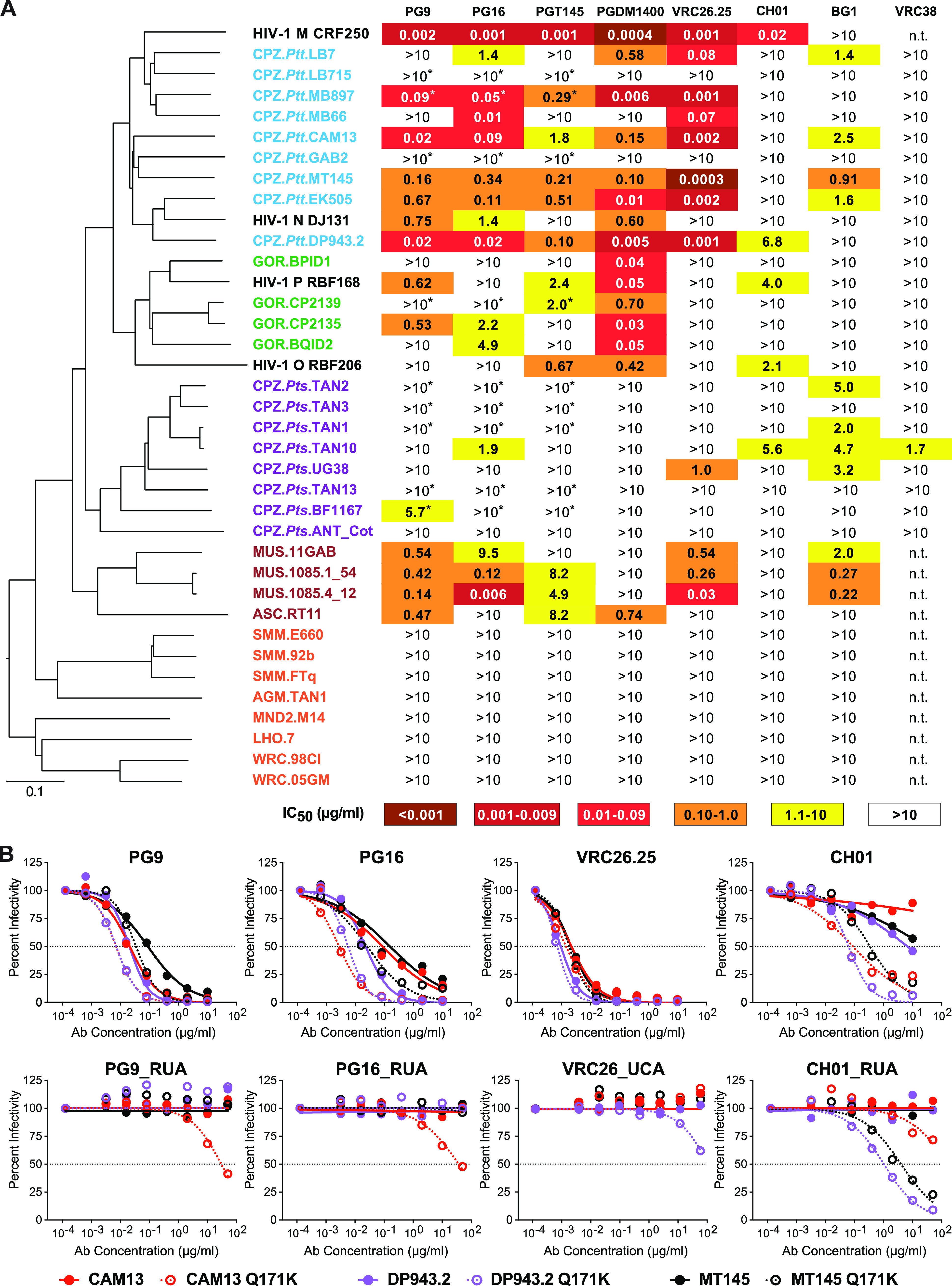
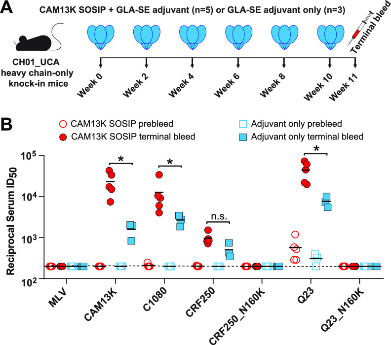
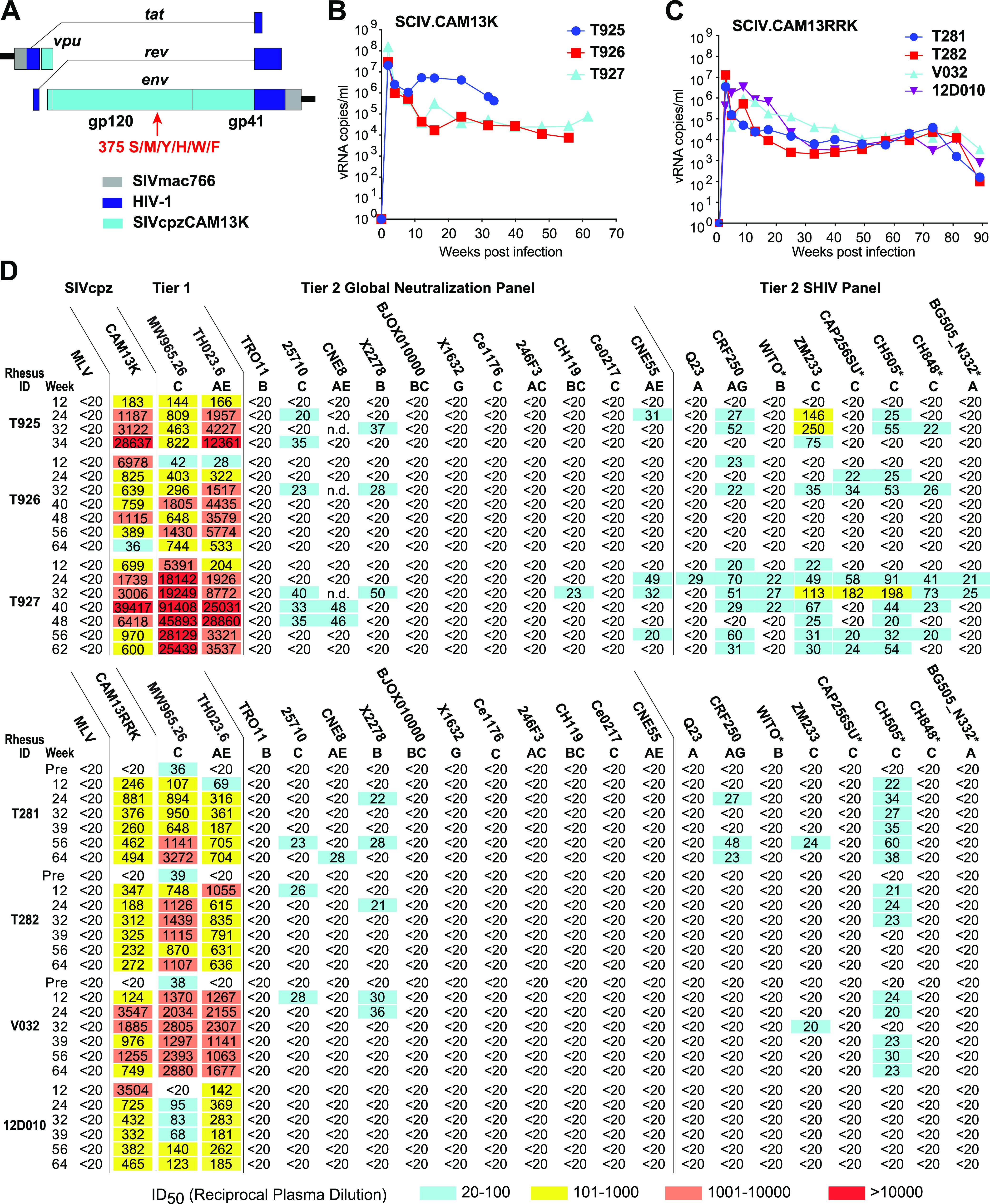
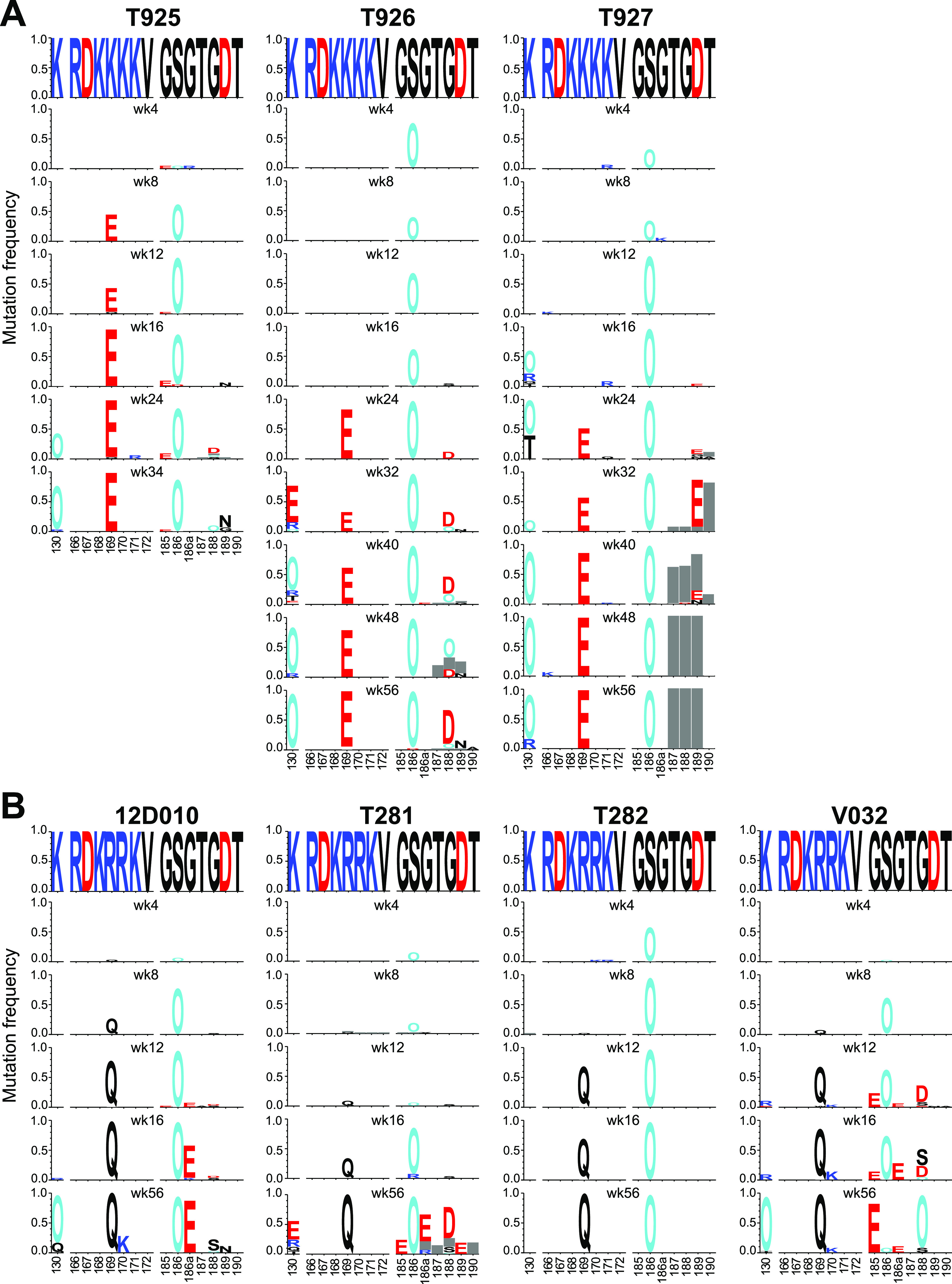
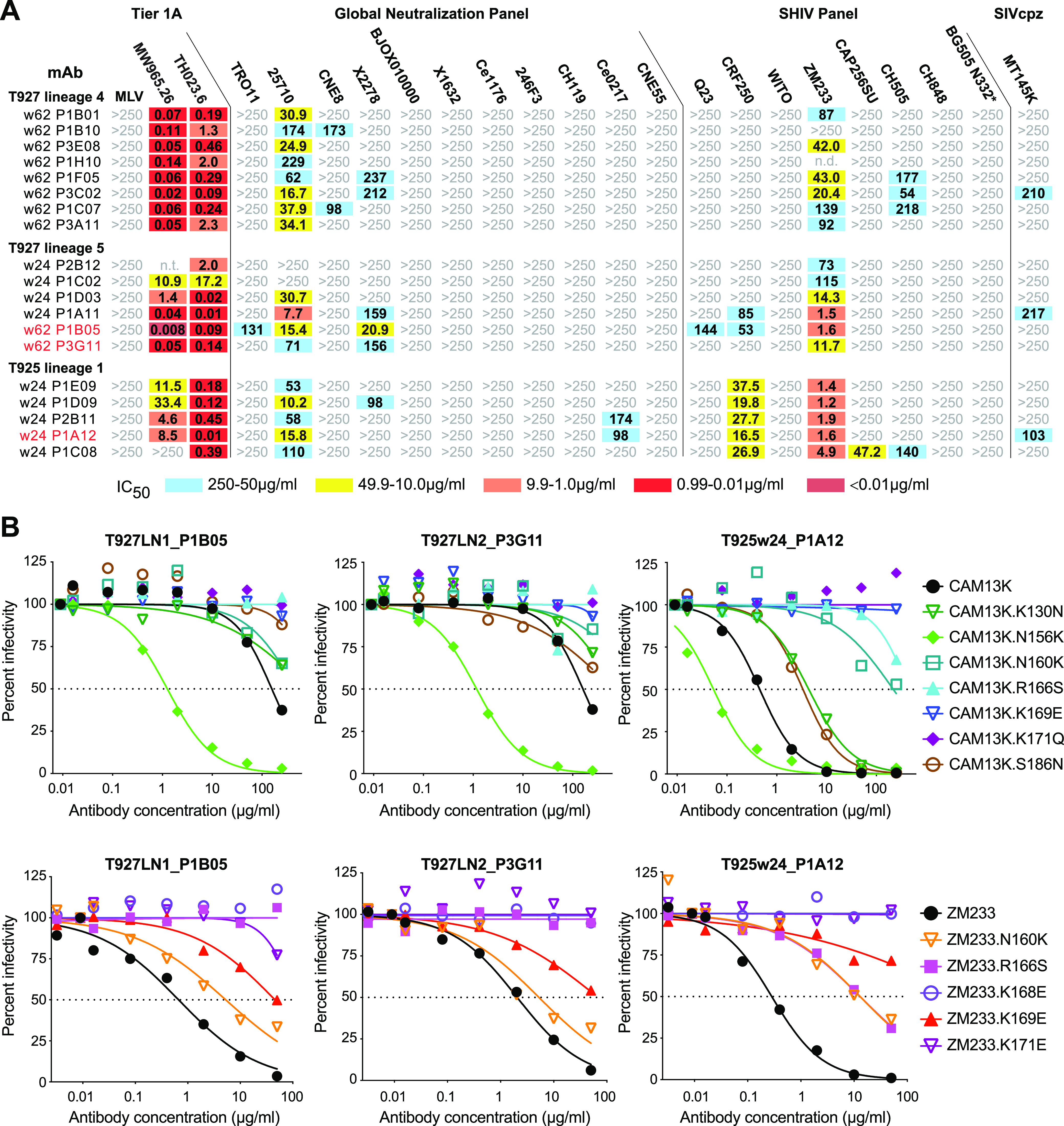
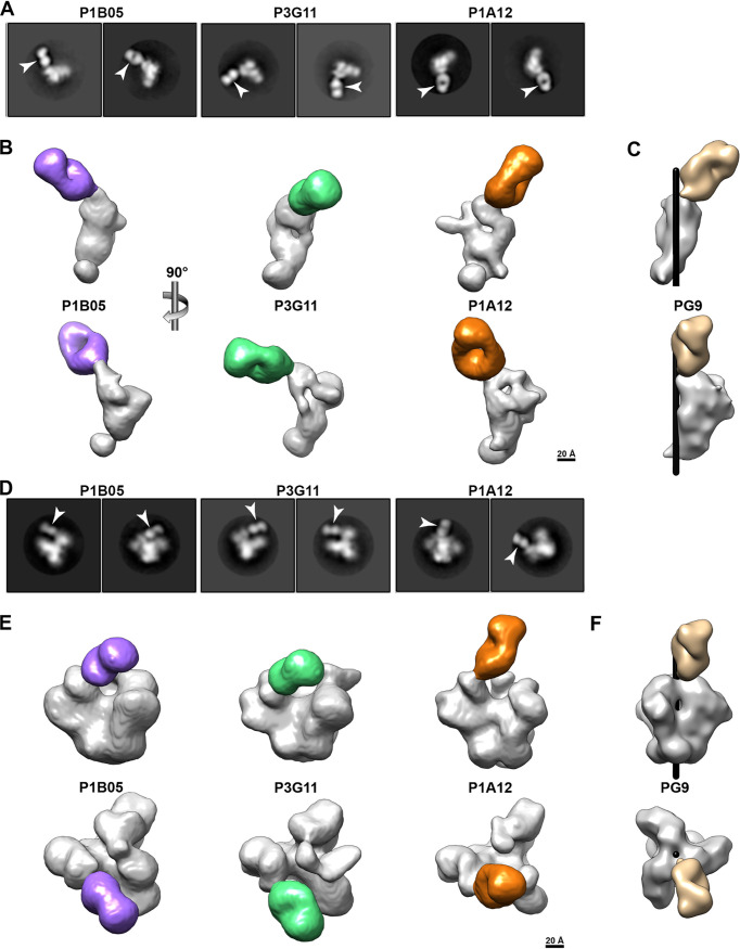
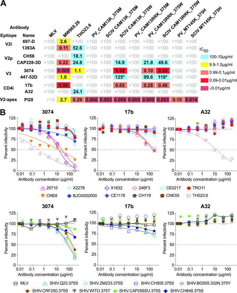
HIV-1 and its SIV precursors share a broadly neutralizing antibody (bNAb) epitope in variable loop 2 (V2) at the envelope glycoprotein (Env) trimer apex. Here, we tested the immunogenicity of germ line-targeting versions of a chimpanzee SIV (SIVcpz) Env in human V2-apex bNAb heavy-chain precursor-expressing knock-in mice and as chimeric simian-chimpanzee immunodeficiency viruses (SCIVs) in rhesus macaques (RMs). Trimer immunization of knock-in mice induced V2-directed NAbs, indicating activation of V2-apex bNAb precursor-expressing mouse B cells. SCIV infection of RMs elicited high-titer viremia, potent autologous tier 2 neutralizing antibodies, and rapid sequence escape in the canonical V2-apex epitope. Six of seven animals also developed low-titer heterologous plasma breadth that mapped to the V2-apex. Antibody cloning from two of these animals identified multiple expanded lineages with long heavy chain third complementarity determining regions that cross-neutralized as many as 7 of 19 primary HIV-1 strains, but with low potency. Negative stain electron microscopy (NSEM) of members of the two most cross-reactive lineages confirmed V2 targeting but identified an angle of approach distinct from prototypical V2-apex bNAbs, with antibody binding either requiring or inducing an occluded-open trimer. Probing with conformation-sensitive, nonneutralizing antibodies revealed that SCIV-expressed, but not wild-type SIVcpz Envs, as well as a subset of primary HIV-1 Envs, preferentially adopted a more open trimeric state. These results reveal the existence of a cryptic V2 epitope that is exposed in occluded-open SIVcpz and HIV-1 Env trimers and elicits cross-neutralizing responses of limited breadth and potency. IMPORTANCE An effective HIV-1 vaccination strategy will need to stimulate rare precursor B cells of multiple bNAb lineages and affinity mature them along desired pathways. Here, we searched for V2-apex germ line-targeting Envs among a large set of diverse primate lentiviruses and identified minimally modified versions of one chimpanzee SIV Env that bound several human V2-apex bNAb precursors and stimulated one of these in a V2-apex bNAb precursor-expressing knock-in mouse. We also generated chimeric simian-chimpanzee immunodeficiency viruses and showed that they elicit low-titer V2-directed heterologous plasma breadth in six of seven infected rhesus macaques. Characterization of this antibody response identified a new class of weakly cross-reactive neutralizing antibodies that target the V2-apex, but only in occluded-open Env trimers. The existence of this cryptic epitope, which in some Env backgrounds is immunodominant, needs to be considered in immunogen design.

Keywords: SCIV; V2-apex; broadly neutralizing antibodies; chimpanzee; germline-targeting; human immunodeficiency virus; immunofocusing; neutralizing antibodies; occluded-open trimer; vaccines.


Conflict of interest statement

The authors declare no conflict of interest.

Figures

FIG 1
FIG 1
Antigenic conservation of the V2-apex among primate lentiviruses. (A) The ability of human V2-apex bNAbs (top) to neutralize pseudoviruses bearing Envs from diverse SIV strains (left) is shown, with numbers indicating IC50 values (μg/mL) as determined in the TZM-bl assay (39). Coloring indicates the relative neutralization potency. Values highlighted by asterisks have previously been reported (19). SIV Envs are color-coded to indicate their species/subspecies origin (central chimpanzees [CPZ.Ptt], blue; western gorillas [GOR], green; eastern chimpanzees [CPZ.Pts], purple; moustached [MUS] and red-tailed [ASC] monkeys, brown; sooty mangabeys [SMM], African green monkeys [AGM], mandrills [MND2], L’Hoest’s monkeys [LHO], and western red colobus [WRC], red), and their phylogenetic relationships are illustrated by a neighbor joining tree of full-length Env protein sequences to the left (the scale bar indicates 0.1 amino acid substitutions per site). HIV-1 strains CRF250 (group M), DJ131 (group N), RBF168 (group O), and RBF206 (group P) are shown for comparison. The highest antibody concentration used was 10 μg/mL (n.t., not tested). (B) Neutralization curves depicting the sensitivity of pseudoviruses containing wild-type (closed circles) and Q171K mutated (open circles) versions of the SIVcpz Envs CAM13 (red), DP943.2 (blue), and MT145 (black) to mature V2-apex bNAbs PG9, PG16, VRC26.25, and CH01 (top) and their inferred precursors (bottom). Dashed lines indicate 50% reduction in virus infectivity (the antibody concentration is shown on the x axis in μg/mL).
FIG 2
FIG 2
CAM13K SOSIP immunization elicits neutralizing antibody responses in V2-apex bNAb precursor expressing knock-in mice. (A) CH01_RUA heavy chain (HC)-only knock-in mice (20) were immunized either with 20 μg CAM13K SOSIP trimer in 5 μg GLA-SE adjuvant (n = 5) or with 5 μg GLA-SE adjuvant alone (n = 3). Time points of immunization and bleeds are indicated. (B) Comparison of serum neutralization titers (reciprocal 50% inhibitory dilutions, ID50) from trimer-immunized (red circles) and adjuvant-only (blue squares) knock-in mice (preimmunization and terminal bleeds are shown as open and filled symbols, respectively). Serum neutralizing activity was tested in the TZM-bl assay (39) against pseudoviruses carrying the immunogen-matched autologous CAM13K Env, three CH01_RUA sensitive HIV-1 Envs (C1080, CRF250, Q23), as well as N160 glycan knockout variants of CRF250 and Q23. MLV Env containing pseudovirus was used for control. Differences between immunized and adjuvant-only groups were assessed using a nonparametric Mann-Whitney test (Prism 9.4.0 GraphPad Software), with asterisks indicating P < 0.05.
FIG 3
FIG 3
SCIV-infected RMs develop low-titer heterologous neutralization breadth. (A) Design scheme of SCIV vectors expressing SIVcpz Env ectodomains. The SIVcpz CAM13K vpu-env region (teal) was cloned into an optimized SHIV vector (21) consisting of a SIVmac766 proviral backbone (gray) and HIV-1-derived tat and rev genes (dark blue). Six isogenic SCIV mutants were generated with serine (S), methionine (M), tyrosine (Y), histidine (H), tryptophan (W) or phenylalanine (F) at position 375 of the CAM13K Env. (B) Plasma vRNA kinetics in three RMs infected with SCIV.CAM13K. Animals were inoculated with equal mixtures of SCIV.CAM13K variants bearing all six Env375 mutants. SCIV.CAM13K.M375W emerged as the predominant strain in all animals. (C) Plasma vRNA kinetics in four RMs infected SCIV.CAM13RRK (generated from SCIV.CAM13K.375W by introducing K169R and K170R mutations). (D) Autologous and heterologous neutralization of longitudinal plasma samples from RMs infected with SCIV.CAM13K (top) or SCIV.CAM13RRK (bottom). Reciprocal 50% inhibitory dilutions (ID50) are shown for autologous (wild-type CAM13K and CAM13RRK encoding 375M) and heterologous (tier 1 and tier 2) viruses representing different HIV-1 subtypes (A, AG, AE, AC, B, C, BC, G; indicated below virus name), with no reactivity observed against a murine leukemia virus (MLV) Env containing control (all ID50 <1:20). Coloring indicates relative neutralization potency. Both Env pseudotypes (CAM13K, CAM13RRK, tier 1 and tier 2 global panel) and replication-competent SHIV strains were tested, all of which encoded the wild-type amino acid at position 375, except for SHIV.BG505_N332, which encoded a tyrosine instead of a serine. SHIVs expressing transmitted founder HIV-1 Envs are denoted by asterisks. For three SCIV.CAM13RRK-infected animals, which were repurposed from prior HIV-1 immunization studies (see methods), preinfection (pre) plasma neutralization titers are also shown.
FIG 4
FIG 4
SCIV infection elicits strong V2-apex directed immune selection. (A, B) Longitudinal Env evolution at the V2-apex is shown over time for the N-linked glycosylation site at position 130, strand C (amino acids 166 to 172), and the hypervariable part of the V2 loop (amino acids 185 to 190) for all SCIV.CAM13K (A) and SCIV.CAM13RRK (B) infected RMs (see Fig. S3 for a similar analysis of the entire Env). Sequences are shown as logo plots, where the height of each amino acid is proportional to its frequency at the respective time point. For each RM, the top logo depicts the infecting SCIV, with subsequent logos representing longitudinal time points denoted in weeks (wk). To highlight mutations, the infecting virus sequence is blanked out for all longitudinal time points. Positively charged amino acids (R, H, K) are colored blue, negatively charged amino acids (D, E) are colored red, potential N-linked glycosylation sites are denoted as “O” and colored cyan, and gray bar indicate deletions. The webtool AnalyzeAlign from the Los Alamos HIV Databases was used for to generate the logos.
FIG 5
FIG 5
SCIV infections elicit low-potency V2-directed antibodies that neutralize HIV-1. (A) Neutralization breadth and potency of SCIV-induced antibodies cloned from RMs T927 and T925 (indicated on the left). Fifty percent inhibitory concentrations (IC50) are shown for representative lineage members (μg/mL) against pseudoviruses and SHIV strains (indicated on top). Only the most potent and cross-reactive lineages are shown (see Fig. S7A for similar results for the remaining lineages). The highest antibody concentration used was 250 μg/mL (coloring indicates relative neutralization potency; n.t., not tested). MAbs analyzed by negative stain electron microscopy are highlighted in red (heavy and light chain variable region sequences are shown in Fig. S8). (B) Epitope mapping of select cross-neutralizing MAbs. Neutralization curves are shown for three monoclonal antibodies (indicated on top and highlighted in red in panel A) against a panel of CAM13K and ZM233 mutant pseudoviruses (indicated on the right). Dashed lines indicate 50% reduction in virus infectivity (corresponding IC50 values are shown in Fig. S7B).
FIG 6
FIG 6
SCIV-induced V2-directed cross-neutralizing antibodies bind occluded-open but not closed trimers. (A) 2D class averages of NSEM images showing Fabs (arrows) bound to single Env protomers. Fabs (denoted on top) were generated for members of the most cross-reactive antibody lineage from RM T927 (P3G11 and P1B05) and RM T925 (P1A12), respectively (also see Fig. 5). Although both ZM197-ZM233 and CAM13K SOSIP preparations were used, only Fab-monomer complexes were observed. (B) 3D reconstructions of Fab-monomer complexes, shown in two orthogonal views, with the Env domain in gray and the Fab in color (P1B05, purple; P3G11, green; P1A12, gold). The scale bar indicates 20 Å. (C) Volume rendering of the PG9 Fab (tan, PDB 3U2S) in complex with a prefusion BG505 trimer (PDB 5FYL), of which only a single protomer (gray) is shown. The Env densities in panel B were aligned with the Env density in panel C and are shown in the same orientation. The black cylinder denotes the position of the 3-fold trimer axis. (D) 2D class averages showing Fabs (arrows) bound to SOSIP trimers lacking the N156 glycan (CH505_N156Q SOSIP). (E) 3D reconstructions of Fabs bound to CH505_N156Q SOSIP trimers. Both side (upper) and top (lower) views are shown, with the Fabs highlighted in color as in panel B. The densities of the CH505_N156Q SOSIP are consistent with an occluded-open trimer. The scale bar indicates 20 Å. (F) Volume rendering of PG9 (tan, PDB 3U2S) in complex with a closed, prefusion BG505 trimer (PDB 5FYL), shown in the same orientation as in panel E. The black cylinder indicates the central 3-fold axis of the Env trimer.
FIG 7
FIG 7
SCIV expressed, but not wild-type SIVcpz Env, as well as some primary HIV-1 Envs adopting a more open conformation. (A) The sensitivity of CAM13K and CAM13RRK Envs to nonneutralizing V2p, V2i, and linear V3 and CD4i antibodies as well as the V2-apex bNAb PG9 (indicated on the left) is shown for pseudovirus (PV) and SCIV constructs encoding either the wild-type methionine (M) or the RM-selected tryptophan (W) at position 375 (indicated on top). Fifty percent inhibitory concentrations (IC50) are shown in μg/mL (coloring indicates relative neutralization potency, with asterisks identifying IC50 values estimated by the Prism software). Also shown are IC50 values for pseudovirus and SCIV-expressed MT145K Env, which encodes a histidine at position 375. MLV and tier 1 HIV-1 (MW965.26, TH023.6) Env pseudoviruses are shown for control. (B) Neutralization curves are shown for nonneutralizing V3 (3074) and CD4i (17b, A32) antibodies (indicated on top) against global panel pseudoviruses (upper panel) and SHIV strains (lower panel; with the amino acid residue at position 375 indicated). Dashed lines indicate 50% reduction in virus infectivity.

References

    1. Plotkin SA. 2010. Correlates of protection induced by vaccination. Clin Vaccine Immunol 17:1055–1065. doi: 10.1128/CVI.00131-10. - DOI - PMC - PubMed
    1. Kong R, Duan H, Sheng Z, Xu K, Acharya P, Chen X, Cheng C, Dingens AS, Gorman J, Sastry M, Shen C-H, Zhang B, Zhou T, Chuang G-Y, Chao CW, Gu Y, Jafari AJ, Louder MK, O'Dell S, Rowshan AP, Viox EG, Wang Y, Choi CW, Corcoran MM, Corrigan AR, Dandey VP, Eng ET, Geng H, Foulds KE, Guo Y, Kwon YD, Lin B, Liu K, Mason RD, Nason MC, Ohr TY, Ou L, Rawi R, Sarfo EK, Schön A, Todd JP, Wang S, Wei H, Wu W, Mullikin JC, Bailer RT, Doria-Rose NA, Karlsson Hedestam GB, Scorpio DG, Overbaugh J, NISC Comparative Sequencing Program , et al. 2019. Antibody lineages with vaccine-induced antigen-binding hotspots develop broad HIV neutralization. Cell 178:567–584.e19. doi: 10.1016/j.cell.2019.06.030. - DOI - PMC - PubMed
    1. Lee JH, Crotty S. 2021. HIV vaccinology: 2021 update. Semin Immunol 51:101470. doi: 10.1016/j.smim.2021.101470. - DOI - PMC - PubMed
    1. Haynes BF, Wiehe K, Borrrow P, Saunders KO, Korber B, Wagh K, McMichael AJ, Kelsoe G, Hahn BH, Alt F, Shaw GM. 2022. Strategies for HIV-1 vaccines that induce broadly neutralizing antibodies. Nat Rev Immunol. doi: 10.1038/s41577-022-00753-w. - DOI - PMC - PubMed
    1. Gray ES, Madiga MC, Hermanus T, Moore PL, Wibmer CK, Tumba NL, Werner L, Mlisana K, Sibeko S, Williamson C, Abdool Karim SS, Morris L, Team CS, CAPRISA002 Study Team . 2011. The neutralization breadth of HIV-1 develops incrementally over four years and is associated with CD4+ T cell decline and high viral load during acute infection. J Virol 85:4828–4840. doi: 10.1128/JVI.00198-11. - DOI - PMC - PubMed

Publication types

LinkOut - more resources