. 2023 Jan 3;6(1):e2250613.
doi: 10.1001/jamanetworkopen.2022.50613.

Revision of the Melanocytic Pathology Assessment Tool and Hierarchy for Diagnosis Classification Schema for Melanocytic Lesions: A Consensus Statement

Affiliations

Revision of the Melanocytic Pathology Assessment Tool and Hierarchy for Diagnosis Classification Schema for Melanocytic Lesions: A Consensus Statement

Raymond L Barnhill et al. JAMA Netw Open. .

Abstract

Importance: A standardized pathology classification system for melanocytic lesions is needed to aid both pathologists and clinicians in cataloging currently existing diverse terminologies and in the diagnosis and treatment of patients. The Melanocytic Pathology Assessment Tool and Hierarchy for Diagnosis (MPATH-Dx) has been developed for this purpose.

Objective: To revise the MPATH-Dx version 1.0 classification tool, using feedback from dermatopathologists participating in the National Institutes of Health-funded Reducing Errors in Melanocytic Interpretations (REMI) Study and from members of the International Melanoma Pathology Study Group (IMPSG).

Evidence review: Practicing dermatopathologists recruited from 40 US states participated in the 2-year REMI study and provided feedback on the MPATH-Dx version 1.0 tool. Independently, member dermatopathologists participating in an IMPSG workshop dedicated to the MPATH-Dx schema provided additional input for refining the MPATH-Dx tool. A reference panel of 3 dermatopathologists, the original authors of the MPATH-Dx version 1.0 tool, integrated all feedback into an updated and refined MPATH-Dx version 2.0.

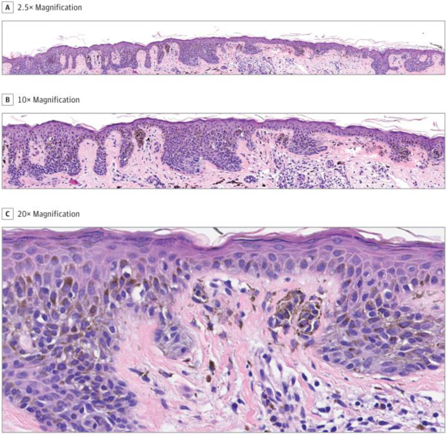
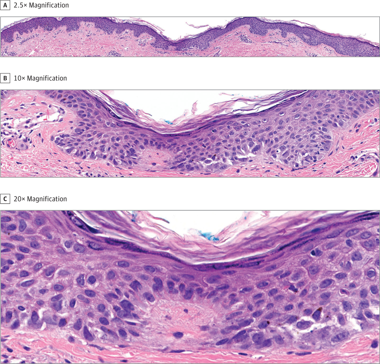
Findings: The new MPATH-Dx version 2.0 schema simplifies the original 5-class hierarchy into 4 classes to improve diagnostic concordance and to provide more explicit guidance in the treatment of patients. This new version also has clearly defined histopathological criteria for classification of classes I and II lesions; has specific provisions for the most frequently encountered low-cumulative sun damage pathway of melanoma progression, as well as other, less common World Health Organization pathways to melanoma; provides guidance for classifying intermediate class II tumors vs melanoma; and recognizes a subset of pT1a melanomas with very low risk and possible eventual reclassification as neoplasms lacking criteria for melanoma.

Conclusions and relevance: The implementation of the newly revised MPATH-Dx version 2.0 schema into clinical practice is anticipated to provide a robust tool and adjunct for standardized diagnostic reporting of melanocytic lesions and management of patients to the benefit of both health care practitioners and patients.


Figures

Figure 1.
Figure 1.
Melanocytic Pathology Assessment Tool and Hierarchy for Diagnosis (MPATH-Dx) Version 2.0 Class I: Compound Nevus With Low-grade Atypia Note disordered junctional architecture at scanning magnification (A and B). At high magnification, at least 5 melanocytes in junctional nests contain nuclei less than 1.5 times the size of adjacent resting basal keratinocytes (C). This lesion was originally associated with discordant interpretations of mild and moderate atypia by the expert panel and by consensus classified as MPATH-Dx version 1.0 class II: compound nevus with moderate atypia. Based on use of cytological and morphological criteria in Table 2, the lesion is reclassified by consensus as MPATH-Dx version 2.0 class I by the expert consensus panel.
Figure 2.
Figure 2.
Melanocytic Pathology Assessment Tool and Hierarchy for Diagnosis (MPATH-Dx) Version 2.0 Class II: Compound Melanocytic Lesion, Probable Compound Dysplastic Nevus With High-grade Atypia, Uncertain The lesion is poorly defined and measures 4 mm in diameter (A). The junctional and dermal components are paucicellular. The junctional component comprises scant basilar single cells and disordered junctional nesting with relatively sparse pagetoid spread of melanocytes mostly confined to the lower half of the epidermis (B). Rare melanocytes (2–3 cells) reach the granular layer. No effacement of the epidermis is noted, and the epidermal rete-oriented pattern of melanocytic proliferation is maintained. Slight solar elastosis is present. At high magnification, at least 5 junctional melanocytes contain nuclei more than 1.5 times the size of surrounding resting basal keratinocytes (C). Because of scant pagetoid spread and conspicuous cytological atypia, there is concern for melanoma in situ in this lesion. Rare single atypical melanocytes in the dermis raise the possibility of focal invasive melanoma. However, a number of findings argue against clear-cut melanoma in situ or invasive melanoma. The lesion was originally associated with discordant interpretations of moderate and severe atypia by the expert panel and by consensus classified as MPATH-Dx version 1.0 class II: compound nevus with moderate atypia, but with some uncertainty about its biological nature (suspicion for melanoma in situ). Based on the use of cytological and morphological criteria in Table 2, this lesion is reclassified by consensus as MPATH-Dx version 2.0 class II, but again with uncertainty by the expert consensus panel. This lesion illustrates how morphological criteria may not be conclusive for the definitive interpretation of many melanocytic lesions in this intermediate spectrum and particularly for high-grade lesions. Uncertainty about such lesions exists and should be communicated in diagnostic reports.

References

    1. Barnhill RL. Criteria for Melanocytic Lesions: An Introduction. Institut Curie; 2022.
    1. Radick AC, Reisch LM, Shucard HL, et al. Terminology for melanocytic skin lesions and the MPATH-Dx classification schema: a survey of dermatopathologists. J Cutan Pathol. 2021;48(6):733–738. doi:10.1111/cup.13873 - DOI - PMC - PubMed
    1. Duray PH, DerSimonian R, Barnhill R, et al. An analysis of interobserver recognition of the histopathologic features of dysplastic nevi from a mixed group of nevomelanocytic lesions. J Am Acad Dermatol. 1992;27(5 Pt 1): 741–749. doi:10.1016/0190-9622(92)70248-E - DOI - PubMed
    1. Duncan LM, Berwick M, Bruijn JA, Byers HR, Mihm MC, Barnhill RL. Histopathologic recognition and grading of dysplastic melanocytic nevi: an interobserver agreement study. J Invest Dermatol. 1993;100(3):318S–321S. doi: 10.1038/jid.1993.55 - DOI - PubMed
    1. Corona R, Mele A, Amini M, et al. Interobserver variability on the histopathologic diagnosis of cutaneous melanoma and other pigmented skin lesions. J Clin Oncol. 1996;14(4):1218–1223. doi:10.1200/JCO.1996.14.4.1218 - DOI - PubMed

Publication types