. 2023 Jan 13;9(2):eadd4623.
doi: 10.1126/sciadv.add4623. Epub 2023 Jan 11.

Peptide-guided lipid nanoparticles deliver mRNA to the neural retina of rodents and nonhuman primates

Affiliations

Peptide-guided lipid nanoparticles deliver mRNA to the neural retina of rodents and nonhuman primates

Marco Herrera-Barrera et al. Sci Adv. .

Abstract

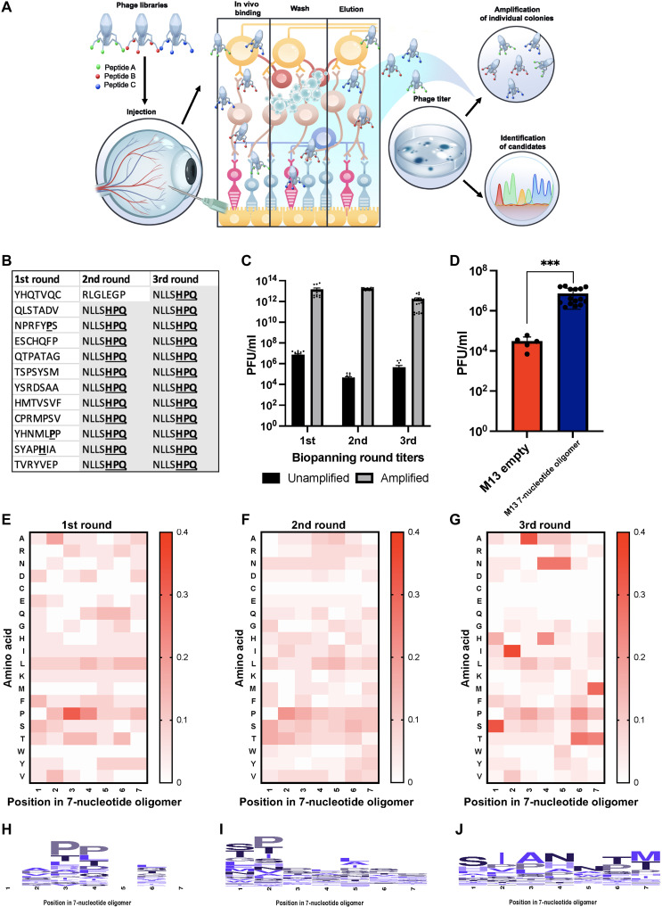
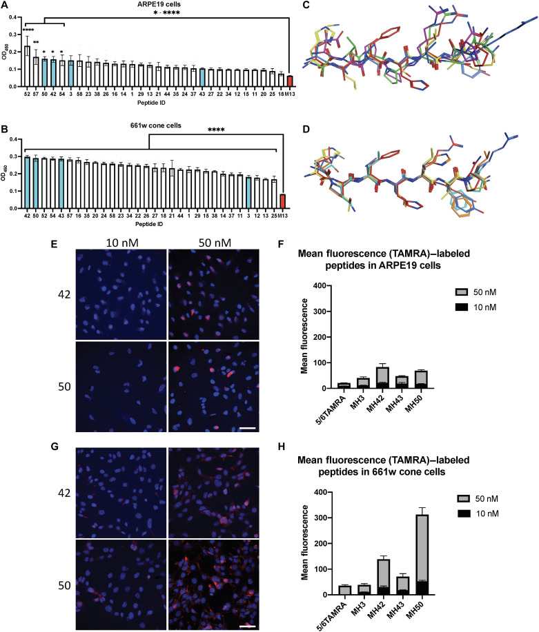
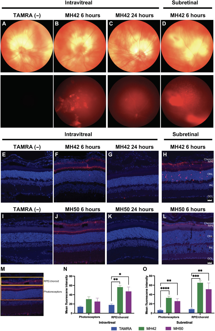
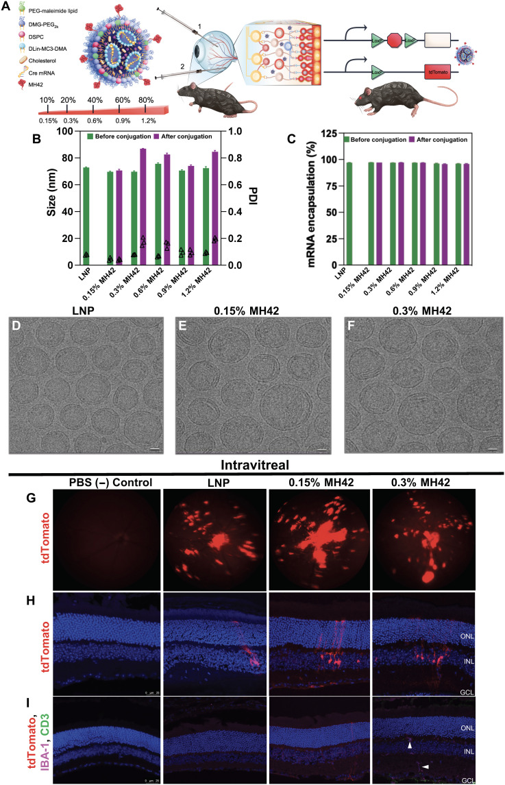
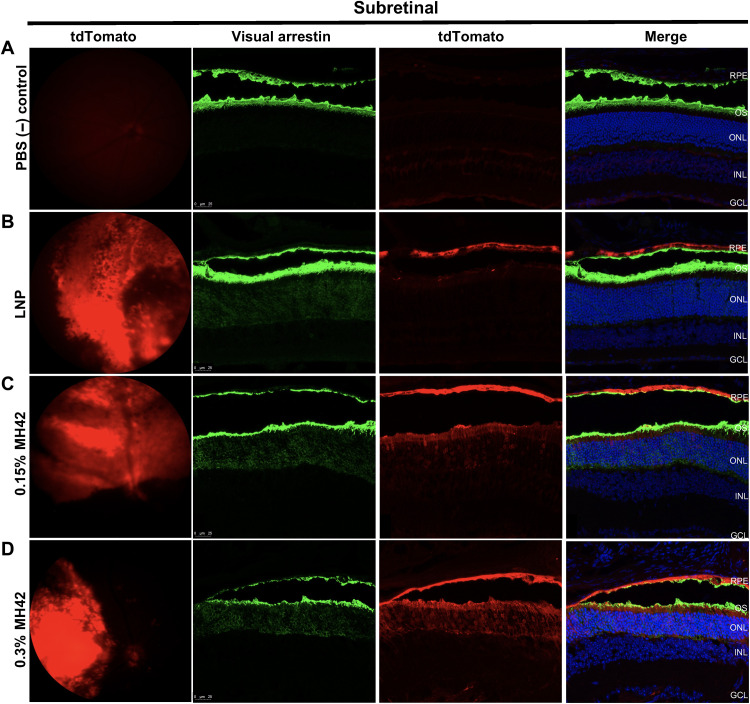
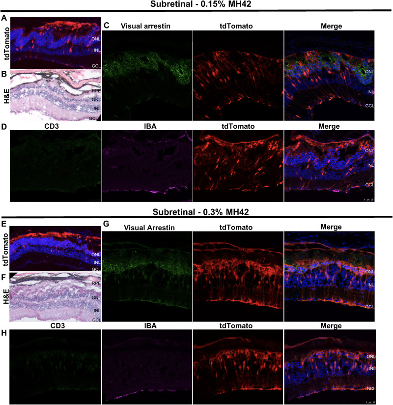
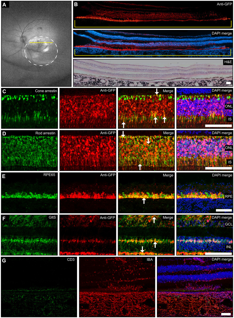
Lipid nanoparticle (LNP)-based mRNA delivery holds promise for the treatment of inherited retinal degenerations. Currently, LNP-mediated mRNA delivery is restricted to the retinal pigment epithelium (RPE) and Müller glia. LNPs must overcome ocular barriers to transfect neuronal cells critical for visual phototransduction, the photoreceptors (PRs). We used a combinatorial M13 bacteriophage-based heptameric peptide phage display library for the mining of peptide ligands that target PRs. We identified the most promising peptide candidates resulting from in vivo biopanning. Dye-conjugated peptides showed rapid localization to the PRs. LNPs decorated with the top-performing peptide ligands delivered mRNA to the PRs, RPE, and Müller glia in mice. This distribution translated to the nonhuman primate eye, wherein robust protein expression was observed in the PRs, Müller glia, and RPE. Overall, we have developed peptide-conjugated LNPs that can enable mRNA delivery to the neural retina, expanding the utility of LNP-mRNA therapies for inherited blindness.


Figures

Fig. 1.
Fig. 1.. In vivo development of peptide phage display biopanning.
(A) In vivo schematic. Heptameric phage libraries were injected intravitreally into BALB/c mice and exposed for 6 hours, followed by retina extraction. Loosely bound phages were subjected to washes and elution to be used in titering and amplification rounds for subsequent biopanning rounds. The process was carried out three times and enriched specific phage-peptide binders were isolated, followed by DNA extraction and Sanger sequencing. (B) Streptavidin-targeted phage biopanning positive control. (C) In vivo phage titers after each round of biopanning. (D) In vivo peptide-targeting validation. Empty M13 bacteriophages were used as a negative control compared to peptide-containing library at the same concentration injected intravitreally in mice using an ordinary one-way analysis of variance (ANOVA). n = 3; means ± SEM. ***P ≤ 0.001. (E to G) Heatmaps depicting phage biopanning enrichment after rounds 1 to 3 in vivo of peptide sequences isolated for each round. For biopanning rounds, n = 3 with at least five technical replicates. (H to J) Consensus sequence for each amino acid in the heptameric sequence isolated after biopanning rounds 1 to 3 in vivo.
Fig. 2.
Fig. 2.. In vitro binding, structural analysis, and internalization of in vivo–isolated peptide candidates.
Single phage-peptide differential binding properties were elucidated using cell-based ELISA against model cell lines of desired target tissues in neural retina. Three high-performing hits and a low binder were selected from ELISA results for further analysis and validation across both cell lines. (A and B) Cell-based ELISA results for ARPE19 cells and 661w cells, respectively, with selected hits highlighted in cyan. Empty M13 bacteriophage used as negative control (red). (C and D) Molecular operating environment (MOE) structural superposition of selected candidates for ARPE19 and 661w cells, respectively. (E and G) Confocal microscopy images of TAMRA-labeled peptides 42 and 50 cell internalization after 30-min incubation with ARPE19 and 661w cells, respectively. Scale bars, 50 μm. (F and H) Mean fluorescence intensity quantification of confocal images. ELISA binding experiments performed in duplicate with six technical replicates; means ± SEM. An ordinary one-way ANOVA, with Tukey’s correction for multiple comparisons test was used for comparisons between treatments. *P ≤ 0.05, **P ≤ 0.01, and ****P ≤ 0.0001.
Fig. 3.
Fig. 3.. In vivo injections of TAMRA-conjugated peptide candidates in BALB/c mice.
Representative images of the clearance kinetics and in vivo targeting of TAMRA-labeled peptides injected intravitreally and subretinally into BALB/c mice and extracted at specific time points after injection. (A to D) In vivo fundus images of peptide MH42 intravitreally and subretinally delivered at the corresponding time points. Top: Bright-field images of the eye. Bottom: Red fluorescence demonstrating localization of the peptide. (E to H and I to L) Confocal images of 12-μm cryosections following intravitreal and subretinal injections of peptides MH42 and MH50, respectively, to validate peptide targeting. Scale bars, 25 μm. (M) Schematic identifying the RPE/choroid and PR layers used for quantification. For the analysis, these layers were manually segmented in ImageJ. (N) Intravitreal and (O) subretinal mean fluorescence intensity quantification of confocal images for localized fluorescence in PR or RPE/choroid layers. An ordinary one-way ANOVA, with Tukey’s correction for multiple comparisons test was used for comparisons between groups. n = 4 to 8 eyes per group; means ± SEM. *P ≤ 0.05, **P ≤ 0.01, ***P ≤ 0.001, and ****P ≤ 0.0001. TAMRA dye injected as negative control. GCL, ganglion cell layer; INL, inner nuclear layer; ONL, outer nuclear layer.
Fig. 4.
Fig. 4.. Peptide-conjugated LNP characterization and in vivo injections in Ai9-tdTomato mice with increasing peptide surface density.
Particle characterization and representative confocal images of MH42 peptide–conjugated, Cre mRNA–loaded LNPs intravitreally administered to Ai9-Rosa mice. (A) Schematic of LNP formulation and conjugation with peptide via maleimide-thiol chemistry and Cre mouse model depicting both routes of administration trialed. (B and C) Graphs depicting size, polydispersity, and mRNA encapsulation efficiency of the LNPs with varying ratios of MH42 peptide conjugation. % refers to molar percent of peptide-functionalized PEG in the formulation. (D to F) Cryo–transmission EM images of the unconjugated control LNP, 0.15% MH42, and 0.3% MH42-LNP. Scale bars, 20 nm. (G) Representative fundus images showing in vivo tdTomato expression after intravitreal delivery of unconjugated and conjugated LNPs. (H) Confocal microscopy images of tdTomato expression following intravitreal uptake and translation of Cre mRNA LNPs. (I) Confocal images showing no T cell infiltration (CD3) or microglia activation (IBA-1) associated with intravitreal delivery and expression. Microglia were restricted to the plexiform layers (arrowheads). Confocal images taken at ×20. n = 6 eyes per group. Scale bars, 50 μm.
Fig. 5.
Fig. 5.. MH42-conjugated LNPs mediate PR expression after subretinal administration.
Representative fundus images showing in vivo tdTomato expression combined with ×40 confocal images of retinal cross sections expressing tdTomato (red) and stained with visual arrestin (green; rods and cones) and DAPI (blue) for (A) phosphate-buffered saline (PBS), (B) untargeted LNPs (n = 2), (C) 0.15% MH42 LNPs (n = 6), and (D) 0.3% MH42 LNPs (n = 4).
Fig. 6.
Fig. 6.. Retinal toxicity associated with subretinal administration of MH42 LNPs.
Top: Representative images from retinas injected with 0.15% MH42 LNPs (n = 6). Bottom: Representative images from retinas injected with 0.3% MH42 LNPs (n = 4). (A and E) Confocal images at ×10 containing tdTomato expression (red) and labeled with DAPI (blue). (B and F) Hematoxylin and eosin (H&E) images show the retinal morphology in some areas of tdTomato expression. (C and G) Confocal images at ×40 demonstrating tdTomato expression (red) and stained with visual arrestin (green) and DAPI (blue). (D and H) Confocal images at ×40 showing tdTomato expression (red) and stained with CD3 (green; T cells), IBA-1 (microglia; magenta), and DAPI (blue).
Fig. 7.
Fig. 7.. MH42-conjugated LNPs mediate expression in the neural retina after subretinal administration in the NHP.
(A) Wide-field fundus autofluorescence imaging 48 hours after subretinal delivery of MH42 LNPs (n = 1). Circle demarks the location of the bleb, and yellow line indicates cross section that corresponds to immunohistochemistry. (B) Montaged ×10 confocal images of primate retinal cross sections labeled with anti-GFP antibody (red) and DAPI (blue). A montaged ×10 H&E image shows the retinal morphology in areas of GFP expression. (C to F) Confocal images (×40) of retinal cross sections costained with anti-GFP and cell-specific antibodies cone arrestin (cones), rod arrestin (rods and s-cones), RPE65 (RPE), and glutamine synthetase (Müller glia). Arrowheads represent co-localization. (C to G) Confocal cross sections (×20) labeled with CD3 (T cells; green) and IBA-1 (microglia; red) to observe the elicited immune response. GltS, glutamine synthetase.Scale bars represent 100μM.

References

    1. N. Chaudhary, D. Weissman, K. A. Whitehead, mRNA vaccines for infectious diseases: Principles, delivery and clinical translation. Nat. Rev. Drug Discov. 20, 817–838 (2021). - PMC - PubMed
    1. A. V. Garafalo, A. V. Cideciyan, E. Héon, R. Sheplock, A. Pearson, C. WeiYang Yu, A. Sumaroka, G. D. Aguirre, S. G. Jacobson, Progress in treating inherited retinal diseases: Early subretinal gene therapy clinical trials and candidates for future initiatives. Prog. Retin. Eye Res. 77, 100827 (2020). - PMC - PubMed
    1. RetNet–Retinal Information Network (14 February 2022).
    1. S. Veleri, C. H. Lazar, B. Chang, P. A. Sieving, E. Banin, A. Swaroop, Biology and therapy of inherited retinal degenerative disease: Insights from mouse models. Dis. Model. Mech. 8, 109–129 (2015). - PMC - PubMed
    1. S. Makin, Four technologies that could transform the treatment of blindness. Nature 10.1038/d41586-019-01107-8 (2019). - PubMed