Review
. 2023 Jan 10;56(1):14-31.
doi: 10.1016/j.immuni.2022.12.003.

Control of tumor-associated macrophage responses by nutrient acquisition and metabolism

Affiliations
Review

Control of tumor-associated macrophage responses by nutrient acquisition and metabolism

Xian Zhang et al. Immunity. .

Abstract

Metazoan tissue specification is associated with integration of macrophage lineage cells in sub-tissular niches to promote tissue development and homeostasis. Oncogenic transformation, most prevalently of epithelial cell lineages, results in maladaptation of resident tissue macrophage differentiation pathways to generate parenchymal and interstitial tumor-associated macrophages that largely foster cancer progression. In addition to growth factors, nutrients that can be consumed, stored, recycled, or converted to signaling molecules have emerged as crucial regulators of macrophage responses in tumor. Here, we review how nutrient acquisition through plasma membrane transporters and engulfment pathways control tumor-associated macrophage differentiation and function. We also discuss how nutrient metabolism regulates tumor-associated macrophages and how these processes may be targeted for cancer therapy.

Keywords: engulfment; macrophage; metabolism; nutrient; transporter; tumor.


Conflict of interest statement

Declaration of interests The authors declare no competing interests.

Figures

Figure 1.
Figure 1.. TAM subsets and metabolic crosstalk in the tumor microenvironment
A) A simplified schematic depicting healthy and tumorous epithelial tissues with phenotypically distinct resident tissue macrophages (RTMs) and tumor-associated macrophages (TAMs) localized in sub-tissular interstitial and parenchymal niches. Epithelial cell transformation is associated with expansion and phenotypic adaptation of cancer cell-associated parenchymal TAMs (pTAMs), while interstitial TAMs (iTAMs) with or without expression of the scavenger receptor Lyve1 are also adapted in interstitial regions composed predominantly of fibroblasts, endothelial cells, nerves, and acellular extracellular matrix (ECM). B) RTM and TAM differentiation from macrophage progenitors is driven by tissue niche factors, including growth factors and nutrients as discussed in for RTMs. The parenchymal and interstitial niche factors regulate differentiation of pRTMs/pTAMs and iRTMs/iTAMs, respectively. C) Modes of metabolic crosstalk between TAMs, cancer cells and other cell types in the tumor microenvironment. Abundant nutrients are taken up by all cell types with no restriction, while limited nutrients can be cross-fed or competed between TAMs, cancer cells, and other cell types in the tumor stroma, promoting or inhibiting tumor growth, respectively. TAMs can also scavenge irritants that otherwise impair tumor tissue fitness and suppress tumor development. D) A number of animal models have been used to study TAM responses in epithelial cancers. Autochthonous murine tumor models involve transformation of endogenous epithelial lineage cells, and preserve the parenchymal and interstitial tissue architecture with pTAMs and iTAMs differentiated in distinct sub-tissular niches. Transplantable murine tumor models involve inoculation of cancer cell lines propagated in vitro into target tissues, which fails to recapitulate the tumor tissue architecture. In addition, many cancer cell lines derived from epithelial tumors acquire mesenchymal phenotype during in vitro propagation. TAM responses in these models are often associated with acute influx of a large number of inflammatory monocytes with TAM differentiation poorly resembling that induced in human tumor.
Figure 2.
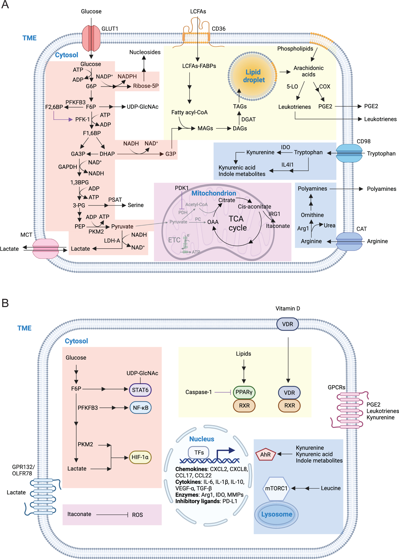
Figure 2.. Macronutrient uptake and metabolism control of TAM responses
A) Macronutrients including glucose, lipids and amino acids are taken up by tumor-associated macrophages (TAMs) in the tumor microenvironment (TME) and are catabolized or converted to biosynthetic intermediates or signaling metabolites to regulate TAM responses. Glucose acquired through glucose transporter 1 (GLUT1) undergoes glycolysis to produce adenosine triphosphate (ATP) and generates metabolic intermediates to support several biosynthetic pathways. Pyruvate is the end-product of glycolysis, and is mainly reduced to lactate in the cytosol, rather than enter the mitochondrion to complete the tricarboxylic acid (TCA) cycle as a likely consequence of low oxygen level in the TME. Instead, itaconate can be converted from the TCA intermediate cis-aconitate with tumor-promoting functions. Long-chain fatty acids (LCFAs) are acquired via CD36-mediated lipid uptake and contribute to lipogenesis and lipid droplet formation in TAMs, while prostaglandin E2 (PGE2) and leukotrienes are converted from phospholipids, and act as bioactive signaling lipids. Amino acids arginine and tryptophan acquired from plasma membrane transporters can also be converted to bioactive metabolites to regulate tumor progression. B) Signaling functions of metabolites in TAMs. Metabolites of the glycolytic pathway can promote activation of transcription factors including signal transducer and activator of transcription 6 (STAT6), nuclear factor kappa B (NF-κB), and hypoxia-inducible factor 1-alpha (HIF-1α). In addition, lactate can be sensed by the G protein-coupled receptors (GPCRs) GPR132 and OLFR78, while the TCA cycle metabolite itaconate suppresses the generation of reactive oxygen species (ROS). Lipid-sensing peroxisome proliferator-activated receptor-γ (PPAR-γ) is subject to caspase-1-mediated cleavage to prevent fatty acid oxidation, while lipid-derived metabolites including vitamin D, PGE2, and leukotrienes can bind to vitamin D receptor (VDR) and GPCR family members to induce cellular signaling. Moreover, cytosolic amino acids such as leucine promotes activation of the metabolic regulator mammalian target of rapamycin complex 1 (mTORC1), while the tryptophan metabolites kynurenine, kynurenic acid, and indole metabolites are sensed by aryl hydrocarbon receptor (AhR) and GPCR family member to regulate TAM responses. 1,3BPG, 1,3-bisphosphoglycerate; 3-PG, 3-phosphoglycerate; 5-LO, 5-lipoxygenase; α-KG, alpha-ketoglutarate; ADP, adenosine diphosphate; Arg1, arginase 1; CAT, cationic amino acid transporter; CCL, chemokine (C-C motif) ligand; COX, cyclooxygenase; CXCL, chemokine (C-X-C motif) ligand; DAGs, diacylglycerols; DGAT, diglyceride acyltransferase; DHAP, dihydroxyacetone phosphate; ETC, electron transport chain; F1,6BP, fructose-1,6-bisphosphate; F2,6BP, fructose-2,6-bisphosphate; F6P, fructose 6-phosphate; FABPs, fatty acid-binding proteins; G3P, glycerol 3-phosphate; G6P, glucose 6-phosphate; GA3P, glyceraldehyde 3-phosphate; GAPDH, glyceraldehyde 3-phosphate dehydrogenase; IL, interleukin; IL4I1, interleukin-4 induced 1; IRG1, immune-responsive gene 1; LDH-A, lactate dehydrogenase-A; MAGs, monoacylglycerols; MCT, monocarboxylate transporter; MGLL, monoacylglycerol lipase; MMPs, matrix metalloproteinase; NAD, nicotinamide adenine dinucleotide; NADP, nicotinamide adenine dinucleotide phosphate; OAA, oxaloacetate; PC, pyruvate carboxylase; PDH, pyruvate dehydrogenase; PDK1, pyruvate dehydrogenase kinase 1; PD-L1, programmed death-ligand 1; PEP, phosphoenolpyruvate; PFK-1, 6-phosphofructokinase-1; PFKFB3, 6-phosphofructo-2-kinase/fructose-2,6-biphosphatase 3; PKM2, pyruvate kinase M2; PSAT, phosphoserine aminotransferase; Ribose-5P, ribose 5-phosphate; RXR, retinoid-X receptor; TAGs, triacylglycerols; TFs, transcription factors; TGF-β, transforming growth factor-beta; UDP-GlcNAc, uridine diphosphate-N-acetylglucosamine; VEGF-α, vascular endothelial growth factor-alpha.
Figure 3.
Figure 3.. Engulfment-mediated nutrient acquisition control of TAM responses
Apoptotic cell as well as lipids, extracellular matrix (ECM) proteins and iron complexes can be phagocytosed or endocytosed in tumor-associated macrophages (TAMs), and nutrients are further generated in the lysosome and exported to the cytosol to support metabolism and signaling responses. Upon ligand binding, efferocytosis receptors such as MER proto-oncogene, tyrosine kinase (MerTK) or lipid-scavenging receptors such as triggering receptor expressed on myeloid cells 2 (Trem2) can activate intracellular kinase signaling cascades and induce expression of anti-inflammatory mediators. The recycled lipids can also induce anti-inflammatory signaling or exported outside of TAMs to promote cross-feeding. Degradation of apoptotic bodies and ECM proteins generate amino acids in the lysosome and may activate the metabolic regulator mammalian target of rapamycin complex 1 (mTORC1). Arginine is a major amino acid generated from ECM degradation and can activate Rac1 through its downstream metabolites. Engulfed apoptotic cells also contain nucleic acids, proper degradation of which is critical to prevent activation of nucleic acid-innate immune sensing pathways. TAMs can either uptake or release iron in both free and complex forms and regulate iron metabolism through competition or crossfeeding in tumor. ABCA1, ATP Binding cassette subfamily A member 1; CD163, CD163 molecule; DNase II, deoxyribonuclease 2, lysosomal; ENT3, equilibrative nucleoside transporter 3; FPN1, ferroportin 1; SLC39A8, solute carrier family 39 member 8; HO-1, heme oxygenase 1; HRG1, heme-responsive gene 1 protein homolog; FCGR1A, Fc gamma receptor Ia; Lcn2, lipocalin 2; LIPA, lipase A, lysosomal acid type A; LRP1, LDL receptor related protein 1; Lyve1, lymphatic vessel endothelial hyaluronan receptor 1; Marco, macrophage receptor with collagenous structure; Mertk, MER proto-Oncogene, tyrosine kinase; Mincle, macrophage-inducible C-type lectin; Mrc1, mannose receptor C-type 1; NPC1, NPC intracellular cholesterol transporter 1; PLA2G15, phospholipase A2 group XV; SLC11A1, solute carrier family 11 member 1; SLC11A2, solute carrier family 11 member 2; SLC22A17, solute carrier family 22 member 17; SLC39A14, solute carrier family 39 member 14; TfR, transferrin receptor; Trem2, triggering receptor expressed on myeloid cells 2.

References

    1. Okabe Y, and Medzhitov R (2016). Tissue biology perspective on macrophages. Nat Immunol 17, 9–17. 10.1038/ni.3320. - DOI - PubMed
    1. Guilliams M, Thierry GR, Bonnardel J, and Bajenoff M (2020). Establishment and Maintenance of the Macrophage Niche. Immunity 52, 434–451. 10.1016/j.immuni.2020.02.015. - DOI - PubMed
    1. Blériot C, Chakarov S, and Ginhoux F (2020). Determinants of Resident Tissue Macrophage Identity and Function. Immunity 52, 957–970. 10.1016/j.immuni.2020.05.014. - DOI - PubMed
    1. Lavin Y, Mortha A, Rahman A, and Merad M (2015). Regulation of macrophage development and function in peripheral tissues. Nat Rev Immunol 15, 731–744. 10.1038/nri3920. - DOI - PMC - PubMed
    1. Jenkins SJ, and Allen JE (2021). The expanding world of tissue-resident macrophages. Eur J Immunol 51, 1882–1896. 10.1002/eji.202048881. - DOI - PubMed

Publication types