. 2023 Jul;72(7):1355-1369.
doi: 10.1136/gutjnl-2022-327448. Epub 2023 Jan 11.

Activated regulatory T-cells promote duodenal bacterial translocation into necrotic areas in severe acute pancreatitis

Affiliations

Activated regulatory T-cells promote duodenal bacterial translocation into necrotic areas in severe acute pancreatitis

Juliane Glaubitz et al. Gut. 2023 Jul.

Abstract

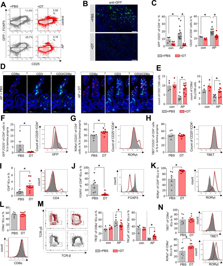
Objective: In acute pancreatitis (AP), bacterial translocation and subsequent infection of pancreatic necrosis are the main risk factors for severe disease and late death. Understanding how immunological host defence mechanisms fail to protect the intestinal barrier is of great importance in reducing the mortality risk of the disease. Here, we studied the role of the Treg/Th17 balance for maintaining the intestinal barrier function in a mouse model of severe AP.

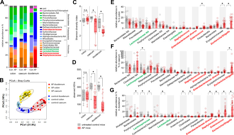
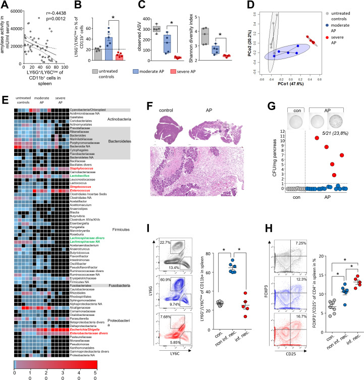
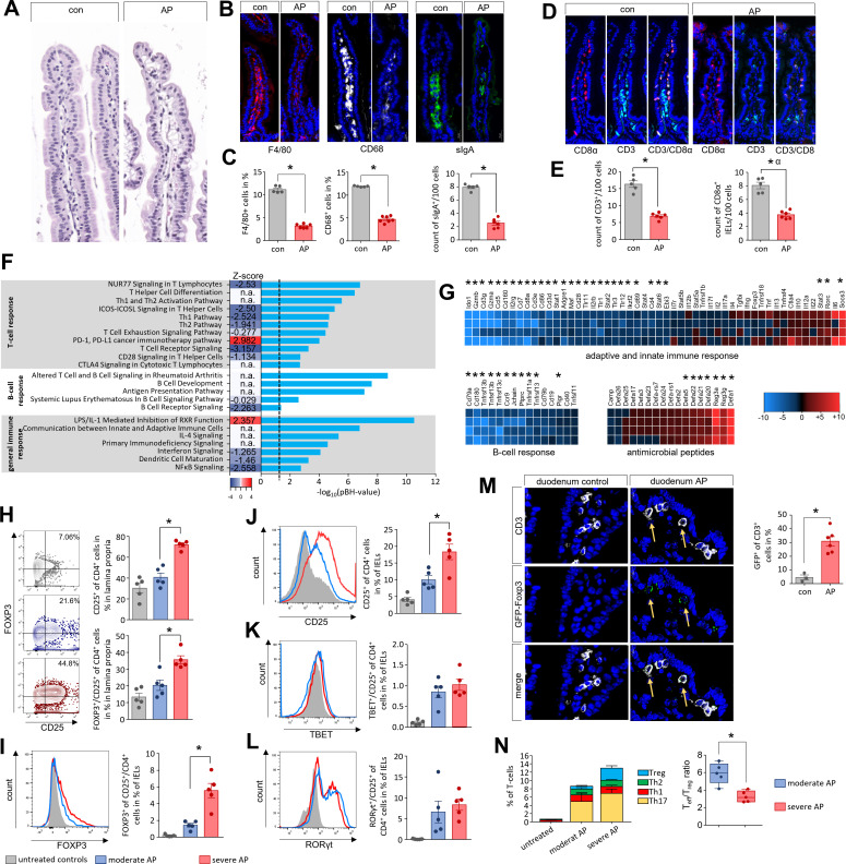
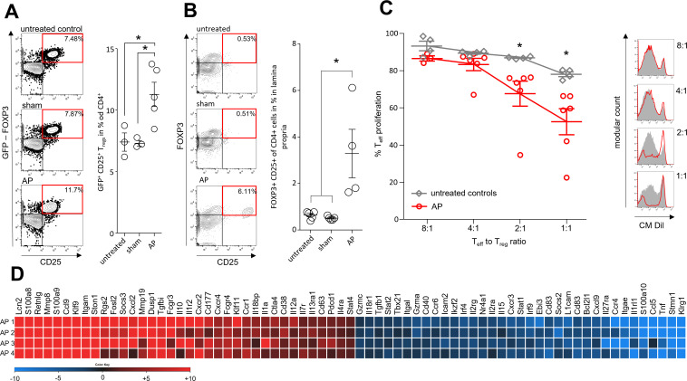
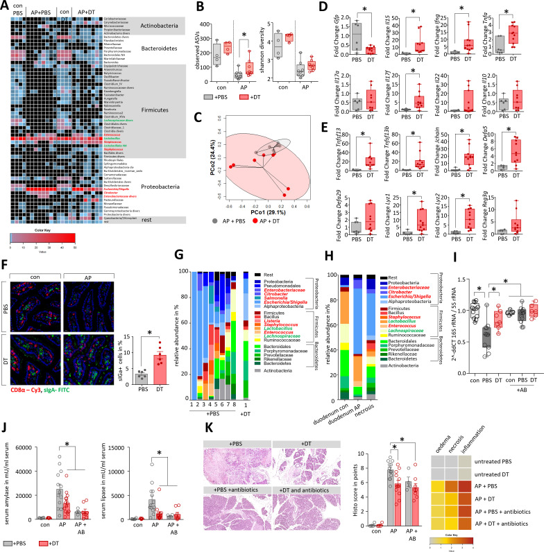
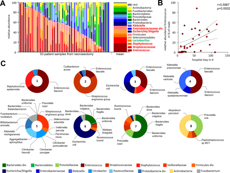
Design: AP was induced by partial duct ligation in C57Bl/6 or DEREG mice, in which regulatory T-cells (Treg) were depleted by intraperitoneal injection of diphtheria toxin. By flow cytometry, functional suppression assays and transcriptional profiling we analysed Treg activation and characterised T-cells of the lamina propria as well as intraepithelial lymphocytes (IELs) regarding their activation and differentiation. Microbiota composition was examined in intestinal samples as well as in murine and human pancreatic necrosis by 16S rRNA gene sequencing.

Results: The prophylactic Treg-depletion enhanced the proinflammatory response in an experimental mouse model of AP but stabilised the intestinal immunological barrier function of Th17 cells and CD8+/γδTCR+ IELs. Treg depleted animals developed less bacterial translocation to the pancreas. Duodenal overgrowth of the facultative pathogenic taxa Escherichia/Shigella which associates with severe disease and infected necrosis was diminished in Treg depleted animals.

Conclusion: Tregs play a crucial role in the counterbalance against systemic inflammatory response syndrome. In AP, Treg-activation disturbs the duodenal barrier function and permits translocation of commensal bacteria into pancreatic necrosis. Targeting Tregs in AP may help to ameliorate the disease course.

Keywords: acute pancreatitis; bacterial infection; experimental pancreatitis; immune response.


Conflict of interest statement

Competing interests: None declared.

Figures

Figure 1
Figure 1
Acute pancreatitis (AP) is associated with changes of the intestinal microbiota composition. (A) Faecal samples from colon, caecum and duodenum were collected from C57Bl/6 mice with AP (n=24) and from untreated control animals Con (n=10). Isolated DNA was analysed by 16S rRNA gene sequencing. The microbiota composition and AP-associated changes of the taxonomic units with the highest abundance are illustrated by a stacked bar graph. (B) Principal coordinate analysis illustrates the changes of gut microbiome between untreated controls and AP mice. (C, D) Shannon-Diversity Index (C) and richness of observed species (D) demonstrate significant impact of AP on the duodenal microbiome composition. (E–G) The bar graph illustrates changes of the most abundant taxa in colonic (E), caecal (F) and duodenal samples (G) of AP (red) and control mice (grey). Facultative pathogenic were marked in red, beneficial commensal bacteria were marked in green. Statistical significance was determined by unpaired Student’s t-test and Kruskal-Wallis test followed by a Dunn’s multiple comparisons test to analyse differentially abundant taxa in colon, caecum and duodenum samples. Significance levels of p<0.05 are marked by an asterisk.
Figure 2
Figure 2
Intestinal microbiota changes correlate with the severity of AP and the systemic immune response. (A) Dot plot illustrates negative correlation of LY6G+/LY6Clow/CD11b+-cells to the activity of serum amylase of mice with AP (n=50), significance of correlation was tested by Spearman’s rank correlation-coefficient. (B) Disease severity in animals was classified according to the percentage of LY6G+/LY6Clow/CD11b+-cells in spleen (n=5/group). (C) Box plots illustrate the duodenal microbiome species richness and Shannon-diversity according to moderate and severe pancreatitis. (D) Principal coordinate analysis illustrates significant differences of duodenal microbiome between these groups (permutational multivariate analysis of variance; control vs severe: p=0.007, R2=59.4%, control vs moderate: p=0.015, R2=24.5% and moderate vs severe: p=0.007, R2=48.4%). (E) Heat-map illustrates 16S rRNA gene sequencing results of the duodenal microbiome analysis, facultative pathogenic were marked in red, beneficial commensal bacteria were marked in green. (F) H&E-staining of pancreatic tissue of healthy and AP mice. (G) Colony forming units (CFU) were counted from necrotic tissue homogenates and exceeded the cut-off level of 1 CFU/mg tissue weight in 5 of 21 (23.8%) animals. (I) From infected (n=5) and non-infected (n=6) groups, we analysed in spleen the percentage of LY6G+/LY6Clow/CD11b+-cells and (H) the percentage of FOXP3+/CD25+/CD4+-cells. Statistical evaluation was done by unpaired Student’s t-test for independent samples and significance levels of p<0.05 are marked by an asterisk. AP, acute pancreatitis.
Figure 3
Figure 3
Imbalance of the Th17/Treg ratio in duodenal mucosa during AP. (A–E) Histological examination of the duodenum illustrates changes of the intestinal mucosa in C57Bl/6 mice after induction of AP. (A–C) In contrast to only slight changes in H&E-staining we detected differences in immunofluorescent labelling of F4/80, CD68 as marker for macrophages and sIgA as marker for plasma cells (B), all cells were significantly decreased in AP mice (C). (D) CD3 as T-cell marker and CD8α as marker of IELs were labelled in small intestine. (E) Bar graphs illustrate significant changes of T-cells and IELs in duodenum. (F) The bar graph shows the -log10(pBH) values of overrepresented pathways from an IPA analysis which was based on differentially expressed transcriptome data of duodenal tissue from AP and control mice. Heatmap illustrates the z-score which indicates activation (positive z-score, red) or inhibition (negative z-score, blue) of this pathway. (G) Heat-map illustrates fold change differences of upregulated (red) and downregulated (blue) genes in duodenal tissue of AP mice compared with untreated controls. (H) Lymphocytes isolated from lamina propria as well as from the epithelial layer were analysed by flow cytometry. In CD4+ T-cells of the lamina propria the increased levels of CD25 and FOXP3 correlated with disease severity. (I–J) The numbers of CD4+/FOXP3+/CD25+-Tregs (I) and CD4+/CD25+-T-cells (J) from IELs were also increased in a severity-dependent manner. (K, L) Th17-cells marked by RORγt+/CD25+ (K) and Th1-cells marked by TBET+/CD25+ (L) did not show a severity dependent increase. (M) GFP-producing Tregs in the duodenum of DEREG-mice were detected by anti-GFP and anti-CD3 labelling and showed a significant increase after onset of AP. (N) AP induced a shift of the Teff/Treg ratio. Tregs showed a clear increase between moderate and severe AP in contrast to Teff cells (Th1, Th2 and Th17). Statistically significant differences were tested by unpaired Student’s t-test for independent samples and significance levels of p<0.05 are marked by an asterisk. AP, acute pancreatitis.
Figure 4
Figure 4
Immune suppressive function of Tregs during AP. AP was induced by partial duct ligation in DEREG- and C57Bl/6 mice. (A) The number of splenic GFP producing Tregs was increased in the AP group (n=5) but not in control or sham operated mice (n=3). (B) The same increase of FOXP3+/CD25+/CD4+ Tregs was observed in the small intestine. (n=5). (C) Suppression assays showed that Tregs from AP mice (red line) have an increased suppressive capacity on Teff cell proliferation. (D) Heat map illustrates fold changes of gene transcription in Tregs from AP mice (n=4) compared with untreated controls (n=4). Statistically significant differences were tested by unpaired Student’s t-test for independent samples and significance levels of p<0.05 are marked by an asterisk, corrected for multiple testing (bonferroni-correction). AP, acute pancreatitis.
Figure 5
Figure 5
Depletion of Tregs stabilises the homoeostasis of CD4+ T cells in the lamina propria as well as of CD8α+ IELs in the duodenum. AP pancreatitis was induced in DEREG mice by partial duct ligation, Tregs were depleted by diphtheria toxin (DT) (n=11), controls receive PBS (n=7). (A) The efficiency of Treg depletion with DT was verified by flow cytometry analysis of splenocytes. (B) Immunofluorescence detection of GFP-producing Tregs was performed in lymph nodes of DT-treated and PBS-treated mice. (C) Bar graphs show the ratios of CD25+ T-cells and GFP+/CD25+ Tregs analysed by flow cytometry of splenocytes. (D, E) Immunofluorescent labelling of CD3 and CD8α showed a significant persistence of the T-cell and IEL population in the absence of Tregs in DT-treated DEREG-mice compared with the PBS-treated group, (E). (F–N) Lymphocytes of the lamina propria and the epithelial layer were isolated from duodenum and analysed by flow cytometry. (F) Following Treg-depletion, no GFP+/CD25+ Tregs were detected within the lamina propria. (G, H) Bar graph show the numbers of RORγt+/CD25+ Th17-cells (G) and TBET+/CD25+ Th1-cells (H). (I) The bar graph shows a higher ratio of CD4hi expressing IELs in DT-treated mice. (J, K) In the CD4hi IELs we observed a shift from GFP+ Tregs (J) to RORγt+ Th17-cells (K). (L) The ratio of CD8α+ IELs was not affected by Treg-depletion. (M) Dot plot and bar graphs illustrate the ratio changes of TCRγδ+ IELs and TCRβ+ after DT-treatment. (N) Within the population of CD8α+ IELs we measured an increase in RORγt producing cells whereas TBET was not affected. Statistically significant differences were tested by unpaired student’s t-test for independent samples and significance levels of p<0.05 are marked by an asterisk. AP, acute pancreatitis; IELs, intraepithelial lymphocytes; PBS, phosphate-buffered saline.
Figure 6
Figure 6
Depletion of Tregs attenuates microbial dysbiosis during pancreatitis and prevents bacterial translocation into pancreatic necroses. (A) Heat-map illustrates the major microbial taxa in the duodenum of DEREG treated with PBS (n=8) or DT (n=8) after induction of AP. Animals without AP referred as healthy controls (0d). Facultative pathogenic were marked in red, beneficial commensal bacteria were marked in green. (B) Box plots illustrate Shannon-Diversity Index and species richness in DT- or PBS-treated DEREG-mice. (C) A principal coordinate analysis illustrates significant differences of the duodenal microbiome between these groups (permutational multivariate analysis of variance; control vs severe: p<0.001, R2=18.2%). (D, E) Box plots show RT-qPCR analysis of duodenal transcriptional changes after depletion of Tregs. (F) Immunofluorescent labelling of sIgA producing cells in the duodenal mucosa from control and AP animals showed significantly decreased numbers of sIgA producing cells in AP mice which received PBS. (G) Staked-bar graphs illustrates bacterial taxa which could be identified in all samples with more than 5.000 clean reads from pancreatic necrosis in DEREG AP-mice (8 PBS treated vs 1 DT treated DEREG-mice). (H) The same bacterial taxa that appear or expand in the duodenum of AP-mice are found in murine pancreatic necrosis samples. The stacked bar graphs show the mean of all mice. (I) We analysed control (con) and duct-ligated DEREG mice with AP (PBS and DT) by 16S rRNA gene RT-qPCR analysis of isolated DNA from pancreatic tissue. Decreased Ct values indicate bacterial infection in the pancreas of mice with AP with a significant greater extent in the PBS-treated group compared with Treg depleted mice. Antibiotic treatment (+AB) significantly reduced the copy number of bacterial 16S rRNA gene in the pancreas and therefore prevented bacterial translocation during AP. (J, K) To evaluate how Tregs and microbiome composition affect the disease severity DEREG mice were treated after AP induction with antibiotics (AB), either in presence (+PBS) or absence (+DT) of Tregs. (J) Disease severity was evaluated by analysis of serum amylase and lipase. (K) Pancreatic histology was analysed by H&E-staining of tissue sections, histology score was evaluated by quantification of oedema, necrosis and leucocyte infiltration. The heatmap illustrates the mean result for all groups. Statistically significant differences were tested by unpaired Student’s t-test for independent samples. For more than two groups, statistical significance was determined by ANOVA one way analysis of variance followed by Bonferroni correction for multiple testing, significance levels of p<0.05 are marked by an asterisk. ANOVA, analysis of variance; DT, diphtheria toxin; RT-qPCR, reverse transcription-quantitative PCR; PBS, phosphate-buffered saline.
Figure 7
Figure 7
Bacterial infection of human pancreatic necrosis. (A) The bar chart represents the major bacterial taxonomic units that were identified by 16S rRNA gene sequencing in 53 samples of human pancreatic necrosis. Facultative pathogenic were marked in red, beneficial commensal bacteria were marked in green. (B) Enterococcus was observed in necrosectomy samples of 33 patients and thus represented the most abundant taxon and showed a positive correlation with the duration of hospitalisation (Spearman correlation p=0.0002, Spearman r=0.599). (C) Bacterial species were analysed using metagenomic-shotgun sequencing of eight selected necrosis samples. Identified commensal gut bacteria include from the phylum Firmicutes Enterococcus faecalis, Enterococcus faecium and Streptococcus anginosus. In addition, Escherichia coli (phylum of Proteobacteria) and Bacteroides dorei, Bacteroides vulgatus, Bacteroides fragilis, Bacteroides uniformis and Prevotella copri as representatives of the phylum Bacteroidetes were detected.

References

    1. Peery AF, Crockett SD, Murphy CC, et al. . Burden and cost of gastrointestinal, liver, and pancreatic diseases in the United States: update 2018. Gastroenterology 2019;156:254–72. 10.1053/j.gastro.2018.08.063 - DOI - PMC - PubMed
    1. van Dijk SM, Hallensleben NDL, van Santvoort HC, et al. . Acute pancreatitis: recent advances through randomised trials. Gut 2017;66:2024–32. 10.1136/gutjnl-2016-313595 - DOI - PubMed
    1. Wolbrink DRJ, Kolwijck E, Ten Oever J, et al. . Management of infected pancreatic necrosis in the intensive care unit: a narrative review. Clin Microbiol Infect 2020;26:18–25. 10.1016/j.cmi.2019.06.017 - DOI - PubMed
    1. Neumann C, Blume J, Roy U, et al. . c-Maf-dependent Treg cell control of intestinal TH17 cells and IgA establishes host-microbiota homeostasis. Nat Immunol 2019;20:471–81. 10.1038/s41590-019-0316-2 - DOI - PubMed
    1. Sendler M, Maertin S, John D, et al. . Cathepsin B activity initiates apoptosis via digestive protease activation in pancreatic acinar cells and experimental pancreatitis. J Biol Chem 2016;291:14717–31. 10.1074/jbc.M116.718999 - DOI - PMC - PubMed

Publication types

Substances