. 2023 Jan 11;10(2):e200081.
doi: 10.1212/NXI.0000000000200081. Print 2023 Mar.

Anti-LGI4 Antibody Is a Novel Juxtaparanodal Autoantibody for Chronic Inflammatory Demyelinating Polyneuropathy

Affiliations

Anti-LGI4 Antibody Is a Novel Juxtaparanodal Autoantibody for Chronic Inflammatory Demyelinating Polyneuropathy

Xu Zhang et al. Neurol Neuroimmunol Neuroinflamm. .

Abstract

Background and objectives: The objective of this study was to discover novel nodal autoantibodies in chronic inflammatory demyelinating polyneuropathy (CIDP).

Methods: We screened for autoantibodies that bind to mouse sciatic nerves and dorsal root ganglia (DRG) using indirect immunofluorescence (IFA) assays with sera from 113 patients with CIDP seronegative for anti-neurofascin 155 and anticontactin-1 antibodies and 127 controls. Western blotting, IFA assays using HEK293T cells transfected with relevant antigen expression plasmids, and cell-based RNA interference assays were used to identify target antigens. Krox20 and Periaxin expression, both of which independently control peripheral nerve myelination, was assessed by quantitative real-time PCR after application of patient and control sera to Schwann cells.

Results: Sera from 4 patients with CIDP, but not control sera, selectively bound to the nodal regions of sciatic nerves and DRG satellite glia (p = 0.048). The main immunoglobulin G (IgG) subtype was IgG4. IgG from these 4 patients stained a 60-kDa band on Western blots of mouse DRG and sciatic nerve lysates. These features indicated leucine-rich repeat LGI family member 4 (LGI4) as a candidate antigen. A commercial anti-LGI4 antibody and IgG from all 4 seropositive patients with CIDP showed the same immunostaining patterns of DRG and cultured rat Schwann cells and bound to the 60-kDa protein in Western blots of LGI4 overexpression lysates. IgG from 3 seropositive patients, but none from controls, bound to cells cotransfected with plasmids containing LGI4 and a disintegrin and metalloprotease domain-containing protein 22 (ADAM22), an LGI4 receptor. In cultured rat Schwann and human melanoma cells constitutively expressing LGI4, LGI4 siRNA effectively downregulated LGI4 and reduced patients' IgG binding compared with scrambled siRNA. Application of serum from a positive patient to Schwann cells expressing ADAM22 significantly reduced the expression of Krox20, but not Periaxin. Anti-LGI4 antibody-positive patients had a relatively old age at onset (mean age 58 years), motor weakness, deep and superficial sensory impairment with Romberg sign, and extremely high levels of CSF protein. Three patients showed subacute CIDP onset resembling Guillain-Barré syndrome.

Discussion: IgG4 anti-LGI4 antibodies are found in some elderly patients with CIDP who present subacute sensory impairment and motor weakness and are worth measuring, particularly in patients with symptoms resembling Guillain-Barré syndrome.


Figures

Figure 1
Figure 1. Screening for Autoantibodies in Anti-neurofascin155 and Anti-contactin1 Antibody-Negative Patients With CIDP Using Mouse Sciatic Nerves and DRG
(A) Double immunostaining of a mouse teased sciatic nerve with a patient's serum and anti-Kv1.2 antibody shows colocalization of the patient's IgG with Kv1.2, a marker of juxtaparanodes. (B) IgG from 4 patients with CIDP, but not from a healthy control, demonstrates a circle-like staining pattern around the ganglion cells (see the magnified images). For case 1, immunostaining of mouse DRG by serum samples obtained before and 16 years after the first IVIg treatment is shown. Scale bar: 50 μm. Kv = potassium channel; CIDP = chronic inflammatory demyelinating polyneuropathy; DRG = dorsal root ganglia; IFA = immunofluorescence assay; IVIg = IV immunoglobulin.
Figure 2
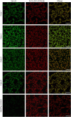
Figure 2. Identification of Anti-LGI4 Antibodies in Sera From Tissue-Based IFA-Positive Patients With CIDP by DRG-Based IFAs
Double immunostaining of mouse DRG with serum from a tissue-based IFA-positive patient with CIDP (case 1 in Table 1) and different commercial antibodies. (A) Immunostaining of glutamine synthetase (red), a specific satellite glial cell marker, colocalized with patient IgG binding (green). (B) Immunostaining of S100β (red), a marker of large DRG neurons and satellite glial cells, partly colocalized with patient IgG binding (green). (C) IgG binding of the patient's serum (green) colocalized with that of the commercial anti-LGI4 antibody (LGI4; red). (D) immunostaining of CASPR1 (red), a paranode marker, which mainly stained sensory neuron somata, barely colocalized with patient IgG binding (green). Scale bar: 50 µm. CIDP = chronic inflammatory demyelinating polyneuropathy; CASPR1 = contactin-associated protein 1; DRG = dorsal root ganglia; IFA = immunofluorescence assay; LGI4 = leucine-rich repeat LGI family member 4.
Figure 3
Figure 3. Cell-Based IFA Using Rat Schwann Cells With Patients' IgG and a Commercial Anti-LGI4 Antibody
Serum samples from 4 seropositive patients with CIDP and a commercial anti-LGI4 antibody all demonstrate the same immunostaining pattern on rat Schwann cells, while a healthy control shows no immunoreactivity. Scale bars: 20 µm. CIDP = chronic inflammatory demyelinating polyneuropathy; IFA = immunofluorescence assay; LGI4 = leucine-rich repeat LGI family member 4.
Figure 4
Figure 4. Cell-Based IFAs Using Commercial Anti-LGI4 Antibodies and Sera of Seropositive Patient With CIDP
(A) LGI4-Flag–cotransfected and ADAM22-HA–cotransfected HEK293T cells stained with anti-Flag and commercial anti-LGI4 antibodies. (B) LGI4-Flag–cotransfected and ADAM22-HA–cotransfected HEK293T cells stained with anti-Flag antibodies and IgG from seropositive patients with CIDP (cases 1 and 4) showed cell surface staining. Scale bar: 50 μm. ADAM22 = a disintegrin and metalloprotease domain–containing protein 22, CIDP = chronic inflammatory demyelinating polyneuropathy; HEK = human embryonic kidney; IFA = immunofluorescence assay; LGI4 = leucine-rich repeat LGI family member 4.
Figure 5
Figure 5. Confirmation of the Autoantibody Specificity for LGI4 Using a Genetic Strategy With Rat Schwann Cells
(A) Assessment of the effect of Lgi4 siRNA transfection on Lgi4 mRNA levels in rat Schwann cells by quantitative real-time PCR. The results are expressed as mean ± SEM. The expression of glyceraldehyde-3-phosphate dehydrogenase was determined as a housekeeping gene. *p = 0.0213. (B) WB analysis of LGI4 protein using rat Schwann cells after Lgi4 siRNA or scrambled siRNA treatment. IgG from 1 representative seropositive patient with CIDP (case 2) and a commercial anti-LGI4 antibody showed decreased signals after Lgi4 siRNA treatment compared with the scrambled siRNA in rat Schwann cells. (C) Cell-based IFA using Lgi4 siRNA or scrambled siRNA-treated rat Schwann cells. Signals in rat Schwann cells from the serum of case 2 and the anti-LGI4 antibody were significantly decreased after Lgi4 siRNA treatment compared with those after scrambled siRNA treatment. (D) Comparison of the MFI from a CIDP patient's IgG between Lgi4 siRNA and scrambled siRNA-treated rat Schwann cells. Nuclei are counterstained with DAPI (blue). The results are expressed as the mean ± SEM. *p = 0.0241. Scale bars: (C) 30 μm. AU = arbitrary units; CIDP = chronic inflammatory demyelinating polyneuropathy; DAPI = 4′,6-diamidino-2-phenylindole; IFA = immunofluorescence assay; LGI4 = leucine-rich repeat LGI family member 4; MFI = mean fluorescence intensity; WB = Western blotting.
Figure 6
Figure 6. Effects of Sera From Anti-LGI4 Antibody-Seropositive Patients With CIDP and Controls on Schwann Cells
Quantitative real-time PCR assessment of Krox20 (A) and Periaxin (B) expression in rat Schwann cells. The results are expressed as the mean ± SEM. *p = 0.03, **p = 0.0045. CIDP = chronic inflammatory demyelinating polyneuropathy; LGI4 = leucine-rich repeat LGI family member 4.

References

    1. Fehmi J, Scherer SS, Willison HJ, Rinaldi S. Nodes, paranodes and neuropathies. J Neurol Neurosurg Psychiatry. 2018;89(1):61-71. doi: 10.1136/jnnp-2016-315480. - DOI - PubMed
    1. Vural A, Doppler K, Meinl E. Autoantibodies against the node of Ranvier in seropositive chronic inflammatory demyelinating polyneuropathy: diagnostic, pathogenic, and therapeutic relevance. Front Immunol. 2018;9:1029. doi: 10.3389/fimmu.2018.01029. - DOI - PMC - PubMed
    1. Devaux JJ, Miura Y, Fukami Y, et al. . Neurofascin-155 IgG4 in chronic inflammatory demyelinating polyneuropathy. Neurology. 2016;86(9):800-807. doi: 10.1212/WNL.0000000000002418. - DOI - PMC - PubMed
    1. Querol L, Nogales-Gadea G, Rojas-Garcia R, et al. . Neurofascin IgG4 antibodies in CIDP associate with disabling tremor and poor response to IVIg. Neurology. 2014;82(10):879-886. doi: 10.1212/WNL.0000000000000205. - DOI - PMC - PubMed
    1. Kawamura N, Yamasaki R, Yonekawa T, et al. . Anti-neurofascin antibody in patients with combined central and peripheral demyelination. Neurology. 2013;81(8):714-722. doi: 10.1212/WNL.0b013e3182a1aa9c. - DOI - PubMed

Publication types