. 2023 Jul;56(7):e13397.
doi: 10.1111/cpr.13397. Epub 2023 Jan 11.

The beneficial effects of chick embryo extract preconditioning on hair follicle stem cells: A promising strategy to generate Schwann cells

Affiliations

The beneficial effects of chick embryo extract preconditioning on hair follicle stem cells: A promising strategy to generate Schwann cells

Sareh Pandamooz et al. Cell Prolif. 2023 Jul.

Abstract

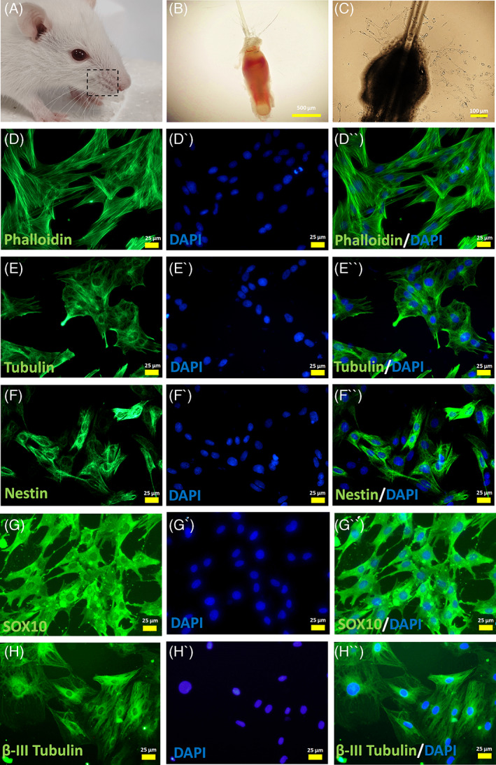
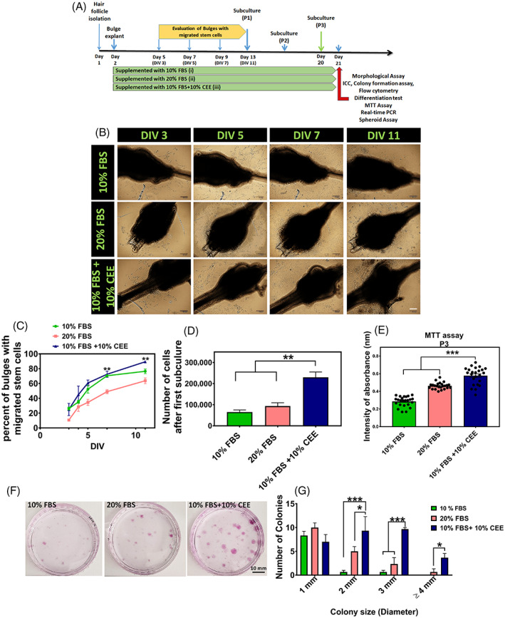
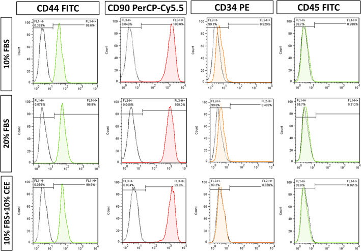
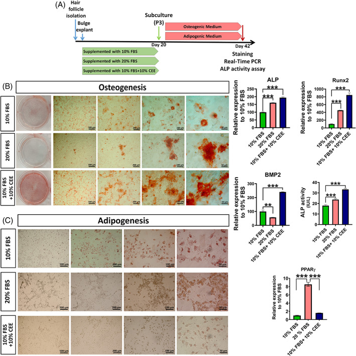
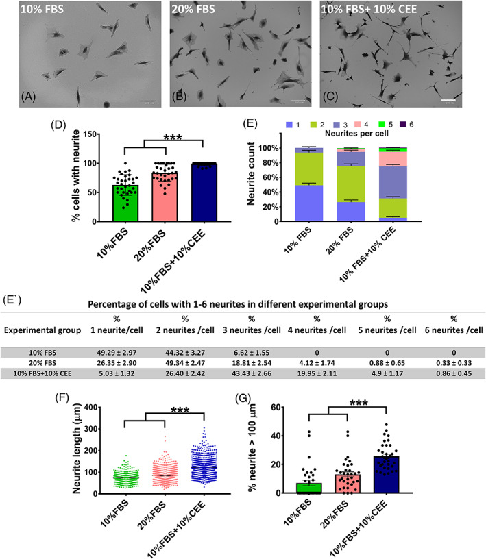
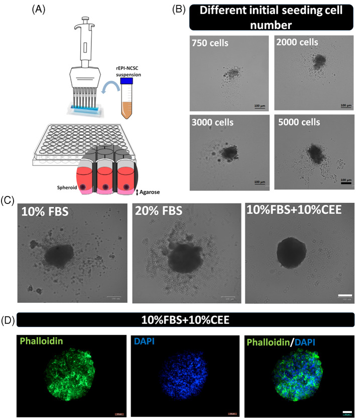
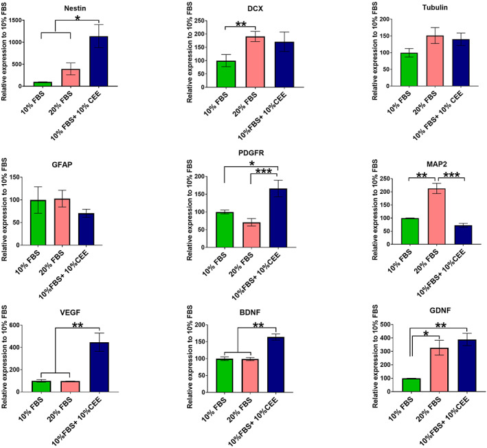
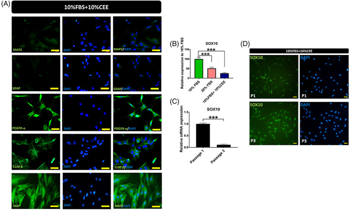
The beneficial effects of hair follicle stem cells in different animal models of nervous system conditions have been extensively studied. While chick embryo extract (CEE) has been used as a growth medium supplement for these stem cells, this is the first study to show the effect of CEE on them. The rat hair follicle stem cells were isolated and supplemented with 10% fetal bovine serum plus 10% CEE. The migration rate, proliferative capacity and multipotency were evaluated along with morphometric alteration and differentiation direction. The proteome analysis of CEE content identified effective factors of CEE that probably regulate fate and function of stem cells. The CEE enhances the migration rate of stem cells from explanted bulges as well as their proliferation, likely due to activation of AP-1 and translationally controlled tumour protein (TCTP) by thioredoxin found in CEE. The increased length of outgrowth may be the result of cyclic AMP response element binding protein (CREB) phosphorylation triggered by active CamKII contained in CEE. Further, CEE supplementation upregulates the expression of vascular endothelial growth factor (VEGF), brain-derived neurotrophic factor and glial cell line-derived neurotrophic factor. The elevated expression of target genes and proteins may be due to CREB, AP-1 and c-Myc activation in these stem cells. Given the increased transcript levels of neurotrophins, VEGF, and the expression of PDGFR-α, S100B, MBP and SOX-10 protein, it is possible that CEE promotes the fate of these stem cells towards Schwann cells.


Conflict of interest statement

The authors declare that they have no competing interests.

Figures

FIGURE 1
FIGURE 1
Isolation and characterization of rat hair follicle stem cells. (A, B) In the currrent study, the whisker pads of rat was dissected to isolate large hair follicles. Then, the bulge region of the hair follicle was carefully microdissected and explanted onto collagen coated plates. (C) Few days later, migrated stem cells were detected around the explanted bulge. (D,E) The immunostaining against phalloidin and β‐Tubulin revealed the general morphology of stem cells. (F‐H) Nestin, SOX‐10 and β‐III Tubulin expression confirmed the identity of migrated cells as hair follicle stem cells. Cell nuclei were counterstained with DAPI. Images are examples of three different assessments for each immunostaining (n = 3)
FIGURE 2
FIGURE 2
Epidermal neural crest stem cells in vitro migration and expansion. (A) Schematic time‐line protocol of the study. (B) In this study, the explanted bulges were supplemented with different concentrations of fetal bovine serum (FBS) and chick embryo extract (CEE). Evaluation of the bulges at DIV: 3, 5, 7 and 11 revealed increasing rates of migrating stem cells around the bulges in all experimental groups, Scale bar: 100 μm. (C) Although the percent of bulges with migrated stem cells increased over the culture time course in all three groups, it was significantly higher in experimental group 3 (10% FBS + 10% CEE) than in group 2 (20% FBS) at DIV: 7 and 11 (**p < 0.01, one‐way ANOVA followed by post hoc Tukey's test). (D) Also, cell count after the first subculture in experimental group 3 was significantly higher compared to the two other groups (**p < 0.01, one‐way ANOVA followed by post hoc Tukey's test). (E) Assessment of cell proliferation using MTT assay at passage 3 in the different experimental groups showed, that growth medium supplemented with 10% FBS + 10% CEE increased significantly the proliferation rate of stem cells (***p < 0.001, one‐way ANOVA followed by post hoc Tukey's test). (F, G) Comparison of the colony‐forming unit efficiency of hair follicle stem cells treated with different growth media demonstrated that treatment with 10% FBS + 10% CEE generated a significantly larger number of colonies over the culture period. In addition, the number of colonies with 2, 3 and 4 mm in diameter was significantly higher in 10% FBS + 10% CEE treated cell cultures (***p < 0.001; *p < 0.05, two‐way ANOVA followed by post hoc Tukey's test).
FIGURE 3
FIGURE 3
Comparison of multipotency of hair follicle stem cells treated with different growth media. Flow cytometric analysis of key mesenchymal stem cell surface markers revealed, that almost all cells in the three experimental groups expressed CD44 and CD90, but not haematopoietic markers (CD34 and CD45).
FIGURE 4
FIGURE 4
Comparison of osteogenic and adipogenic potential of hair follicle stem cells preconditioned with different growth media. (A) Following third subculture, cells of all experimental groups were seeded in 6‐well plates and incubated with induction medium for 21 days. (B) Alizarine Red staining of cells after 21 days in osteogenic medium showed increased bone matrix mineralization in the chick embryo extract (CEE)‐supplemented group. The density of calcification in the 10% fetal bovine serum (FBS) supplemented group was lower than in the CEE treated group, and the presence of calcified nodules was reduced in the 20% FBS supplemented cells. Evaluation of osteogenic markers of ALP, Runx2 and BMP2 showed increased expression of these genes in CEE supplemented group and higher activity of ALP enzyme was detected in this group, as well. (C) Oil Red O staining reveals an increase in the formation of lipid‐droplets in cells supplemented with 20% FBS. In contrast, cells that contained lipid vesicles were rare in cultures treated with 10% FBS + 10% CEE and 10% FBS only. Also, PPAR‐γ transcription was downregulated in 10% FBS + 10% CEE and in 10% FBS treated cells, respectively (***p < 0.001, **p < 0.01, one‐way ANOVA followed by post hoc Tukey's test).
FIGURE 5
FIGURE 5
Morphological evaluation of stem cells in different experimental groups. (A–C) The representative images of the experimental groups taken 12 h after the third passage depict morphological differences between them, scale bar: 100 μm. (D) In this study, the chick embryo extract (CEE) supplemented experimental group yielded a higher percentage of cells with neurites. (E, E′) Of the neurite growing cells in the 10% fetal bovine serum (FBS) group, the majority grew only one or two neurites, but not more than three. In the 20% FBS group, roughly 5% of cells grew more than three neurites, while in the CEE supplemented group cells growing 3 and more neurites dominated. (F) Measurement of neurite length showed that cells in CEE group had significantly longer neurites compared to all other groups. (G) Also, the percentage of cells with neurites longer than 100 μm in this group was significantly greater than in the other two groups (***p < 0.001, one‐way ANOVA followed by post hoc Tukey's test).
FIGURE 6
FIGURE 6
Evaluation of spheroid formation in different experimental groups. (A, B) To define the ability of stem cells to form spheroids on agarose coated 96‐well plates, different numbers of cells were seeded, with 5000 cells per well yielding an appropriate spheroid morphology, scale bar: 100 μm. (C) Next, employing the same initial seeding cell number of 5000 in each of the experimental groups, cells in group 3 (supplemented with 10% fetal bovine serum + 10% chick embryo extract) were found to form a single tight round neurosphere with a minimum of unattached cells around the main core, scale bar: 100 μm. (D) Also, immunostaining against phalloidin and DAPI revealed the shape of spheroid in experimental group 3 by staining of cytoskeletal filaments and nuclei, scale bar: 50 μm.
FIGURE 7
FIGURE 7
The gene expression pattern of hair follicle stem cells. The gene expression level of Nestin increased significantly in chick embryo extract (CEE) treated stem cells. At the same time, cells grown in 20% fetal bovine serum (FBS) supplemented medium expressed higher level of doublecortin transcript. Also, expression level of neuronal (β‐III Tubulin) and glial‐lineages markers (GFAP and PDGFR‐α) remained unchanged in all three experimental groups, while MAP2 expression (neuron‐specific cytoskeletal marker) significantly increased in the 20% FBS group. Interestingly, CEE treatment induced expression of VEGF, BDNF and GDNF in hair follicle stem cells. The expression of target genes was normalized against the housekeeping gene HPRT. Values are mean ± SEM of three independent experiments and one‐way ANOVA and Tukey's post hoc tests were performed to test for statistical differences among the means. *p < 0.05; **p < 0.01; ***p < 0.001
FIGURE 8
FIGURE 8
Protein expression of hair follicle stem cells in presence of chick embryo extract (CEE). To define fate of CEE treated stem cells, immunostaining was carried out against six markers. (A) Here, very low fluorescent intensity was detected after MAP2 and GFAP staining, while strong expression of PDGR‐α, S100 B and MBP was observed in these stem cells, Scale bar: 50 μm. (B) Evaluation of SOX10 gene expression by RT‐PCR revealed the significant reduction of SOX10 transcript in the CEE group. (C) Comparison of SOX10 transcript levels between passage 1 and 3 showed a 90% decrease in SOX‐10 expression in passage 3 (***p < 0.001, one‐way ANOVA followed by post hoc Tukey's test). However, strong expression of SOX10 protein was observed in both passages with quite different expression patterns (***p < 0.001, one‐way ANOVA followed by post hoc Tukey's test). (D) In passage 1, SOX10 was equally distributed between the nucleus and cytoplasm or exclusively in the cytoplasm. While in passage 3, this transcription factor localized mainly to the nucleus, Scale bar: 25 μm. The micrographs are representative images of four independent experiments.

Similar articles

Cited by

References

    1. Jarrige M, Frank E, Herardot E, et al. The future of regenerative medicine: cell therapy using pluripotent stem cells and acellular therapies based on extracellular vesicles. Cell. 2021;10(2):240. - PMC - PubMed
    1. Sivandzade F, Cucullo L. Regenerative stem cell therapy for neurodegenerative diseases: an overview. Int J Mol Sci. 2021;22(4):2153. - PMC - PubMed
    1. Perera SN, Kerosuo L. On the road again: establishment and maintenance of stemness in the neural crest from embryo to adulthood. Stem Cells. 2021;39(1):7‐25. - PMC - PubMed
    1. Chen C‐L, Huang W‐Y, Wang EHC, Tai K‐Y, Lin S‐J. Functional complexity of hair follicle stem cell niche and therapeutic targeting of niche dysfunction for hair regeneration. J Biomed Sci. 2020;27(1):1‐11. - PMC - PubMed
    1. Sieber‐Blum M, Hu Y. Epidermal neural crest stem cells (EPI‐NCSC) and pluripotency. Stem Cell Rev. 2008;4(4):256‐260. - PubMed

Substances