

Candida albicans-induced activation of the TGF-β/Smad pathway and upregulation of IL-6 may contribute to intrauterine adhesion
- PMID: 36631456
- PMCID: PMC9834405
- DOI: 10.1038/s41598-022-25471-0
Candida albicans-induced activation of the TGF-β/Smad pathway and upregulation of IL-6 may contribute to intrauterine adhesion
Abstract
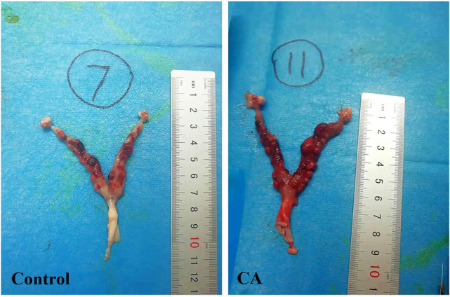
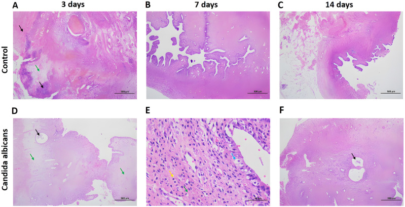
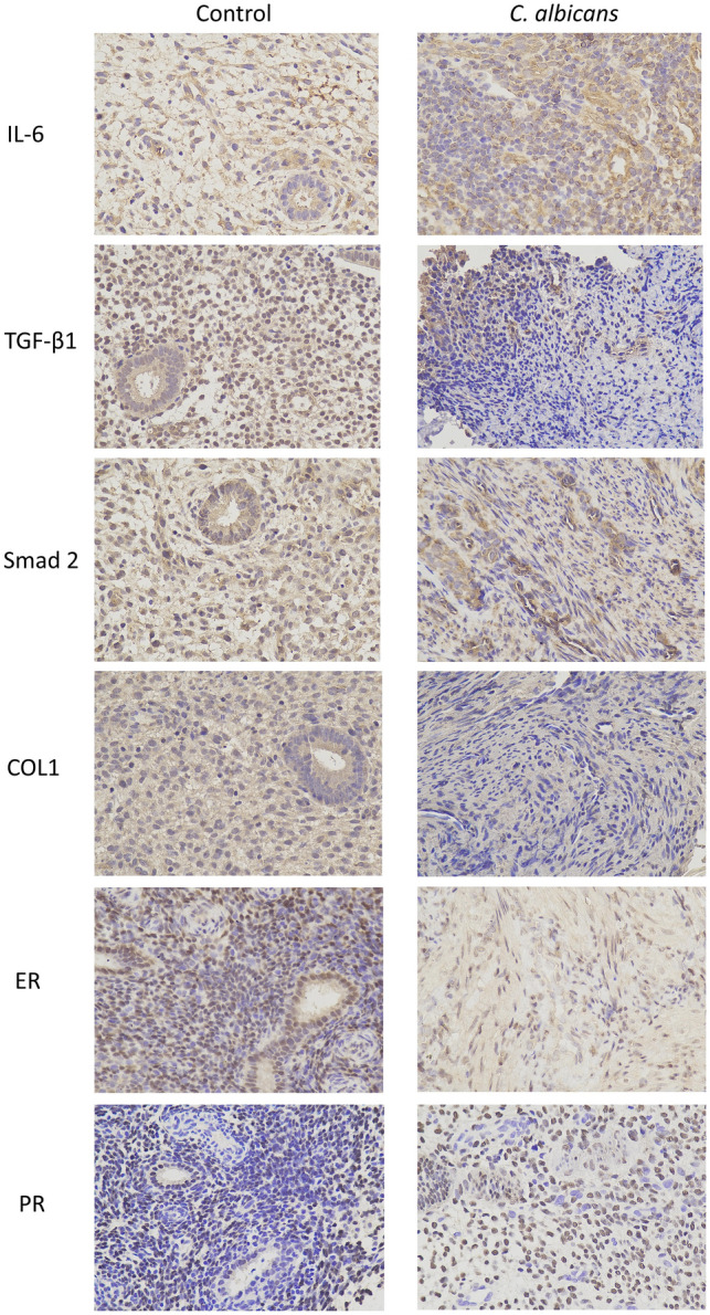
Iatrogenic injury to endometrial tissue is the main cause of intrauterine adhesions (IUA) and infection can also damage the endometrium. The microbiota plays an important role in the health of the female reproductive tract. However, the mechanism is still unclear. In total, 908 patients with IUA and 11,389 healthy individuals were retrospectively selected for this clinical study. Participant information including vaginal microecological results and human papillomavirus (HPV) status were collected. Univariate and multivariate logistic regression analyses were used to identify the factors related to IUA. Next, animal experiments were performed in a curettage-induced IUA rat model. After the procedure, rats in the experimental group received a vaginal infusion of a Candida albicans (C. albicans) fungal solution. On days 3, 7, and 14 after curettage and infusion, the expression levels of IL-6, fibrotic pathway-related factors (TGF-β1, Smad 2, and COL1), and estrogen receptor (ER) and progesterone receptor (PR) in rat endometrial tissues were assessed. Fungal infection of the reproductive tract was found to be an independent risk factor for IUA (P < 0.05). The inflammatory response and degree of fibrosis were greater in rats infected with C. albicans than in the controls. The levels of IL-6, TGF-β1, Smad 2, and COL1 expression in endometrial tissues were significantly higher in the experimental group than in the control group (P < 0.05). However, the ER and PR levels were lower in the IUA group than in the non-IUA group (P < 0.05). C. albicans infection may be related to IUA. C. albicans elicits a strong inflammatory response that can lead to more severe endometrial fibrosis.
© 2023. The Author(s).
Conflict of interest statement
The authors declare no competing interests.
Figures

Similar articles
-
Growth hormone combined with estrogen improves intrauterine adhesion fibrosis by downregulating endometrial microbial citraconic acid to target β-catenin protein.mSystems. 2025 Jul 22;10(7):e0169224. doi: 10.1128/msystems.01692-24. Epub 2025 Jun 5. mSystems. 2025. PMID: 40470939 Free PMC article.
-
Apelin-13 alleviates intrauterine adhesion by inhibiting epithelial-mesenchymal transition of endometrial epithelial cells and promoting angiogenesis.Hum Cell. 2024 Nov;37(6):1613-1623. doi: 10.1007/s13577-024-01117-3. Epub 2024 Aug 19. Hum Cell. 2024. PMID: 39158615
-
Human embryonic stem cell-derived immunity-and-matrix-regulatory cells promote endometrial repair and fertility restoration in IUA rats.Stem Cell Res Ther. 2025 Apr 23;16(1):204. doi: 10.1186/s13287-025-04298-2. Stem Cell Res Ther. 2025. PMID: 40269948 Free PMC article.
-
Recent Advances in Understandings Towards Pathogenesis and Treatment for Intrauterine Adhesion and Disruptive Insights from Single-Cell Analysis.Reprod Sci. 2021 Jul;28(7):1812-1826. doi: 10.1007/s43032-020-00343-y. Epub 2020 Oct 30. Reprod Sci. 2021. PMID: 33125685 Free PMC article. Review.
-
Transforming growth factor-β1 in intrauterine adhesion.Am J Reprod Immunol. 2020 Aug;84(2):e13262. doi: 10.1111/aji.13262. Epub 2020 Jun 3. Am J Reprod Immunol. 2020. PMID: 32379911 Review.
Cited by
-
Gut Microbiota in Women with Eating Disorders: A New Frontier in Pathophysiology and Treatment.Nutrients. 2025 Jul 14;17(14):2316. doi: 10.3390/nu17142316. Nutrients. 2025. PMID: 40732941 Free PMC article. Review.
-
HPV infection and vaginal microecological disorders in women with intrauterine adhesion: cross-sectional study in a Chinese population.BMC Infect Dis. 2023 Nov 27;23(1):836. doi: 10.1186/s12879-023-08659-1. BMC Infect Dis. 2023. PMID: 38012631 Free PMC article.
-
Luliconazole-niacinamide lipid nanocarrier laden gel for enhanced treatment of vaginal candidiasis: in vitro, ex vivo, in silico and preclinical insights.RSC Adv. 2025 Feb 19;15(8):5665-5680. doi: 10.1039/d4ra08397k. eCollection 2025 Feb 19. RSC Adv. 2025. PMID: 39980997 Free PMC article.
-
Exploration of Perturbed Liver Fibrosis-Related Factors and Collagen Type I in Animal Model of Non-Alcoholic Fatty Liver Disease.Appl Biochem Biotechnol. 2024 Jun;196(6):3260-3273. doi: 10.1007/s12010-023-04694-5. Epub 2023 Aug 30. Appl Biochem Biotechnol. 2024. PMID: 37646888
-
Advances in human umbilical cord mesenchymal stem cells-derived extracellular vesicles and biomaterial assemblies for endometrial injury treatment.World J Stem Cells. 2025 Jan 26;17(1):97905. doi: 10.4252/wjsc.v17.i1.97905. World J Stem Cells. 2025. PMID: 39866901 Free PMC article. Review.
References
Publication types
MeSH terms
Substances
LinkOut - more resources
Full Text Sources
Medical
Molecular Biology Databases
Research Materials
