. 2023 Jan 11;8(1):3.
doi: 10.1038/s41536-023-00276-5.

PDGF inhibits BMP2-induced bone healing

Affiliations

PDGF inhibits BMP2-induced bone healing

Sanja Novak et al. NPJ Regen Med. .

Abstract

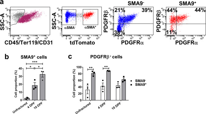
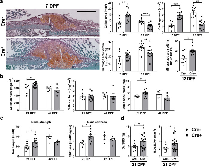
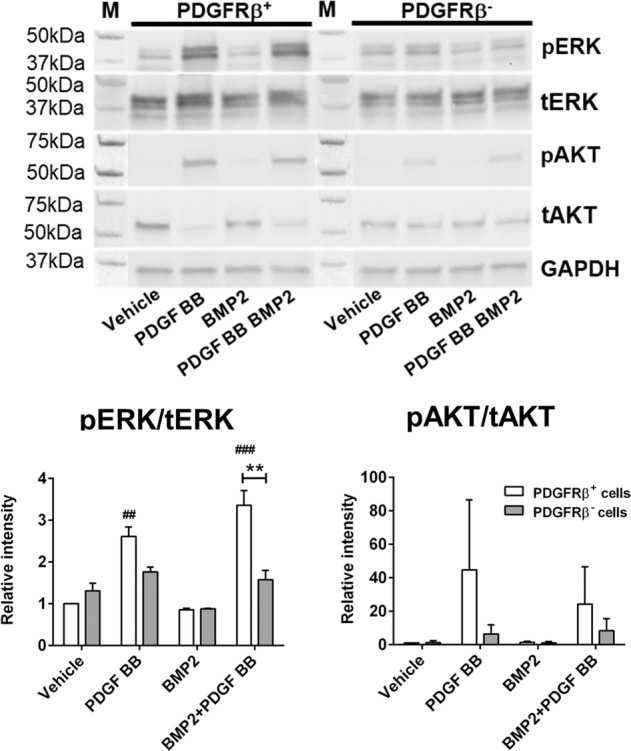
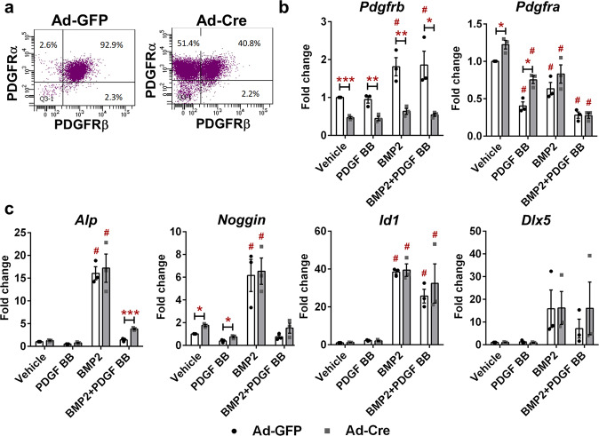
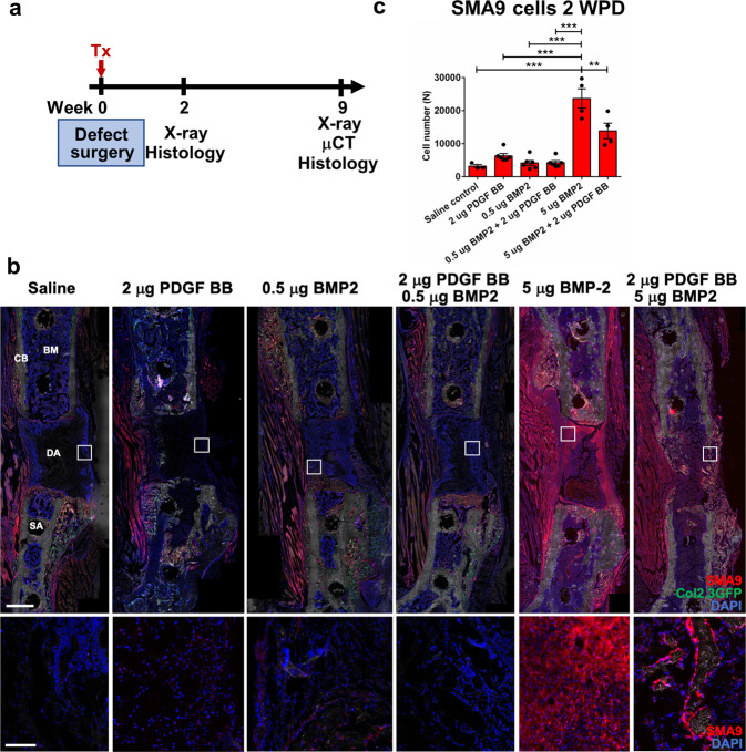
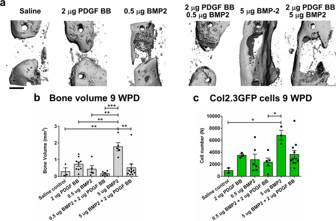
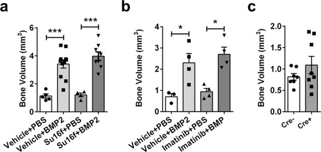
Bone regeneration depends on a pool of bone/cartilage stem/progenitor cells and signaling mechanisms regulating their differentiation. Using in vitro approach, we have shown that PDGF signaling through PDGFRβ inhibits BMP2-induced osteogenesis, and significantly attenuates expression of BMP2 target genes. We evaluated outcomes of treatment with two anabolic agents, PDGF and BMP2 using different bone healing models. Targeted deletion of PDGFRβ in αSMA osteoprogenitors, led to increased callus bone mass, resulting in improved biomechanical properties of fractures. In critical size bone defects BMP2 treatment increased proportion of osteoprogenitors, while the combined treatment of PDGF BB with BMP2 decreased progenitor number at the injury site. BMP2 treatment induced significant bone formation and increased number of osteoblasts, while in contrast combined treatment with PDGF BB decreased osteoblast numbers. This is in vivo study showing that PDGF inhibits BMP2-induced osteogenesis, but inhibiting PDGF signaling early in healing process does not improve BMP2-induced bone healing.


Conflict of interest statement

Recombinant human BMP2 and Adsorbable collagen sponge—Infuse provided by Medtronic to I.K. The remaining authors declare no competing interests.

Figures

Fig. 1
Fig. 1. SMA9-labeled mesenchymal progenitor cells express PDGFRβ during fracture healing.
Tibia fractures were created in 8 to 10-week-old SMA9 mice. Tamoxifen was injected on −1 and 0 DPF, and samples were collected for flow cytometry on 0 (unfractured periosteum), 4 and 10 DPF (fractured tibias). Two intact or fractured tibias were pooled for each sample, n = 3 for each group. a Representative dot plots for SMA9 cells and PDGFRβ+ cells gating in periosteal callus 4 DPF by flow cytometry. b SMA9 and c PDGFRs expression was analyzed within live, non-hematopoietic (CD45/Ter119/CD31) cells. Values are expressed as mean ± s.e.m. *p < 0.05, **p < 0.01, ***p < 0.001. Statistical test: One-way ANOVA with Tukey’s post hoc test (b) and two-way ANOVA with Tukey’s post hoc test (c).
Fig. 2
Fig. 2. Deletion of PDGFRβfl/fl in progenitor cells improves fracture healing.
Deleting of PDGFRβ in αSMA osteoprogenitors was induced by injecting tamoxifen on the day of femur fracture and 2 DPF. a Representative safranin O images of Cre and Cre+ fractured femurs 7 DPF. Increased callus size with increased cartilage area is present in Cre+ animals. Safranin O staining sections were analyzed on 7 and 12 DPF and von Kossa staining on 12 DPF. ImageJ was used to evaluate callus, cartilage and mineralized area. Cre n = 7, Cre+ n = 10 for day 7, and Cre n = 9, Cre+ n = 11 for day 12. Scale bar 1 mm. b PDGFRβ deletion in αSMA osteoprogenitors leads to changes in callus bone mass (Cre n = 6, Cre+ n = 10) and (c) stiffness 21 DPF (Cre n = 6, Cre+ n = 8). At 42 DPF there are no differences in structural or biomechanical femur properties (Cre n = 5, Cre+ n = 5). d Oc.S/BS and N.Oc/B.Pm are increased 21 DPF in Cre+ healing femurs (Cre n = 8, Cre+ n = 17). Values are expressed as mean ± s.e.m. *p < 0.05, **p < 0.01 ***p < 0.001. Statistical test: an unpaired two-tailed Students t-test. Oc.S/BS osteoclast surface per bone surface, N.Oc/B.Pm osteoclast number per bone perimeter.
Fig. 3
Fig. 3. Deletion of PDGFRβ blocks ERK1/2 signaling upon PDGF BB treatment.
Cultured periosteal cells from PDGFRβfl/fl mice, transduced with Ad-GFP or Ad-Cre. Ad-GFP were sorted for PDGFRβ+ cells and PDGFRβ cells were sorted from Ad-Cre transduced cells. Cell lysates were immunoblotted for pERK1/2, tERK, pAKT, tAKT or GAPDH. Values are expressed as mean + s.e.m. n = 2. **p < 0.001, #p < 0.05, ##p < 0.001 different from Ad-GFP vehicle. M – protein ladder. Colorimetric image is stitched to original blot to visualize protein size. Statistical test: Two-way ANOVA with Tukey’s post hoc test.
Fig. 4
Fig. 4. Effect of PDGFRβ deletion in periosteal cells on BMP2 downstream signaling.
Ad-Cre deletion of PDGFRβ showed decrease in expression at (a) protein and (b) mRNA level. c BMP2 induced expression of Alp, Noggin, Id1 and Dlx5, while PDGF BB treatment blocked their upregulation in combined PPDGF BB + BMP2 treatment. Deletion of PDGFRβ using Ad-Cre partially restored Alp expression upon combined BMP2 + PDGF BB treatment. n = 3. Values are expressed as mean ± s.e.m. *p < 0.05, **p < 0.01, ***p < 0.001, # different from its vehicle treatment control. Statistical test: Two-way ANOVA with Tukey’s post hoc test.
Fig. 5
Fig. 5. PDGF BB inhibits SMA9 cell expansion induced by BMP2 during femoral defect healing.
a Experimental design. Femoral defects were performed on SMA9/Col2.3GFP male mice. Tamoxifen was administrated on the day of surgery and treatment combination were delivered in adsorbable collagen scaffold. X-ray was taken once per week to monitor defect healing process. b Representative images of the femoral defect 2 WPD surgery. Boxes demarcate magnified images to show SMA9+ and SMA9 cells within the healing defect. Scale bar of defect images is 1 mm, and magnified image 250 μm. n = 3–6. c SMA9+ progenitor cells were assessed 2-weeks post defect (WPD) surgery in the defect area ± 30% of the defect to capture periosteal and bone marrow response to healing process. All results are expressed as mean ± s.e.m. **p < 0.01, ***p < 0.001. Statistical test: One-way ANOVA with Tukey’s post hoc test.
Fig. 6
Fig. 6. PDGF BB inhibits BMP2-induced osteogenesis in critical femoral defects.
a Representative 3D images of μCT morphometry of healing defect 9 WPD with bone bridging. b Evaluation of bone volume within the defect with highest bone volume in group treated with 5 µg BMP2. c Histological evaluation of Col2.3GFP+ cells within defect area 9 WPD surgery. Scale bar of defect images is 1 mm. Saline control n = 2, n = 3–10 mice per experimental group. All the results are expressed as mean ± s.e.m. *p < 0.05, **p < 0.01, ***p < 0.001. Statistical test: One-way ANOVA with Tukey’s post hoc test.
Fig. 7
Fig. 7. PDGFRβ inhibition does not positively affect critical femoral defect healing.
a Femoral defects performed on C57BL/6J male mice treated with 10 mg/ml Su16f or vehicle in a period of two days before defect surgery till two days post, by oral gavage. Adsorbable collagen sponge was loaded with 5 μg of BMP2 or PBS on a day of surgery. Femurs were evaluated by μCT 9 WPD (Vehicle + PBS n = 5, Vehicle + BMP2 n = 11, Su16f + PBS n = 4, Su16f + BMP2 n = 8). b Imatinib (50 mg/kg/day, ip) was used to inhibit PDGFRβ from two days before surgery till 2 days post-surgery, femurs were evaluated by μCT 9 WPD (Vehicle + PBS n = 3, Vehicle + BMP2 n = 4, Imatinib + PBS n = 4, Imatinib + BMP2 n = 4). c PDGFRβ deletion did not affect critical size femoral defect healing in αSMACrePDGFRβfl/fl animals (tamoxifen induced deletion at −3/0/3 days post defect, Cre- n = 7, Cre+ n = 8). All the results are expressed as mean ± s.e.m. *p < 0.05, ***p < 0.001. Statistical test: One-way ANOVA with Tukey’s post hoc test.

Similar articles

Cited by

References

    1. Thomas, J. D. & Kehoe, J. L. In StatPearls (2021).
    1. Colnot C. Skeletal cell fate decisions within periosteum and bone marrow during bone regeneration. J. Bone Min. Res. 2009;24:274–282. doi: 10.1359/jbmr.081003. - DOI - PMC - PubMed
    1. Matthews, B. G. et al. Heterogeneity of murine periosteum progenitors involved in fracture healing. Elife10, 10.7554/eLife.58534 (2021). - PMC - PubMed
    1. Grcevic D, et al. In vivo fate mapping identifies mesenchymal progenitor cells. Stem Cells. 2012;30:187–196. doi: 10.1002/stem.780. - DOI - PMC - PubMed
    1. Salazar VS, Gamer LW, Rosen V. BMP signalling in skeletal development, disease and repair. Nat. Rev. Endocrinol. 2016;12:203–221. doi: 10.1038/nrendo.2016.12. - DOI - PubMed

LinkOut - more resources