

Comprehensive study upon physicochemical properties of bio-ZnO NCs
- PMID: 36631546
- PMCID: PMC9834250
- DOI: 10.1038/s41598-023-27564-w
Comprehensive study upon physicochemical properties of bio-ZnO NCs
Abstract
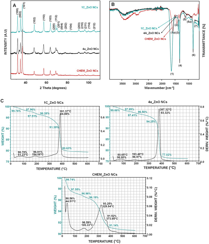
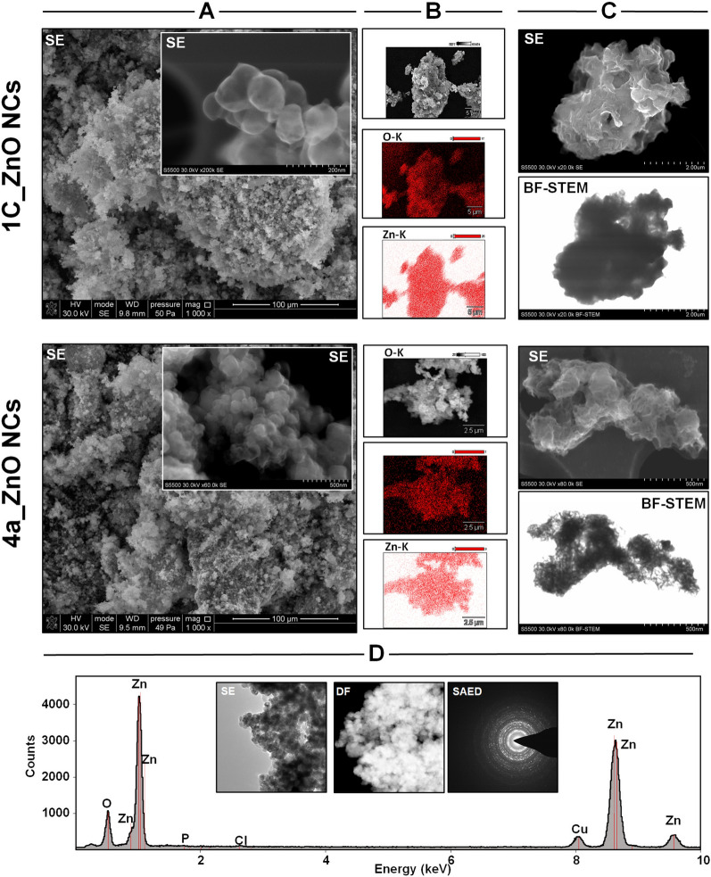
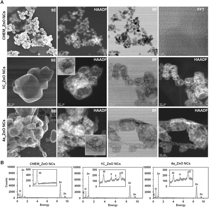
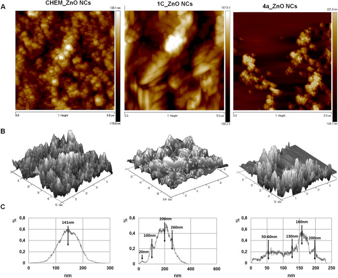
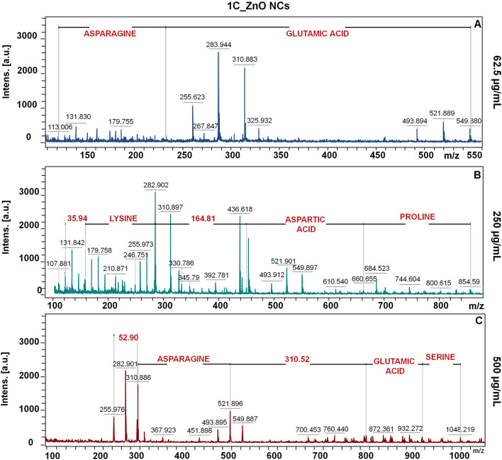
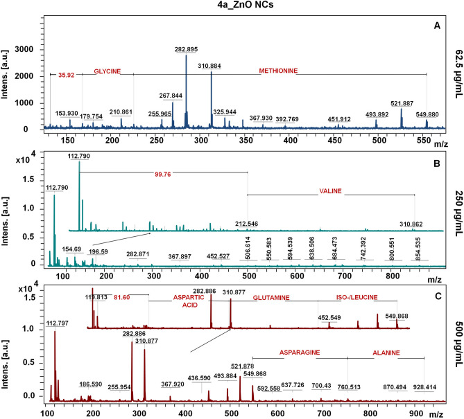
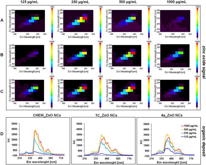
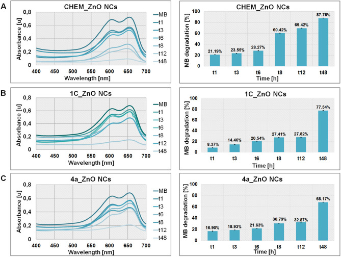
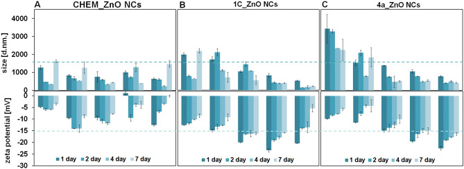
In this study, for the first time, the comparison of commercially available chemical ZnO NCs and bio-ZnO NCs produced extracellularly by two different probiotic isolates (Latilactobacillus curvatus MEVP1 [OM736187] and Limosilactobacillus fermentum MEVP2 [OM736188]) were performed. All types of ZnO formulations were characterized by comprehensive interdisciplinary approach including various instrumental techniques in order to obtain nanocomposites with suitable properties for further applications, i.e. biomedical. Based on the X- ray diffraction analysis results, all tested nanoparticles exhibited the wurtzite structure with an average crystalline size distribution of 21.1 nm (CHEM_ZnO NCs), 13.2 nm (1C_ZnO NCs) and 12.9 nm (4a_ZnO NCs). The microscopy approach with use of broad range of detectors (SE, BF, HAADF) revealed the core-shell structure of bio-ZnO NCs, compared to the chemical one. The nanoparticles core of 1C and 4a_ZnO NCs are coated by the specific organic deposit coming from the metabolites produced by two probiotic strains, L. fermentum and L. curvatus. Vibrational infrared spectroscopy, photoluminescence (PL) and mass spectrometry (LDI-TOF-MS) have been used to monitor the ZnO NCs surface chemistry and allowed for better description of bio-NCs organic coating composition (amino acids residues). The characterized ZnO formulations were then assessed for their photocatalytic properties against methylene blue (MB). Both types of bio-ZnO NCs exhibited good photocatalytic activity, however, the effect of CHEM_ZnO NCs was more potent than bio-ZnO NCs. Finally, the colloidal stability of the tested nanoparticles were investigated based on the zeta potential (ZP) and hydrodynamic diameter measurements in dependence of the nanocomposites concentration and investigation time. During the biosynthesis of nano-ZnO, the increment of pH from 5.7 to around 8 were observed which suggested possible contribution of zinc aquacomplexes and carboxyl-rich compounds resulted in conversion of zinc tetrahydroxy ion complex to ZnO NCs. Overall results in present study suggest that used accessible source such us probiotic strains, L. fermentum and L. curvatus, for extracellular bio-ZnO NCs synthesis are of high interest. What is important, no significant differences between organic deposit (e.g. metabolites) produced by tested strains were noticed-both of them allowed to form the nanoparticles with natural origin coating. In comparison to chemical ZnO NCs, those synthetized via microbiological route are promising material with further biological potential once have shown high stability during 7 days.
© 2023. The Author(s).
Conflict of interest statement
The authors declare no competing interests.
Figures

References
-
- Jamkhande PG, Ghule NW, Bamer AH, Kalaskar MG. Metal nanoparticles synthesis: An overview on methods of preparation, advantages and disadvantages, and applications. J. Drug Deliv. Sci. Technol. 2019;53:101174. doi: 10.1016/j.jddst.2019.101174. - DOI
-
- Khan Z. Recent Trends in Nanomaterials. Synthesis and Properties. Singapore: Springer; 2017.
-
- Liu C, Xiao C, Li W. Zinc oxide nanoparticles as electron transporting interlayer in organic solar cells. J. Mater. Chem. C. 2021;9:14093–14114. doi: 10.1039/D1TC03434K. - DOI
Publication types
MeSH terms
Substances
LinkOut - more resources
Full Text Sources
Miscellaneous
