Review
. 2023 Mar;21(3):178-194.
doi: 10.1038/s41579-022-00839-1. Epub 2023 Jan 11.

Innate immune evasion strategies of SARS-CoV-2

Affiliations
Review

Innate immune evasion strategies of SARS-CoV-2

Judith M Minkoff et al. Nat Rev Microbiol. 2023 Mar.

Abstract

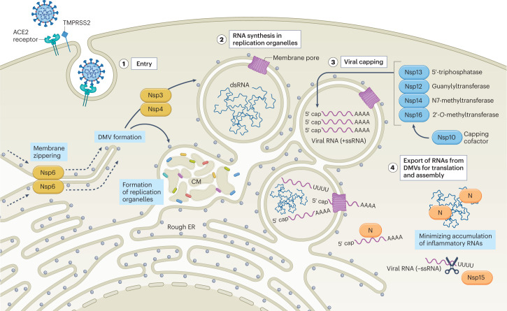
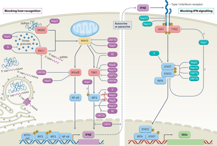
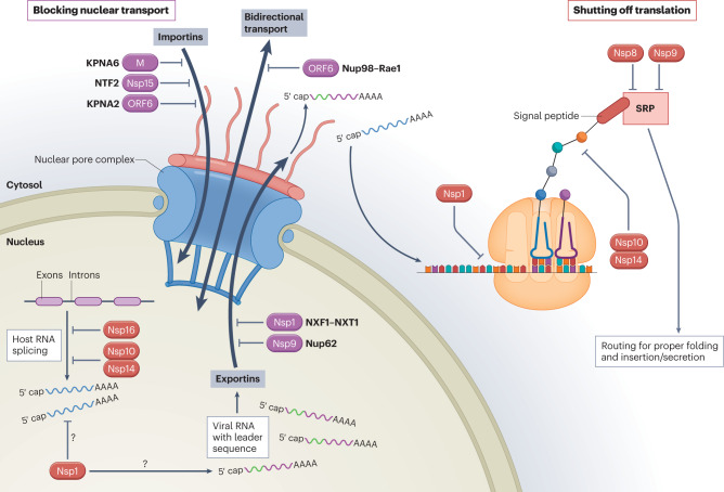
SARS-CoV-2, the virus responsible for the COVID-19 pandemic, has been associated with substantial global morbidity and mortality. Despite a tropism that is largely confined to the airways, COVID-19 is associated with multiorgan dysfunction and long-term cognitive pathologies. A major driver of this biology stems from the combined effects of virus-mediated interference with the host antiviral defences in infected cells and the sensing of pathogen-associated material by bystander cells. Such a dynamic results in delayed induction of type I and III interferons (IFN-I and IFN-III) at the site of infection, but systemic IFN-I and IFN-III priming in distal organs and barrier epithelial surfaces, respectively. In this Review, we examine the relationship between SARS-CoV-2 biology and the cellular response to infection, detailing how antagonism and dysregulation of host innate immune defences contribute to disease severity of COVID-19.


Conflict of interest statement

The authors declare no competing interests.

Figures

Fig. 1
Fig. 1. SARS-CoV-2 strategies to minimize host detection.
SARS-CoV-2 enters into a host cell by binding angiotensin-converting enzyme 2 (ACE2) on the cell surface, a process that can be facilitated by transmembrane protease serine 2 (TMPRSS2), which provides proteolytic cleavage of the viral spike (S) protein to promote virus–host fusion. Following internalization of the viral particle, the capped and polyadenylated genomic viral RNA is released into the cytoplasm where it can be directly translated (stage 1). Initial translation of viral genomic RNA results in the production of the ORF1a and ORF1ab polyproteins (pp1a and pp1ab) that are subsequently processed by viral proteases to form the replicase and non-structural proteins (Nsps; depicted in yellow) necessary to establish replication organelles (ROs). Nsp3 and Nsp4 mediate the modification of endoplasmic reticulum (ER) membranes into convoluted membranes (CM) and double-membrane vesicles (DMVs) that make up ROs, whereas Nsp6 forms a zippered molecular tether between ROs and the ER that enables the flow of lipids (stage 2). Nascent viral RNA is modified by Nsp enzymes (depicted in blue) to mimic host transcripts and minimize the ability of the cell to induce a defence. First, Nsp13 (a 5′ RNA triphosphatase) removes the phosphate from the 5′ end of the viral RNA. This is followed by the transfer of a guanosine monophosphate to the 5′ end by Nsp12 (a guanylyltransferase) to yield the cap core. Subsequently, Nsp14 (an N7-methyltransferase) and Nsp16 (a 2′-O-methyltransferase) assisted by the Nsp10 capping cofactor catalyse the final methylation steps necessary to complete the viral cap (stage 3). As viral replication proceeds, negative-sense RNA (−ssRNA) and double-stranded RNA (dsRNA) intermediates are sequestered inside ROs to prevent host detection. In parallel, the positive-sense, single-stranded genomic and subgenomic RNAs (+ssRNA) needed for translation of viral proteins and de novo virion assembly are chaperoned from the replication organelles. As replication intensifies, viral RNAs accumulate outside of ROs, and are masked and/or minimized by the SARS-CoV-2 nucleocapsid protein (N) and/or Nsp15, depicted in orange (stage 4).
Fig. 2
Fig. 2. SARS-CoV-2-mediated interference of cellular innate immune signalling.
Virus infection generates replication intermediates and/or induces the formation of stress granules that serve as platforms for RIG-I-like receptor (RIG-I or MDA5) activation. Host recognition of viral pathogen-associated molecular patterns, such as single-stranded RNA (ssRNA) with an exposed 5′-triphosphate or double-stranded RNA (dsRNA), promotes the assembly of a mitochondria-localized signalling hub orchestrated by mitochondrial antiviral signalling protein (MAVS), and culminates in the activation of host kinases IKKα, IKKβ and TBK1. Kinase activation induces the production of interferon-β (IFNβ) through cooperative engagement of the ATF2–JUN, interferon regulatory factor 3 (IRF3) and nuclear factor-κB (NF-κB) transcription factors. Secreted IFNβ functions in an autocrine or paracrine manner to promote an antiviral state in cells. On binding of IFNβ, the type I IFN receptor subunits on the cell surface dimerize, bringing together the receptor-associated kinases, Janus kinase 1 (JAK1) and tyrosine kinase 2 (TYK2), which subsequently activate each other via transphosphorylation and promote the recruitment and phosphorylation of the signal transducer and activator of transcription (STAT) molecules, STAT1 and STAT2. Phosphorylated STAT1 and STAT2 form a stable complex with interferon regulatory factor 9 (IRF9) that translocates into the nucleus, where it promotes the transcription of IFN-stimulated genes (ISGs). Each of these processes is the target of SARS-CoV-2 interference, as illustrated here and further described in Table 1. Viral proteins that inhibit aspects of host recognition and the associated signalling pathways are shown in purple, whereas those that block components of the IFN signalling pathway are depicted in teal. Owing to the ability of Nsp1 to more generally inhibit protein synthesis, its role in specifically blocking elements of these pathways remains uncertain (question marks). M, membrane protein; N, nucleocapsid protein; P, phosphorylation; S, spike protein.
Fig. 3
Fig. 3. SARS-CoV-2-mediated interference of general host cell biology.
Cellular induction of an antiviral response is dependent on bidirectional trafficking through the nuclear pore complex (NPC). With the aid of nuclear transport receptors (importins), cellular transcription factors that are induced in response to infection translocate through the NPC into the nucleus and bind sequences within antiviral genes to drive their expression. Following de novo transcription and capping of the nascent messenger RNA (mRNA), the host spliceosome assembles at RNA splicing sites and promotes intron excision to yield translationally competent mature transcripts. These transcripts associate with nuclear transport receptors (exportins) and are exported through the NPC into the cytoplasm, where they are translated by host ribosomes and routed for proper folding and cellular localization based on recognition of their signal peptide by the signal recognition particle (SRP). SARS-CoV-2 encodes several proteins that block nuclear transport (depicted in purple), including non-structural proteins (Nsps), the open reading frame 6 (ORF6) accessory protein and the membrane structural protein (M). This inhibition is facilitated by interactions with the host proteins indicated (KPNA2, karyopherin subunit α2; KPNA6, karyopherin subunit α6; NTF2, nuclear transport factor 2; Nup62, nucleoporin 62; Nup98, nucleoporin 98; NXF1, nuclear RNA export factor 1; NXT1, nuclear transport factor 2-like export factor 1; Rae1, ribonucleic acid export factor 1). SARS-CoV-2 also encodes proteins that ultimately shut off translation (depicted in red) by inhibiting host RNA splicing, preferentially blocking host RNAs for nuclear export in favour of viral RNAs, interfering with ribosomal function and preventing protein trafficking. As Nsp1 is also capable of more generally inhibiting protein synthesis, its role in preferential targeting of viral RNAs remains uncertain (question marks).

Similar articles

Cited by

References

    1. Kawai T, Akira S. The roles of TLRs, RLRs and NLRs in pathogen recognition. Int. Immunol. 2009;21:317–337. doi: 10.1093/intimm/dxp017. - DOI - PMC - PubMed
    1. Ma Z, Ni G, Damania B. Innate sensing of DNA virus genomes. Annu. Rev. Virol. 2018;5:341–362. doi: 10.1146/annurev-virology-092917-043244. - DOI - PMC - PubMed
    1. Deretic V, Saitoh T, Akira S. Autophagy in infection, inflammation and immunity. Nat. Rev. Immunol. 2013;13:722–737. doi: 10.1038/nri3532. - DOI - PMC - PubMed
    1. Mogensen TH. Pathogen recognition and inflammatory signaling in innate immune defenses. Clin. Microbiol. Rev. 2009;22:240–273. doi: 10.1128/CMR.00046-08. - DOI - PMC - PubMed
    1. tenOever BR. The evolution of antiviral defense systems. Cell Host Microbe. 2016;19:142–149. doi: 10.1016/j.chom.2016.01.006. - DOI - PubMed

Publication types