. 2023 Feb 22;51(3):1034-1049.
doi: 10.1093/nar/gkac1246.

Structural snapshots of base excision by the cancer-associated variant MutY N146S reveal a retaining mechanism

Affiliations

Structural snapshots of base excision by the cancer-associated variant MutY N146S reveal a retaining mechanism

Merve Demir et al. Nucleic Acids Res. .

Abstract

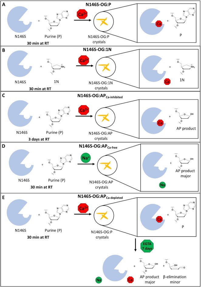
DNA glycosylase MutY plays a critical role in suppression of mutations resulted from oxidative damage, as highlighted by cancer-association of the human enzyme. MutY requires a highly conserved catalytic Asp residue for excision of adenines misinserted opposite 8-oxo-7,8-dihydroguanine (OG). A nearby Asn residue hydrogen bonds to the catalytic Asp in structures of MutY and its mutation to Ser is an inherited variant in human MUTYH associated with colorectal cancer. We captured structural snapshots of N146S Geobacillus stearothermophilus MutY bound to DNA containing a substrate, a transition state analog and enzyme-catalyzed abasic site products to provide insight into the base excision mechanism of MutY and the role of Asn. Surprisingly, despite the ability of N146S to excise adenine and purine (P) in vitro, albeit at slow rates, N146S-OG:P complex showed a calcium coordinated to the purine base altering its conformation to inhibit hydrolysis. We obtained crystal structures of N146S Gs MutY bound to its abasic site product by removing the calcium from crystals of N146S-OG:P complex to initiate catalysis in crystallo or by crystallization in the absence of calcium. The product structures of N146S feature enzyme-generated β-anomer abasic sites that support a retaining mechanism for MutY-catalyzed base excision.


Figures

Graphical Abstract
Graphical Abstract
An X-ray structure of N146S Geobacillus stearothermophilus MutY bound to a substrate duplex was captured due to inhibition via Ca2+ ion within the active site. Removal of Ca2+ during crystallization or in crystallo, allowed for capturing the abasic (AP) site in the β–configuration.
Figure 1.
Figure 1.
Active site structure and mechanism of MutY. (A) Retaining mechanism of adenine excision by DNA glycosylase MutY is an SN1-like mechanism with formation of a covalent MutY-DNA intermediate (7). (B) The hydrogen bond network between the nucleotide and active site residues shows the interaction between Asn146 and Asp144 (TSAC-OG:1N, PDB ID 6U7T) (7). (C) The structure of substrate purine (P) and pyrrolidine transition state analog (1N) used in this work.
Scheme 1.
Scheme 1.
Minimal kinetic scheme of glycosylase activity.
Figure 2.
Figure 2.
Glycosylase activity and substrate affinity of Asn-to-Ser MutY variants. (A) Representative plots of a single trial used to determine the adenine excision rate, k2, of each variant with OG:A or OG:P substrates using enzyme concentrations of 120 nM, 100 nM, 40 and 100 nM for WT Gs, N146S Gs, WT Ec and N140S Gs MutY, respectively. (B) A zoomed in view of Panel A with time shown up to 5 min. (C) Representative plots of a single trial used to determine the dissociation constant, KD, of WT14 and N140S Ec MutY with OG:FA containing DNA substrate at 25°C. The KD values for WT and N140S Ec MutY were representing an average of at least three trials were 0.12 ± 0.02 (53) and 0.14 ± 0.03, respectively.
Figure 3.
Figure 3.
Crystallization Strategies. (A) N146S-OG:P, (B) N146S-OG:1N, (C) and N146S-OG:APCa-inhibited crystallized with calcium containing crystallization solution with their corresponding incubation time and temperature. (D) Crystallization conditions containing sodium instead of calcium for obtaining N146S-OG:APCa-free. (E) The N146S-OG:P crystals soaked in EGTA containing solution for 7 days to remove calcium from active site to initiate the reaction in N146S-OG:APCa-depleted structures.
Figure 4.
Figure 4.
Structural overview of N146S Gs MutY-OG:P complex. (A) The final refinement map of N146S-OG:P. A Ca2+ ion coordination is observed with purine base and active site residues in N146S-OG:P complex structure. (B) Fe-S cluster with alternate conformations The 2|Fo| – |Fc| map (gray) was calculated to the 1.68 Å resolution limit and contoured at 1.0 rmsd. The |Fo| - |Fc| map (green, positive features; red, negative) was contoured at 3.5 rmsd. The ANOM map contoured to 5.0 rmsd (gold). (C) N146S-OG:P complex structure with purine coordinated to Ca2+ and disengaged from Glu43. (D) Fluorinated Lesion Recognition Complex (FLRC) showing the interaction of active site residues and adenine (PDB ID:3G0Q, gray) (6). (E) Lesion Recognition Complex (LRC) showing adenine disengaged from Glu43 (PDB ID:1RRQ, light pink) (39). (F) The overlay of N146S-OG:P (cyan) with FLRC (PDB ID:3G0Q, gray) (6) and LRC (PDB ID:1RRQ, light pink) (39) shows differences in base engagement within active site.
Figure 5.
Figure 5.
The structural overview of N146S-OG:1N. (A) Electron density maps for N146S-OG:1N showing chain A and C. The 2|Fo| - |Fc| map (gray) was calculated to the 1.96 Å resolution limit and contoured at 1.0 rmsd. The |Fo| – |Fc| map (green, positive features; red, negative) was contoured at 3.5 rmsd. (B) The electron density map of iron–sulfur cluster with ANOM map contoured to 5.0 rmsd (gold).
Figure 6.
Figure 6.
Structural overview of the three AP product structures. (A) The active site view of N146S-OG: APCa-inhibited shows Ca2+ coordination and β anomer AP product. The 2|Fo| – |Fc| map (gray) was calculated to the 2.36 Å resolution limit and contoured at 1.0 rmsd. The |Fo| – |Fc| map (green, positive features; red, negative) was contoured at 3.5 rmsd. (B) Electron density features in the |Fo| – |Fc| map indicate the α anomer is incompatible with measured diffraction data. (C) The active site view of N146S-OG:APCa-free with AP site product. (D) β- and δ-elimination products shown with alternate conformations of Q48 coordinating to the phosphate backbone conformations of G (2|Fo| – |Fc| map contoured to 1.0 rmsd and |Fo| – |Fc| map contoured to 3.5 rmsd). (E) N146S-OG:APCa-depleted structures with the β anomer AP product (2|Fo| – |Fc| map contoured to 1.0 rmsd and |Fo| – |Fc| map contoured to 3.8 rmsd) and (F) chemical structures of AP site, β-elimination and β- and δ-elimination products.

References

    1. Michaels M.L., Miller J.H.. The GO system protects organisms from the mutagenic effect of the spontaneous lesion 8-hydroxyguanine (7, 8-dihydro-8-oxoguanine). J. Bacteriol. 1992; 174:6321–6325. - PMC - PubMed
    1. Brieba L.G., Eichman B.F., Kokoska R.J., Doublié S., Kunkel T.A., Ellenberger T.. Structural basis for the dual coding potential of 8-oxoguanosine by a high-fidelity DNA polymerase. EMBO J. 2004; 23:3452–3461. - PMC - PubMed
    1. Al-Tassan N., Chmiel N.H., Maynard J., Fleming N., Livingston A.L., Williams G.T., Hodges A.K., Davies D.R., David S.S., Sampson J.R.et al. .. Inherited variants of MYH associated with somatic G:C→T:A mutations in colorectal tumors. Nat. Genet. 2002; 30:227–232. - PubMed
    1. Raetz A.G., David S.S.. When you’re strange: unusual features of the MUTYH glycosylase and implications in cancer. DNA Repair (Amst). 2019; 80:16–25. - PMC - PubMed
    1. McCann J.A.B., Berti P.J.. Adenine release is fast in MutY-catalyzed hydrolysis of G:a and 8-Oxo-G:a DNA mismatches*. J. Biol. Chem. 2003; 278:29587–29592. - PubMed

Publication types