. 2022;17(4):233-248.
doi: 10.1080/17469899.2022.2100762. Epub 2022 Sep 6.

Hyalocyte origin, structure, and imaging

Affiliations

Hyalocyte origin, structure, and imaging

Peter Wieghofer et al. Expert Rev Ophthalmol. 2022.

Abstract

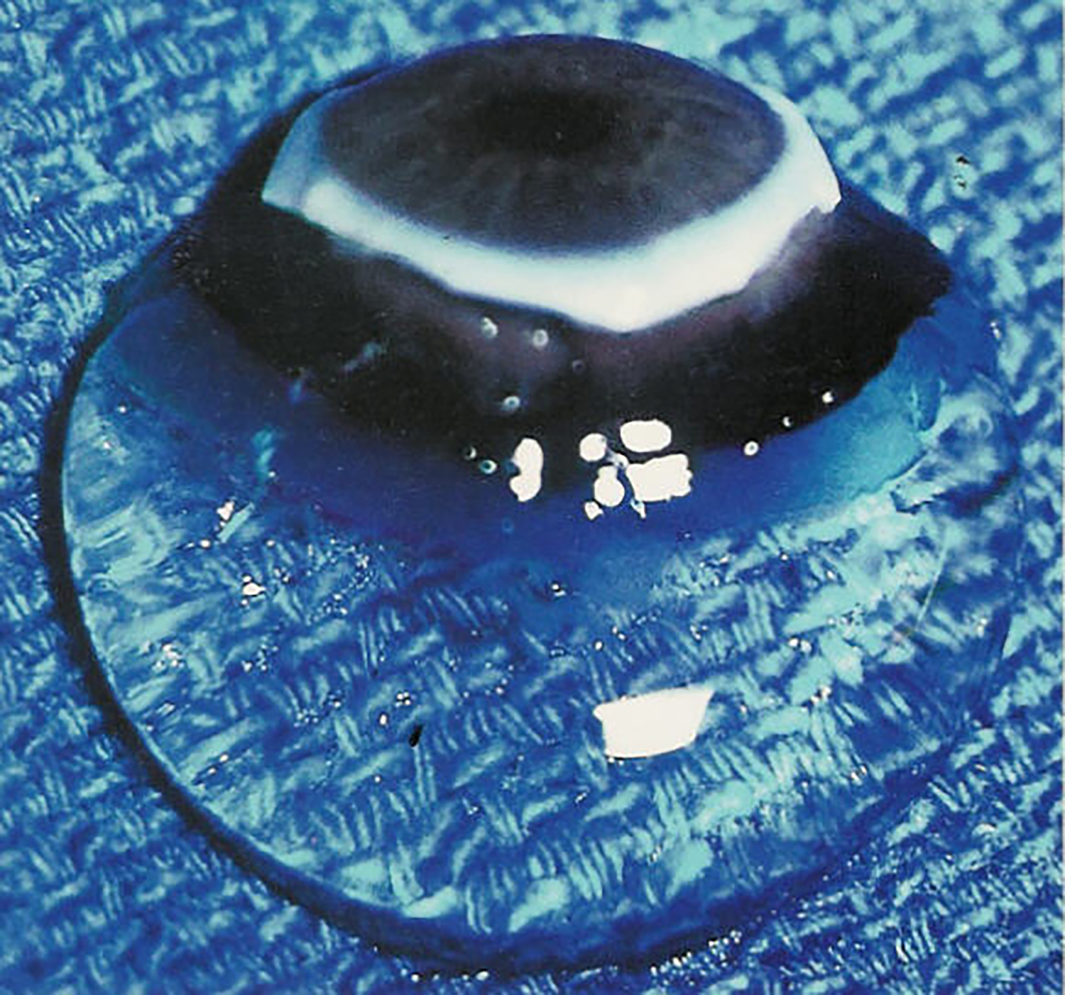
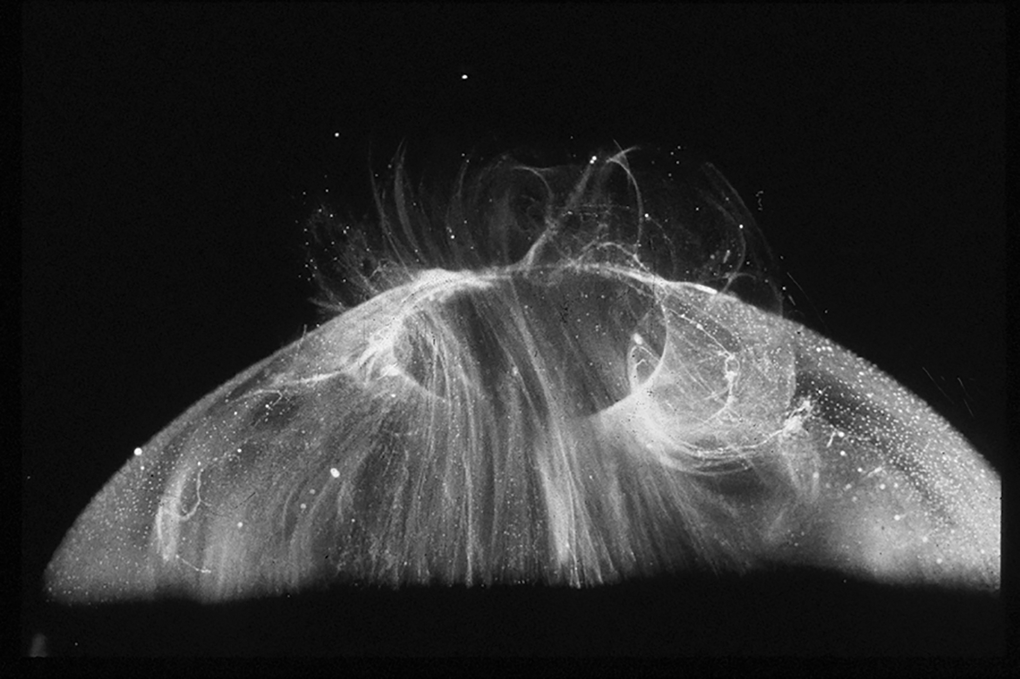
Introduction: Hyalocytes have been recognized as resident tissue macrophages of the vitreous body since the mid-19th century. Despite this, knowledge about their origin, turnover, and dynamics is limited.

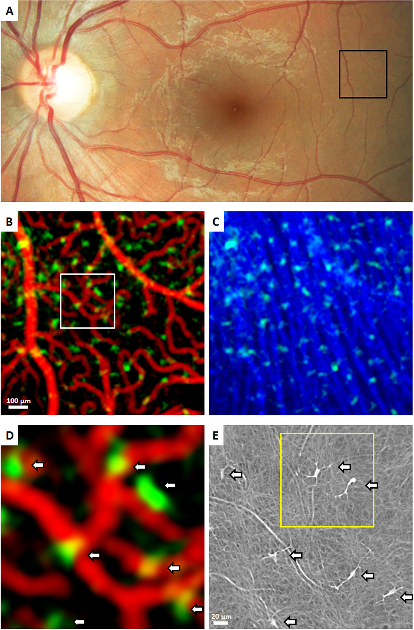
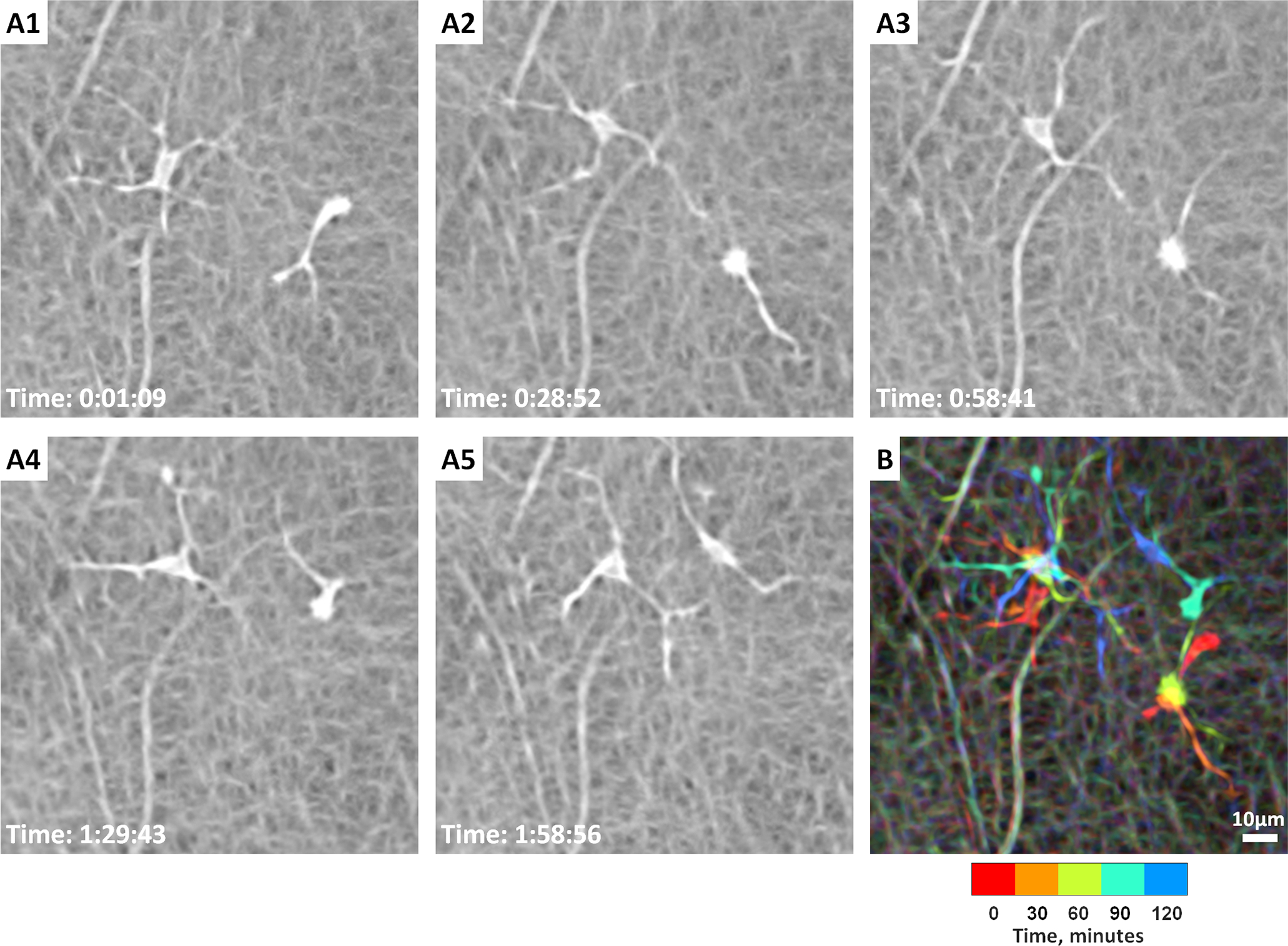
Areas covered: Historically, initial studies on the origin of hyalocytes used light and electron microscopy. Modern investigations across species including rodents and humans will be described. Novel imaging is now available to study human hyalocytes in vivo. The shared ontogeny with retinal microglia and their eventual interdependence as well as differences will be discussed.

Expert opinion: Owing to a common origin as myeloid cells, hyalocytes and retinal microglia have similarities, but hyalocytes appear to be distinct as resident macrophages of the vitreous body.

Keywords: Hyalocytes; Imaging; Macrophages; Monocytes; Origin; Turnover; Vitreous.


Figures

Figure 1.
Figure 1.. Human vitreous body.
This sclera, choroid, and retina were dissected off this specimen from a 9-month-old child. The vitreous body remains attched to the anterior segment and maintains in gel turgenscence in spit of being situtated onto a surgical towel in room air. Image courtesy of New England Eye Bank, Boston, MA; reproduced with permission from [2], © 1989 Springer-Verlag New York Inc. and [121], © 2014 Springer Science+Business Media New York.
Figure 2.
Figure 2.. Human hyalocytes.
LEFT: Dark-field slit microscopy of whole vitreous body from a 59-year-old male reveals hyalocytes embedded within the posterior vitreous cortex, appearing as the white dots. RIGHT: Phase contrast microscopy of flat mount preparation of hyalocytes in the vitreous cortex from the eye of an 11-year-old girl, obtained at autopsy (courtesy of the New England Eye Bank, Boston, MA). No stains or dyes were used in this preparation. (A) Monoculear cells are distributed in a single layer within the vitreous cortex (x 115). (B) Higher magnification (x 290) demonstrates the mononuclear, round appearance of these cells. Pseudopodia are present in some cells. Reproduced with permission from [2], © 1989 Springer-Verlag New York Inc.
Figure 3.
Figure 3.. Transmission electron microspcipy of human hyalocyte.
A mononuclear cell is seen embedded within the dense collagen fibril (black C) network of the vitreous cortex. There is a lobulated nucleus (N) with dense marginal chromatin (white C). In the cytoplasm there are mitochondria (M), dense granules (arrows), vacuoles (V), and microvilli (Mi) (X 11,670). Image courtesy of JL Craft, DM Albert and DG Cogan (Laboratory of Ophthalmic Pathology, Harvard Medical School, Boston, MA). Reproduced with permission from [2], © 1989 Springer-Verlag New York Inc.
Figure 4.
Figure 4.. Confocal microscopy of fluorescently labeled hyalocytes.
TOP: A preretinal hyalocyte close to the inner limiting membrane is shown. The hyalocyte was identified by its location via differential interference contrast (DIC) and a nuclear counterstain with DAPI to investigate the expression of the ferritin light chain (FTL). BOTTOM: A free hyalocyte with filopodia is shown. The hyalocyte was identified by its location via differential interference contrast and a nuclear counterstain with DAPI to investigate the expression of CD74. GCL = ganglion cell layer, INL = inner nuclear layer, ONL = outer nuclear layer, RPE = retinal pigment epithelium. Reproduced from [30], licensed under CC-BY 4.0 (http://creativecommons.org/licenses/by/4.0/)
Figure 5.
Figure 5.. In vitro cultured human hyalocytes.
Primary hyalocytes were purified by flow cytometry after vitrectomy, cultured in vitro and stained via immunofluorescence. Hyalocytes are labeled with the macrophage marker IBA1, alpha smooth muscle actin and phalloidin labeling the cytoskeleton protein actin. Scale Bar = 50 μm. Reproduced from [32], licensed under CC-BY 4.0 (http://creativecommons.org/licenses/by/4.0/)
Figure 6.
Figure 6.. Spectral domain OCT imaging of human hyalocytes in situ.
Enhanced vitreous imaging with the Heidelberg Spectralis demonstrates hyalocytes overlying the superior arcade (A) and the peripapillary retina (B) in a 52-year-old male with central serous chorioretinopathy. Images courtesy of author M Engelbert.
Figure 7.
Figure 7.. Swept source OCT imaging of human hyalocytes in situ.
Swept-Source OCT visualizes not only the Bursa Premacularis of Worst, but other lacunae of liquefied vitreous in the vitreous body in proximity to the retina. Hyalocytes appear as hyperreflective dots (A). En face imaging demonstrates a greater abundance of hyalocytes in a 20 μm slab just over the retinal surface (B) than in an equivalent slab 100 μm away from the retina (C). Images courtesy of author M Engelbert.
Figure 8.
Figure 8.. Imaging human hyalocytes in vivo.
A 32-year-old male was imaged with clinical OCT & Adaptive Optics Scanning Light Ophthalmoscopy (AOSLO). A: Color fundus photo, black box indicates a region imaged using clinical OCT. B & C: OCT reflectance and OCT angiography (OCTA) color overlays of the black box in (A). Clinical OCT color overlays of (B) superficial retinal vascular network (red) and hyalocytes (green), and (C) hyalocytes (green) seen anterior to the retinal nerve fiber bundles (blue) show the spatial relationships among structures. D: Magnified color overlay of the superficial retinal vascular network (red) and hyalocytes (green) of the white box in (B). White arrows indicate seven hyalocytes imaged within this region of interest. E: Corresponding AOSLO image also revealed the same number of hyalocytes (white arrows) with better visibility of their cell somas and processes. Hyalocyte locations appear to match between imaging modalities, but the cell size and shape were different. Yellow box indicates a region imaged over 2 hours in Figure 9. Images courtesy of authors TYP Chui and RB Rosen.
Figure 9.
Figure 9.. Coronal plane imaging of human hyalocytes in vivo.
In vivo imaging of hyalocyte morphology and dynamics in a 32-year-old male using non-confocal quadrant-detection adaptive optics scanning light ophthalmoscopy. A1-A5: Two ramified hyalocytes show noticeable differences in shape and movement over 2 hours. Time of acquisition in the lower-left corner of each image denotes the hrs:mins:secs. B: A chromo-temporal map composite of the 5 time points (each approximately 30 minutes apart) demonstrates the movements of cell somas and processes over 2 hours. While the more ramified cell (left) displays relatively stationary soma, the less ramified cell (right) shows relatively greater movement of the soma as evidenced by the long chromatic trail in the chromo-temporal map. The processes of both cells, however, appear to move significantly over 2 hours. See Movie 1 for the AOSLO time-lapse video of the two hyalocytes and their processes movement over 2 hours. Images courtesy of authors TYP Chui and RB Rosen.

References

    1. Sebag J Vitreous: the resplendent enigma. British Journal of Ophthalmology. 2009;93:989–991. - PubMed
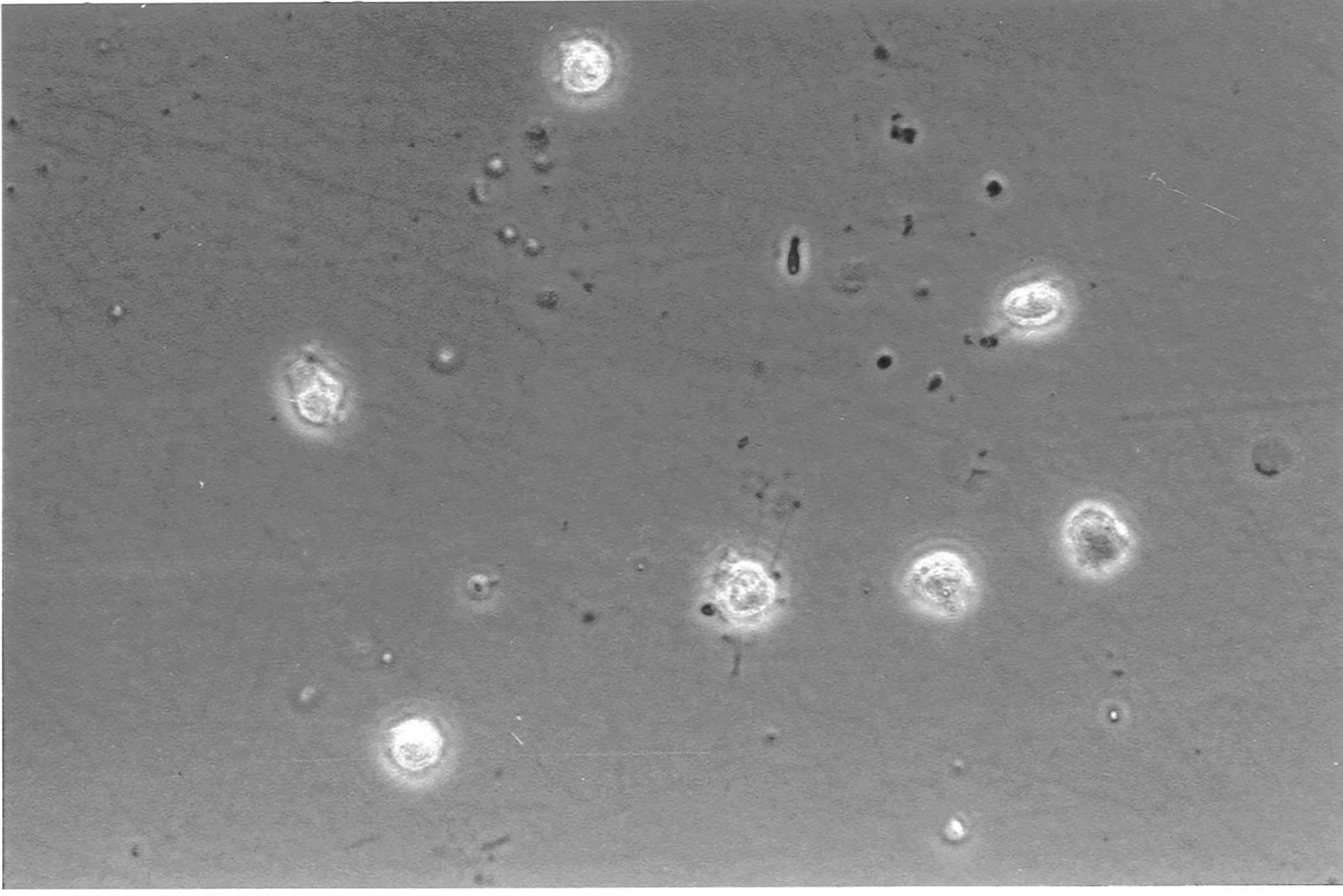
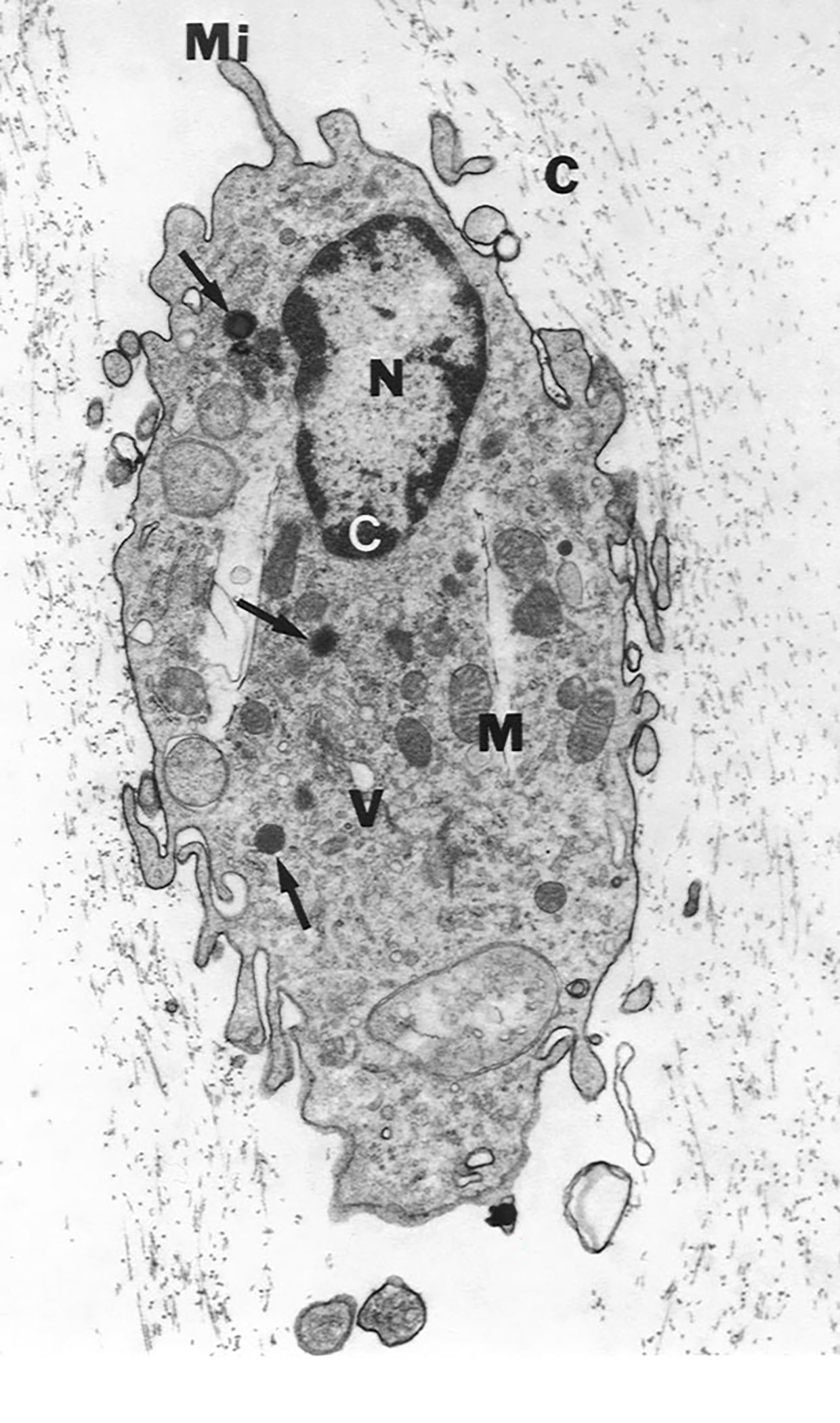
    1. Sebag J Structure of the Vitreous. In: Sebag J, editor. The Vitreous: Structure, Function, and Pathobiology [Internet]. New York, NY: Springer; 1989. [cited 2022 Feb 23]. p. 35–58. Available from: 10.1007/978-1-4613-8908-8_4.

      * This publication features phase contrast microscopy of unstained human hyalocytes in situ, as well as transmission electron microscopy of human hyalocytes.

    1. Ginhoux F, Greter M, Leboeuf M, et al. Fate mapping analysis reveals that adult microglia derive from primitive macrophages. Science. 2010;330:841–845. - PMC - PubMed
    1. Schulz C, Gomez Perdiguero E, Chorro L, et al. A lineage of myeloid cells independent of Myb and hematopoietic stem cells. Science. 2012;336:86–90. - PubMed
    1. Gomez Perdiguero E, Klapproth K, Schulz C, et al. Tissue-resident macrophages originate from yolk-sac-derived erythro-myeloid progenitors. Nature. 2015;518:547–551. - PMC - PubMed