Review
. 2023 Jan 4;24(1):2156257.
doi: 10.1080/14686996.2022.2156257. eCollection 2023.

Advanced materials and technologies for oral diseases

Affiliations
Review

Advanced materials and technologies for oral diseases

Hao Cui et al. Sci Technol Adv Mater. .

Abstract

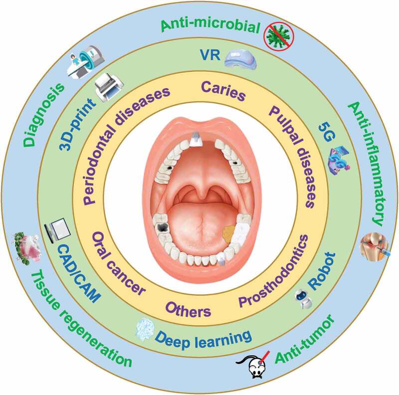
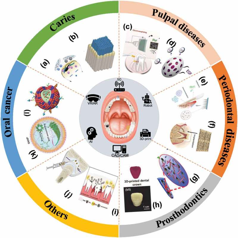
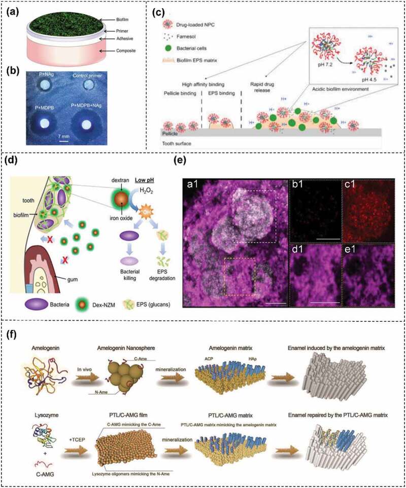
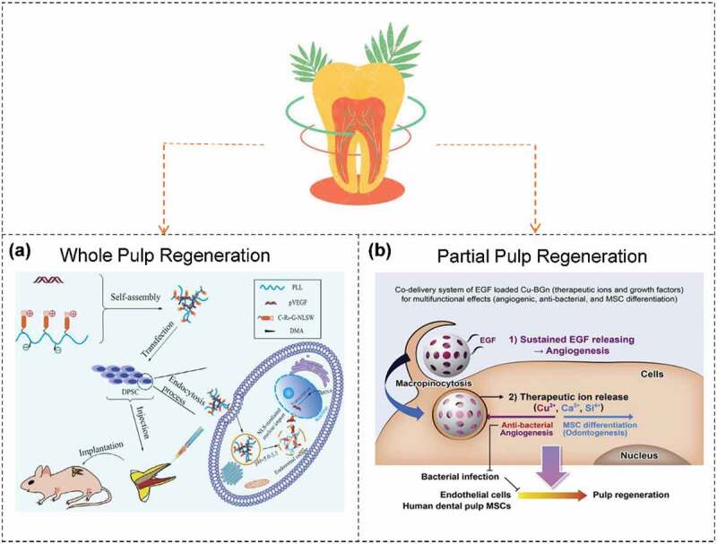
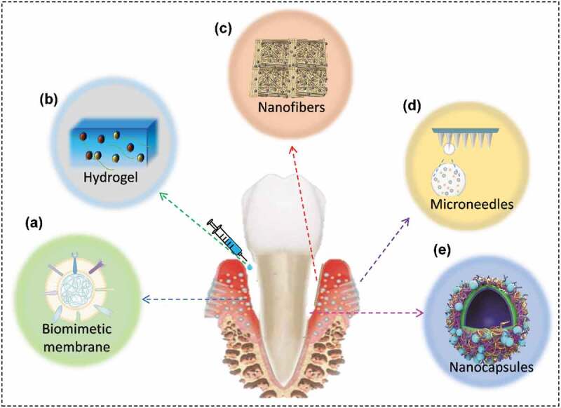
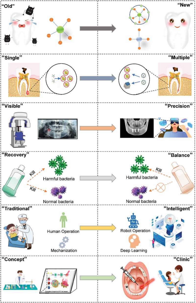
Oral disease, as a class of diseases with very high morbidity, brings great physical and mental damage to people worldwide. The increasing burden and strain on individuals and society make oral diseases an urgent global health problem. Since the treatment of almost all oral diseases relies on materials, the rapid development of advanced materials and technologies has also promoted innovations in the treatment methods and strategies of oral diseases. In this review, we systematically summarized the application strategies in advanced materials and technologies for oral diseases according to the etiology of the diseases and the comparison of new and old materials. Finally, the challenges and directions of future development for advanced materials and technologies in the treatment of oral diseases were refined. This review will guide the fundamental research and clinical translation of oral diseases for practitioners of oral medicine.

Keywords: Oral diseases; advanced technology; antibacterial; nanomaterial; tissue engineering.


Conflict of interest statement

No potential conflict of interest was reported by the authors.

Figures

None
Graphical abstract
Figure 1.
Figure 1.
Advanced materials and technologies in oral diseases. (a) Nano anti-caries material. Reproduced by permission from [17], copyright 2020, Elsevier. (b) Dental enamel regeneration materials. Reproduced by permission from [18], copyright 2020, Wiley. (c) Anti-dentin-sensitive materials. Reproduced by permission from [19], copyright 2021, Dovepress. (d) Dental pulp regeneration materials. Reproduced by permission from [20], copyright 2020, Elsevier. (e) Periodontal drug delivery carriers. Reproduced by permission from [21], copyright 2022, Cell Press. (f) Scaffold materials for tissue engineering. Reproduced by permission from [22], copyright 2021, Wiley. (g) Nanorobot system for antibacterial implants. Reproduced by permission from [23], copyright 2022, American Chemical Society. (h) Nano Ceramic Crown. Reproduced by permission from [24], copyright 2021, Wiley. (i) Nanosensors for oral gas detection. Reproduced by permission from [25], copyright 2020, Wiley. (j) Temporomandibular joint cartilage regeneration material. Reproduced by permission from [26], copyright 2019, Wiley. (k) Chemotherapy drug delivery materials. Reproduced by permission from [27], copyright 2015, American Chemical Society. (l) Hybrid membrane bionanomaterials. Reproduced by permission from [28], copyright 2021, Springer Nature.
Figure 2.
Figure 2.
(a,b) Synergistic antibacterial mechanism of MDPB and AgNps. Reproduced by permission from [33], copyright 2013, Elsevier. (c) Proposed mode of action of pH-responsive nanoparticles. Reproduced by permission from [41], copyright 2015, American Chemical Society. (d) Patterns of pH-responsive dextran-coated iron oxide nanoparticles targeting bacterial biofilms. (e) Representative image of a Dex-NZM treated S. mutans biofilm (a1) before addition of H2O2; dashed white and yellow boxed indicate selected areas for localized antibiofilm effects of Dex-NZM. Close-up views of bacteria and exopolysaccharides before H2O2 exposure (panels b1 and c1, respectively) and 100 min after H2O2 exposure (panels d1 and e1). Reproduced by permission from [42], copyright 2019, American Chemical Society. (f) Schematic demonstration of amelogenin and the PTL/C-AMG matrix (mimicking the N-Ame and C-Ame) to mediate the transition from ACP to HAP on enamel for in situ remineralization. Reproduced by permission from [18], copyright 2018, Wiley.
Figure 3.
Figure 3.
Different strategies in pulp regeneration. (a) the strategy of whole pulp regeneration. Reproduced by permission from [97], copyright 2021, Elsevier. (b) the strategy of partial pulp regeneration. Reproduced by permission from [20], copyright 2020, Elsevier.
Figure 4.
Figure 4.
Drug carriers suitable for periodontal drug delivery. (a) Biomimetic cell membrane. Reproduced by permission from [141], copyright 2021, Elsevier. (b) Hydrogel. (c) Nanofibers. Reproduced by permission from [22], copyright 2022, Wiley. (d) MNs. Reproduced by permission from [21], copyright 2021, Elsevier. (e) Nanocapsules. Reproduced by permission from [142], copyright 2019, Wiley.
Figure 5.
Figure 5.
(a) Schematic illustration of CeO2@Ce6 nanocomposite in synthesis, the antibacterial mechanism, and modulating the polarization of macrophages for the treatment of periodontitis. The enhanced antibacterial efficacy of CeO2@Ce6 could rely on the generation of ROS by aPDT and the innate antibacterial activity of CeO2. (b) In vitro fluorescence images of ROS generation via different nanoparticles by using an in vitro imaging system. The color change indicates that the combination of CeO2@ce6 with light can generate ROS resulting in antimicrobial effect and then remove the ROS to avoid damage to normal tissue. (c) the result of this study suggest that photo-therapy can be used as an adjunct to periodontal therapy, however, a rational design is needed to address its side effects. Reproduced by permission from [174], copyright 2020, Elsevier.
Figure 6.
Figure 6.
Different scaffold materials in periodontal tissue engineering. (a) Schematic diagram demonstrating the role of a double-layered scaffold for periodontal regeneration. Reproduced by permission from [185], copyright 2020, Elsevier. (b) MEW setup to fabricate fibrous scaffolds of distinct fiber configuration and highly-ordered architectures. Representative SEM images of the various MEW PCL scaffolds show melt electrowritten polymeric (i.e. poly(ε-caprolactone) scaffolds with tissue-specific attributes such as fiber morphology (random vs. aligned) and highly-ordered (0◦/90◦ crosshatch pattern) architecture with distinct strand spacings (small 250 μm and large 500 μm). Reproduced by permission from [186], copyright 2022, Elsevier. (c) Schematic illustration of hierarchical-structured mineralized nanofiber (HMF) scaffold for enhanced alveolar bone defect repair. Reproduced by permission from [22], copyright 2021, Wiley.
Figure 7.
Figure 7.
Advanced materials and technologies in dental implants. (a,b) Human-robot collaboration interaction of human-robot collaborative dental implant system (HRCDIS). Reproduced by permission from [209], copyright 2022, Wiley. (c – e) Schematic representation of oral antibiofilm activity of photoactive Fe3O4@PEI/BiVO4 magnetic microrobots. Reproduced by permission from [23], copyright 2022, American Chemical Society. (f) One-step synthesis of versatile antimicrobial nano-architected implant coatings for hard and soft tissue healing. Reproduced by permission from [210], copyright 2021, American Chemical Society. (g) Optical and scanning electron microscopy (SEM) images of different implant types studied. Reproduced by permission from [211], copyright 2013, Wiley. (h,i) Application of nano-modification technology in the field of promoting osseointegration of implants. Reproduced by permission from [212], copyright 2020, Frontiers.
Figure 8.
Figure 8.
Advanced materials and technologies for dental crowns. (a) Application of CAD/CAM technology in the field of dental implant crown design. Reproduced by permission from [225], copyright 2021, Wiley. (b) Morphology and fracture behavior of lithium disilicate dental crowns designed by human and knowledge-based AI. Reproduced by permission from [226], copyright 2022, Elsevier. (c,d) Additive manufacturing technologies in the field crown design. Reproduced by permission from [227], copyright 2021, American College of Prosthodontists. (e) Design of PEEK based telescopic crowns. Reproduced by permission from [228], copyright 2021, Elsevier. (f) Construction process of the multi-scale highly aligned HAP nanorod structures. Reproduced by permission from [24], copyright 2021, Wiley.
Figure 9.
Figure 9.
Development of materials and technologies in the treatment of oral cancer in 2006–2022.
Figure 10.
Figure 10.
Advanced materials and technologies in oral cancer. (a) Application of AI technology in rapid diagnosis of oral cancer. (b) Application of NIR-II window-based nanoprobes in monitoring of the immune cell population in vivo to effectively assess the tumor progression. Reproduced by permission from [234], copyright 2019, American Chemical Society. (c) Self-loading nanoparticles for chemotherapy drug delivery. Reproduced by permission from [235], copyright 2015, American Chemical Society. (d) Schematic illustration of the NIR-II dye SQ890 and the EGFR-targeting nanoparticle SQ890 NPs-Pep for photoacoustic/nir-II fluorescence dual-modality imaging-guided PTT of oral cancer. Reproduced by permission from [236], copyright 2022, Springer. (e) Platelet-facilitated photothermal tumor therapy (PLT-PTT). Reproduced by permission from [237], copyright 2018, Wiley. (f) Application of hybridized membrane technology for drug delivery. Reproduced by permission from [28], copyright 2021, BMC.
Figure 11.
Figure 11.
(a) Schematic diagram of a ZnO-PDMS mouthguard. Reproduced by permission from [25], copyright 2020, Wiley. (b) an illustration of the cellular delivery of HAS2 to synoviocytes using nanoparticles. Reproduced by permission from [280], copyright 2019, Wiley.
Figure 12.
Figure 12.
The development of materials and technologies from ‘past’ to ‘future’.

Similar articles

Cited by

References

    1. Peres MA, Macpherson LMD, Weyant RJ, et al. Oral diseases: a global public health challenge. Lancet. 2019;394(10194):249–42. - PubMed
    1. Collaborators GBDOD, Bernabe E, Marcenes W, et al. Global, regional, and national levels and trends in burden of oral conditions from 1990 to 2017: a systematic analysis for the Global Burden of Disease 2017 study. J Dent Res. 2020;99(4):362–373. - PMC - PubMed
    1. Hugo FN, Kassebaum NJ, Marcenes W, et al. Role of dentistry in global health: challenges and research priorities. J Dent Res. 2021;100(7):681–685. - PMC - PubMed
    1. Nakamura T, Zou K, Shibuya Y, et al. Oral dysfunctions and cognitive impairment/dementia. J Neurosci Res. 2020;99(2):518–528. - PubMed
    1. Mahardawi B, Rochanavibhata S, Jiaranuchart S, et al. Autogenous tooth bone graft material prepared chairside and its clinical applications: a systematic review. Int J Oral Maxillofac Surg. 2022. S0901-5027(22)00193-X. 10.1016/j.ijom.2022.04.018 - DOI - PubMed

LinkOut - more resources