

Arthroscopic Treatment of Bone Cyst of Anterior Half of the Talar Body
- PMID: 36632384
- PMCID: PMC9827032
- DOI: 10.1016/j.eats.2022.08.026
Arthroscopic Treatment of Bone Cyst of Anterior Half of the Talar Body
Abstract
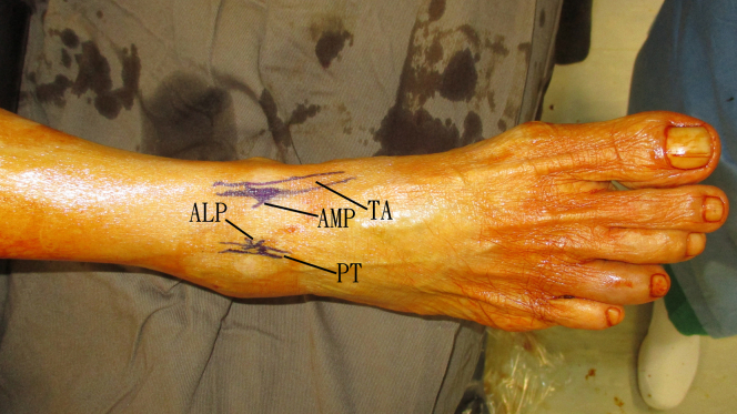
Large talar bone cyst can cause pathologic fracture and damage to the articular cartilage, resulting in persistent swelling and pain of the subtalar joint and ankle joint. For a symptomatic cyst not responding to conservative treatment, surgery can be considered. Open debridement and bone grafting frequently require extensive soft-tissue dissection or even different types of malleolar osteotomy for proper access to the lesion. Arthroscopic treatment of talar bone cyst is a feasible alternative minimally invasive approach to reduce surgical trauma and eliminate the need for osteotomy. Bone cyst of the anterior part of the talar body can be debrided via a bone window of the talar neck, which is normally devoid of cartilage. The purpose of this Technical Note is to describe the technique of arthroscopic treatment of bone cyst of anterior half of the talar body. This minimally invasive approach does not disrupt the normal articular cartilage of the talar dome.
© 2022 The Authors.
Figures


Similar articles
-
Management of Bone Cyst of Talar Body by Endoscopic Curettage, Nanofracture, and Bone Graft Substitute.Arthrosc Tech. 2021 Jul 20;10(8):e1985-e1993. doi: 10.1016/j.eats.2021.04.026. eCollection 2021 Aug. Arthrosc Tech. 2021. PMID: 34401244 Free PMC article.
-
Arthroscopic Curettage and Bone Grafting of Bone Cysts of the Talar Body.Arthrosc Tech. 2017 Jan 2;6(1):e7-e13. doi: 10.1016/j.eats.2016.08.029. eCollection 2017 Feb. Arthrosc Tech. 2017. PMID: 28373933 Free PMC article.
-
Novel Surgical Approach for Large Intraosseous Subchondral Cysts of Talus: A Case Report and Technical Innovation.Cureus. 2024 Jan 11;16(1):e52078. doi: 10.7759/cureus.52078. eCollection 2024 Jan. Cureus. 2024. PMID: 38344643 Free PMC article.
-
Percutaneous osteoplasty for the treatment of a painful osteochondral lesion of the talus: a case report and literature review.Pain Physician. 2012 Sep-Oct;15(5):E743-8. Pain Physician. 2012. PMID: 22996869 Review.
-
Limitations of accessibility of the talar dome with different open surgical approaches.Knee Surg Sports Traumatol Arthrosc. 2021 Apr;29(4):1304-1317. doi: 10.1007/s00167-020-06113-2. Epub 2020 Jun 29. Knee Surg Sports Traumatol Arthrosc. 2021. PMID: 32596777
Cited by
-
Uniportal Endoscopic Intramedullary Debridement for the Management of Tibial Osteomyelitis.Arthrosc Tech. 2025 Apr 16;14(7):103555. doi: 10.1016/j.eats.2025.103555. eCollection 2025 Jul. Arthrosc Tech. 2025. PMID: 40822155 Free PMC article.
-
Biportal Endoscopic Intramedullary Debridement for Management of Tibial Osteomyelitis.Arthrosc Tech. 2025 Apr 24;14(7):103556. doi: 10.1016/j.eats.2025.103556. eCollection 2025 Jul. Arthrosc Tech. 2025. PMID: 40822184 Free PMC article.
References
-
- Zhu X., Yang L., Duan X. Arthroscopically assisted anterior treatment of symptomatic large talar bone cyst. J Foot Ankle Surg. 2019;58:151–155. - PubMed
-
- Lui T.H. Arthroscopic bone grafting of talar bone cyst using posterior ankle arthroscopy. J Foot Ankle Surg. 2013;52:529–532. - PubMed
-
- Lui T.H. Endoscopic curettage and bone grafting of huge talar bone cyst with preservation of cartilaginous surfaces: Surgical planning. Foot Ankle Surg. 2014;20:248–252. - PubMed
LinkOut - more resources
Full Text Sources
