. 2022 Nov 17;11(12):e2265-e2270.
doi: 10.1016/j.eats.2022.08.035. eCollection 2022 Dec.

Arthroscopic Biologic Tuberoplasty for Irreparable Rotator Cuff Tears: An Expedited Technique

Affiliations

Arthroscopic Biologic Tuberoplasty for Irreparable Rotator Cuff Tears: An Expedited Technique

Misty Suri et al. Arthrosc Tech. .

Abstract

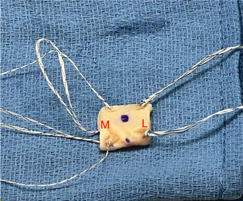
Massive irreparable rotator cuff tears in patients for whom arthroplasty is not an option can be a challenging clinical scenario for shoulder surgeons to manage. To achieve the best patient outcomes, a myriad of options has been presented in the literature, including debridement with biceps tenotomy or tenodesis, various tendon transfer procedures, superior capsular reconstruction, biceps tendon rerouting, bursal acromion resurfacing, balloon spacers, and tuberoplasty. While debridement with biceps tenotomy and superior capsular reconstruction have historically provided improvements in patient-reported outcomes, high rates of arthritis progression and failure of graft healing have been noted with these techniques, respectively. The superior capsular reconstruction has also proven to be technically challenging. The biologic tuberoplasty procedure was developed after several studies noted a lack of correlation between graft healing and improvement in patient-reported outcomes in superior capsular reconstructions, as long as the tuberosity remained covered with the graft. We present a technically efficient and expedited technique using an acellular human dermal allograft.


Figures

Fig 1
Fig 1
Graft site measurement (anterior to posterior). To measure the graft site, use a probe or SCR measuring guide.
Fig 2
Fig 2
Final suture preparation of the graft. Medially, use suture passer to pass 0.9-mm suture tape. FiberLink sutures in the anterior and posterior medial corners. The loops should be placed on the. undersurface of the graft. Bring the loop closer to the graft (as this will be later used to shuttle. the repair suture from knotless FiberTak through the graft). Laterally, use suture passer to pass 1.0 × 1.3 SutureTape FiberLink and 1.0 × 1.3 SutureTape TigerLink in opposite corners of the graft laterally in a luggage tag configuration. The blue ink is shown on the superior side of graft. M, medial; L, lateral.
Fig 3
Fig 3
Arthroflex graft presented at the lateral side on the lateral shoulder. Note the anteromedial. and posteromedial 2.6 self-punching knotless FiberTaks have been inserted already at the corners. of the graft site. Next, through the lateral portal (12 mm × 3 cm passport), use a crab-claw grasper. to grasp the repair (blue) and the round shuttle loop from the anteromedial FiberTak anchor out. of the lateral portal. Clip these 2 limbs on the anteromedial (a) side of the passport. Repeat with. posteromedial anchor and clip the 2 suture limbs on the posteromedial (b) side of the passport cannula. Once the anchors have been inserted, keep the sutures on the relevant sides to prevent suture entanglement. Orientation: Right shoulder lateral position; anterior is left of figure, posterior is right of figure, proximal is bottom of figure, distal is top of figure. Lateral portal is the working portal with the passport cannula (12 mm × 3 cm) in place; and posterior portal is the viewing portal.
Fig 4
Fig 4
Bring the sutured graft on a sterile towel closer to the passport cannula with correct. orientation (medial side facing patients head). Use the anteromedial and posteromedial FiberLinks. to individually shuttle the repair suture (blue) from the knotless FiberTak anchors through the. Arthroflex graft. Note, the knotless mechanism is not yet “converted”.
Fig 5
Fig 5
Once the repair sutures are passed through the graft, use the shuttle loop (on the same. sides) to “convert” the knotless mechanism. All repair stitches (blue) have been converted. through their respective knotless tensionable mechanism (anterior to anterior and posterior to. posterior). Now, the graft can be shuttled through the 12 mm × 3 cm passport cannula.
Fig 6
Fig 6
Posterior (A) and lateral (B) views of completed arthroscopic biologic tuberoplasty. M, medial; L, lateral; A, anterior; P, posterior.

References

    1. Neer C.S., 2nd, Craig E.V., Fukuda H. Cuff-tear arthropathy. J Bone Joint Surg Am. 1983;65:1232–1244. - PubMed
    1. Liem D., Lengers N., Dedy N., et al. Arthroscopic debridement of massive irreparable rotator cuff tears. Arthroscopy. 2008;24:743–748. - PubMed
    1. Walch G., Edwards T.B., Boulahia A., et al. Arthroscopic tenotomy of the long head of the biceps in the treatment of rotator cuff tears: clinical and radiographic results of 307 cases. J Shoulder Elbow Surg. 2005;14:238–246. - PubMed
    1. Elhassan B.T., Sanchez-Sotelo J., Wagner E.R. Outcome of arthroscopically assisted lower trapezius transfer to reconstruct massive irreparable posterior-superior rotator cuff tears. J Shoulder Elbow Surg. 2020;29:2135–2142. - PubMed
    1. Gerber C., Rahm S.A., Catanzaro S., Farshad M., Moor B.K. Latissimus dorsi tendon transfer for treatment of irreparable posterosuperior rotator cuff tears: Long-term results at a minimum follow-up of ten years. J Bone Joint Surg Am. 2013;95:1920–1926. - PubMed

LinkOut - more resources