

A complex presentation of an uncommon disease: Gas-forming pyogenic liver abscess complicated by septic pulmonary emboli and muscle abscesses, a case report and review of the literature
- PMID: 36632483
- PMCID: PMC9827024
- DOI: 10.1016/j.idcr.2022.e01673
A complex presentation of an uncommon disease: Gas-forming pyogenic liver abscess complicated by septic pulmonary emboli and muscle abscesses, a case report and review of the literature
Abstract
Background: Pyogenic liver abscess (PLA) is the most common type of visceral abscess. Its variable clinical presentation depends on patient demography, underlying conditions, causative pathogens as well as the size of the abscess. Most cases are secondary to enteric pathogens that cause focal liver disease. Gas-forming pyogenic liver abscess (GFPLA) is a rare subgroup of PLA characterized by the presence of gas within the abscess. The disease is associated with diabetes mellitus (DM) while Klebsiella penumoniae is the most frequently isolated pathogen. Despite appropriate evaluation and management, secondary complications are common with significant morbidity and mortality that necessitate prompt recognition and management.
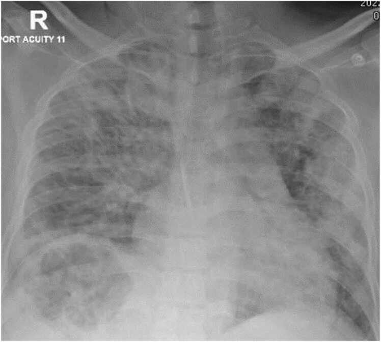
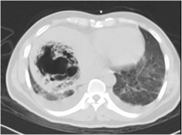
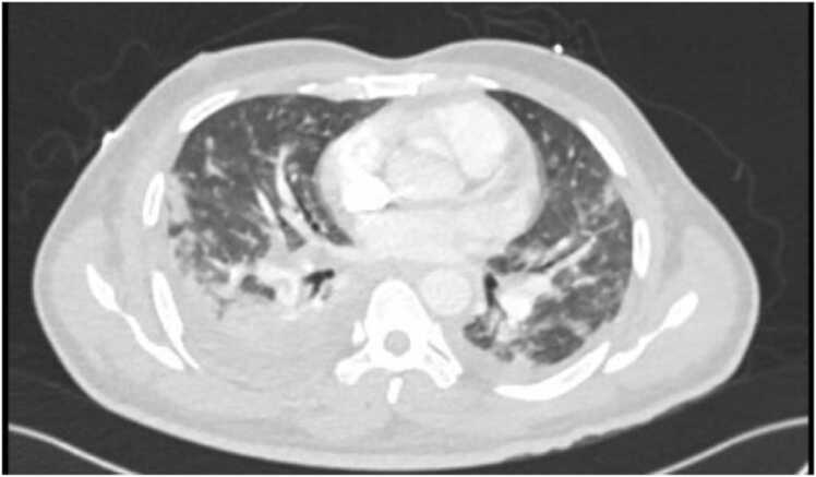
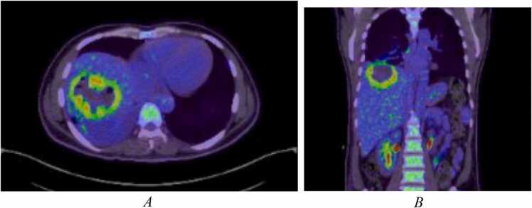
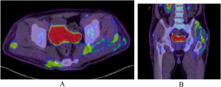
Case presentation: We present a case of a 46-year-old gentleman from Bangladesh who presented to the emergency department with fever, chills, and right upper quadrant abdominal discomfort. Evaluation revealed elevated inflammatory markers with high blood glucose and a subdiaphragmatic lucency on a plain chest radiograph. The suspected underlying visceral infection was confirmed by abdominal ultrasonography and computed tomography which demonstrated an emphysematous abscess of 8 cm in diameter in the right liver lobe.Because of clinical instability, the patient was admitted to the medical intensive care unit (MICU) where he received appropriate supportive management with antimicrobials and percutaneous drainage of the abscess. Cultures collected from blood, the abscess, and urine grew a sensitive strain of Klebsiella pneumoniae. During his stay in the MICU, he complained of dyspnea. A CT pulmonary angiography was suggestive of septic emboli. A few days later, the patient started to complain of left gluteal pain and an US revealed a deep left gluteal abscess which required drainage. Cultures of the pus grew the same sensitive strain of Klebsiella pneumoniae. After receiving 6 weeks of parenteral antimicrobial therapy a repeated US revealed complete resolution of the abscess in the liver. Outpatient follow up showed favorable recovery.
Conclusion: Gas-forming pyogenic liver abscess (GFPLA) is a rare manifestation of pyogenic liver abscess that usually occurs in patients with poorly controlled DM. Despite appropriate evaluation, morbidity remains high therefore timely recognition and anticipation of complications is important.
Keywords: ALT, alanine aminotransferase; AST, aspartate transaminase; CTPA, computed tomography pulmonary angiogram; DM; GFPLA, Gas-forming pyogenic liver abscess; Gas forming; HIV, human immunodeficiency virus; HbA1c, glycated hemoglobin; Klebsiella; Liver abscess; PLA, Pyogenic liver abscess; Septic emboli.
© 2022 The Authors.
Conflict of interest statement
The authors declare that they have no known competing financial interests or personal relationships that could have appeared to influence the work reported in this paper.
Figures
References
-
- Mohsen A.H., Green S.T., Read R.C., McKendrick M.W. Liver abscess in adults: ten years experience in a UK centre. QJM. 2002;95:797–802. [[PubMed] [Google Scholar] [Accessed August 2022]] - PubMed
-
- Chou F.F., Sheen-Chen S.M., Chen Y.S., Lee T.Y. The comparison of clinical course and results of treatment between gas-forming and non-gas-forming pyogenic liver abscess. Arch Surg. 1995;130:401–405. [[PubMed] [Google Scholar] [Accessed August 2022]] - PubMed
-
- Yang C.C., Chen C.Y., Lin X.Z., Chang T.T., Shin J.S., Lin C.Y. Pyogenic liver abscess in Taiwan: emphasis on gas-forming liver abscess in diabetics. Am J Gastroenterol. 1993;88:1911–1915. [Accessed August 2022] - PubMed
Publication types
LinkOut - more resources
Full Text Sources
