

Spatial orientation - a stable marker for vascular cognitive impairment?
- PMID: 36632487
- PMCID: PMC9826950
- DOI: 10.1016/j.cccb.2022.100155
Spatial orientation - a stable marker for vascular cognitive impairment?
Abstract
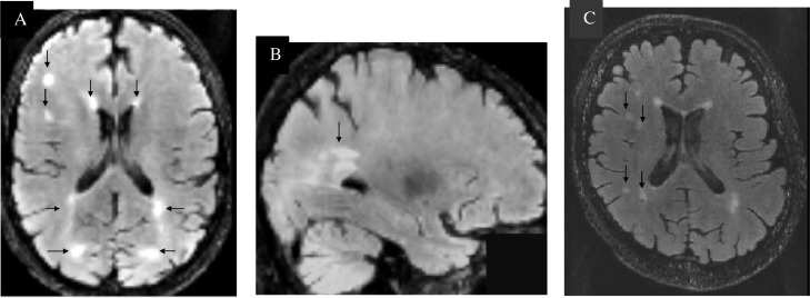
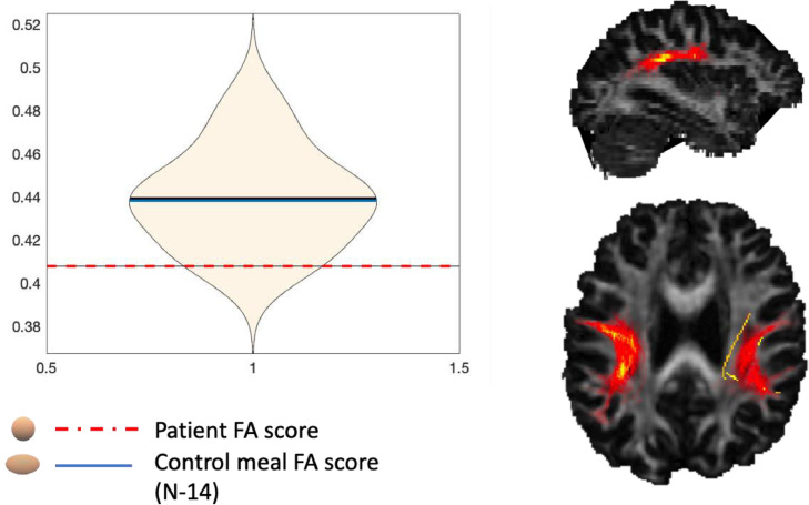
Vascular cognitive impairment (VCI) is the second most prevalent form of dementia, but little is known about the early cognitive and neuroimaging markers. Spatial navigation deficits are an emerging marker for Alzheimer's disease (AD), yet less is known about spatial orientation deficits sensitive to VCI. This case report follows up on the first VCI patient identified to have an egocentric orientation deficit. The study aimed to examine the patient's spatial deficits three years on and gain insights from the addition of the patient's MRI brain scan. A battery of spatial navigation tasks were administered following neuropsychological assessment. Results continue to show spatial orientation deficits. Critically, these changes appear stable and are sensitive to novel spatial tests. Whereas conventional screening tools demonstrate patient recovery. MRI DTI analysis indicates a non-significant trend towards loss of structural integrity to the posterior tracts of the longitudinal superior fasciculus (SLF), while the medial temporal lobe, typically implicated in spatial navigation, is unaffected. This finding potentially reflects reduced network connectivity in posterior to anterior white matter tracts co-existing with spatial orientation deficits. Findings have clinical utility and show spatial orientation as a potential sensitive cognitive marker for VCI.
Keywords: COT, Clock Orientation Test; Egocentric orientation; SHQ, Sea Hero Quest; Spatial navigation; VST, Virtual Supermarket Test; Vascular cognitive impairment; Vascular dementia; Vascular dementia case report; WMH, white matter hyperintensities.
Crown Copyright © 2022 Published by Elsevier B.V.
Conflict of interest statement
All of the contributing authors have no competing interests.
Figures
Similar articles
-
Path Integration Changes as a Cognitive Marker for Vascular Cognitive Impairment?-A Pilot Study.Front Hum Neurosci. 2020 Apr 21;14:131. doi: 10.3389/fnhum.2020.00131. eCollection 2020. Front Hum Neurosci. 2020. PMID: 32372934 Free PMC article.
-
Diagnostic relevance of spatial orientation for vascular dementia: A case study.Dement Neuropsychol. 2018 Jan-Mar;12(1):85-91. doi: 10.1590/1980-57642018dn12-010013. Dement Neuropsychol. 2018. PMID: 29682239 Free PMC article.
-
Cognitive and neuroimaging markers for preclinical vascular cognitive impairment.Cereb Circ Cogn Behav. 2021 Oct 5;2:100029. doi: 10.1016/j.cccb.2021.100029. eCollection 2021. Cereb Circ Cogn Behav. 2021. PMID: 36324708 Free PMC article. Review.
-
Predicting real world spatial disorientation in Alzheimer's disease patients using virtual reality navigation tests.Sci Rep. 2022 Aug 4;12(1):13397. doi: 10.1038/s41598-022-17634-w. Sci Rep. 2022. PMID: 35927285 Free PMC article.
-
Neural correlates of spatial navigation changes in mild cognitive impairment and Alzheimer's disease.Front Behav Neurosci. 2014 Mar 17;8:89. doi: 10.3389/fnbeh.2014.00089. eCollection 2014. Front Behav Neurosci. 2014. PMID: 24672452 Free PMC article. Review.
Cited by
-
Spatial navigation deficits in early Alzheimer's disease: the role of biomarkers and APOE genotype.J Neurol. 2025 Jun 2;272(6):438. doi: 10.1007/s00415-025-13151-8. J Neurol. 2025. PMID: 40456910 Free PMC article.
-
Editorial. CCCB: The journal at age 5.Cereb Circ Cogn Behav. 2024 Sep 7;7:100368. doi: 10.1016/j.cccb.2024.100368. eCollection 2024. Cereb Circ Cogn Behav. 2024. PMID: 39737420 Free PMC article.
References
-
- Gorelick P.B., Scuteri A., Black S.E., Decarli C., Greenberg S.M., Iadecola C., et al. Vascular contributions to cognitive impairment and dementia: a statement for healthcare professionals from the american heart association/american stroke association. Stroke. 2011;42(9):2672–2713. https://www.ncbi.nlm.nih.gov/pubmed/21778438 [Internet]Available from. - PMC - PubMed
-
- Plassman B.L., Langa K.M., Fisher G.G., Heeringa S.G., Weir D.R., Ofstedal M.B., et al. Prevalence of dementia in the United States: the aging, demographics, and memory study. Neuroepidemiology. 2007;29(1–2):125–132. https://www.karger.com/Article/FullText/109998 [Internet]Nov [cited 2021 Dec 9]Available from: - PMC - PubMed
-
- Dichgans M., Leys D. Vascular cognitive impairment. Circ. Res. 2017;120(3):573–591. - PubMed
Publication types
LinkOut - more resources
Full Text Sources
Miscellaneous
