

Copper-cysteamine nanoparticle-mediated microwave dynamic therapy improves cancer treatment with induction of ferroptosis
- PMID: 36632507
- PMCID: PMC9807746
- DOI: 10.1016/j.bioactmat.2022.12.023
Copper-cysteamine nanoparticle-mediated microwave dynamic therapy improves cancer treatment with induction of ferroptosis
Abstract
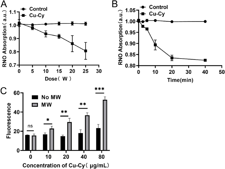
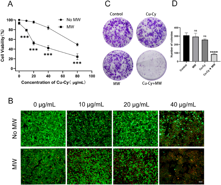
Photodynamic Therapy (PDT) holds a great promise for cancer patients, however, due to the hypoxic characteristics of most solid tumors and the limited penetration depth of light in tissues, the extensive clinical application of PDT is limited. Herein, we report microwave induced copper-cysteamine (Cu-Cy) nanoparticles-based PDT as a promising cancer treatment to overcome cancer resistance in combination with ferroptosis. The treatment efficiency of Cu-Cy-mediated microwave dynamic therapy (MWDT) tested on HCT15 colorectal cancer (CRC) cells via cell titer-blue cell viability assay and live/dead assay reveal that Cu-Cy upon MW irradiation can effectively destroy HCT15 CRC cells with average IC-50 values of 20 μg/mL. The cytotoxicity of Cu-Cy to tumor cells after MW stimulation can be alleviated by ferroptosis inhibitor. Furthermore, Cu-Cy mediated MWDT could deplete glutathione peroxide 4 (GPX4) and enhance lipid peroxides (LPO) and malondialdehyde (MDA). Our findings demonstrate that MW-activated Cu-Cy killed CRC cells by inducing ferroptosis. The superior in vivo antitumor efficacy of the Cu-Cy was corroborated by a HCT15 tumor-bearing mice model. Immunohistochemical experiments showed that the GPX4 expression level in Cu-Cy + MW group was significantly lower than that in other groups. Overall, these findings demonstrate that Cu-Cy nanoparticles have a safe and promising clinical application prospect in MWDT for deep-seated tumors and effectively inhibit tumor cell proliferation by inducing ferroptosis, which provides a potential solution for cancer resistance.
Keywords: Cell death; Colorectal cancer; Cu-Cy; Ferroptosis; Microwave; PDT.
© 2022 The Authors.
Figures


Similar articles
-
Study of copper-cysteamine based X-ray induced photodynamic therapy and its effects on cancer cell proliferation and migration in a clinical mimic setting.Bioact Mater. 2021 May 30;7:504-514. doi: 10.1016/j.bioactmat.2021.05.016. eCollection 2022 Jan. Bioact Mater. 2021. PMID: 34466749 Free PMC article.
-
A New Modality for Cancer Treatment--Nanoparticle Mediated Microwave Induced Photodynamic Therapy.J Biomed Nanotechnol. 2016 Oct;12(10):1835-51. doi: 10.1166/jbn.2016.2322. J Biomed Nanotechnol. 2016. PMID: 29359896
-
Investigation of Copper Cysteamine Nanoparticles as a New Type of Radiosensitiers for Colorectal Carcinoma Treatment.Sci Rep. 2017 Aug 24;7(1):9290. doi: 10.1038/s41598-017-09375-y. Sci Rep. 2017. PMID: 28839163 Free PMC article.
-
A facile method for the synthesis of copper-cysteamine nanoparticles and study of ROS production for cancer treatment.J Mater Chem B. 2019 Nov 14;7(42):6630-6642. doi: 10.1039/c9tb01566c. Epub 2019 Oct 8. J Mater Chem B. 2019. PMID: 31591609
-
The application of graphene oxide and ferroptosis in the diagnosis and treatment of colorectal cancer: a narrative review.J Gastrointest Oncol. 2024 Jun 30;15(3):1297-1308. doi: 10.21037/jgo-23-1016. Epub 2024 Jun 11. J Gastrointest Oncol. 2024. PMID: 38989438 Free PMC article. Review.
Cited by
-
Radiocleavable rare-earth nanoactivators targeting over-expressed folate receptors induce mitochondrial dysfunction and remodel immune suppressive microenvironment in pancreatic cancer.J Nanobiotechnology. 2025 Aug 12;23(1):562. doi: 10.1186/s12951-025-03657-8. J Nanobiotechnology. 2025. PMID: 40797208 Free PMC article.
-
Nanotechnology in cancer treatment: revolutionizing strategies against drug resistance.Front Bioeng Biotechnol. 2025 Apr 30;13:1548588. doi: 10.3389/fbioe.2025.1548588. eCollection 2025. Front Bioeng Biotechnol. 2025. PMID: 40370595 Free PMC article. Review.
-
Role of copper homeostasis and cuproptosis in heart failure pathogenesis: implications for therapeutic strategies.Front Pharmacol. 2025 Jan 9;15:1527901. doi: 10.3389/fphar.2024.1527901. eCollection 2024. Front Pharmacol. 2025. PMID: 39850564 Free PMC article. Review.
-
AKT1 Phosphorylates FDX1 to Promote Cuproptosis Resistance in Triple-Negative Breast Cancer.Adv Sci (Weinh). 2025 May;12(17):e2408106. doi: 10.1002/advs.202408106. Epub 2025 Feb 20. Adv Sci (Weinh). 2025. PMID: 39976173 Free PMC article.
-
Ferroptosis resistance in cancer cells: nanoparticles for combination therapy as a solution.Front Pharmacol. 2024 Jun 19;15:1416382. doi: 10.3389/fphar.2024.1416382. eCollection 2024. Front Pharmacol. 2024. PMID: 38962305 Free PMC article. Review.
References
-
- DeBerardinis R.J. Tumor microenvironment, metabolism, and immunotherapy. N. Engl. J. Med. 2020;382(9):869–871. - PubMed
-
- Petroni G., Buqué A., Coussens L.M., Galluzzi L. Targeting oncogene and non-oncogene addiction to inflame the tumour microenvironment. Nat. Rev. Drug Discov. 2022;21(6):440–462. - PubMed
-
- Bhandari V., Hoey C., Liu L.Y., Lalonde E., Ray J., Livingstone J., Lesurf R., Shiah Y.J., Vujcic T., Huang X., Espiritu S.M.G., Heisler L.E., Yousif F., Huang V., Yamaguchi T.N., Yao C.Q., Sabelnykova V.Y., Fraser M., Chua M.L.K., van der Kwast T., Liu S.K., Boutros P.C., Bristow R.G. Molecular landmarks of tumor hypoxia across cancer types. Nat. Genet. 2019;51(2):308–318. - PubMed
-
- Wan Y., Fu L.H., Li C., Lin J., Huang P. Conquering the hypoxia limitation for photodynamic therapy. Adv. Mater. 2021;33(48) - PubMed
LinkOut - more resources
Full Text Sources
