

Critical periods and Autism Spectrum Disorders, a role for sleep
- PMID: 36632570
- PMCID: PMC9826922
- DOI: 10.1016/j.nbscr.2022.100088
Critical periods and Autism Spectrum Disorders, a role for sleep
Abstract
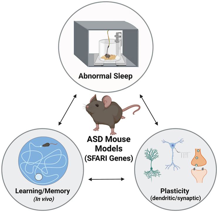
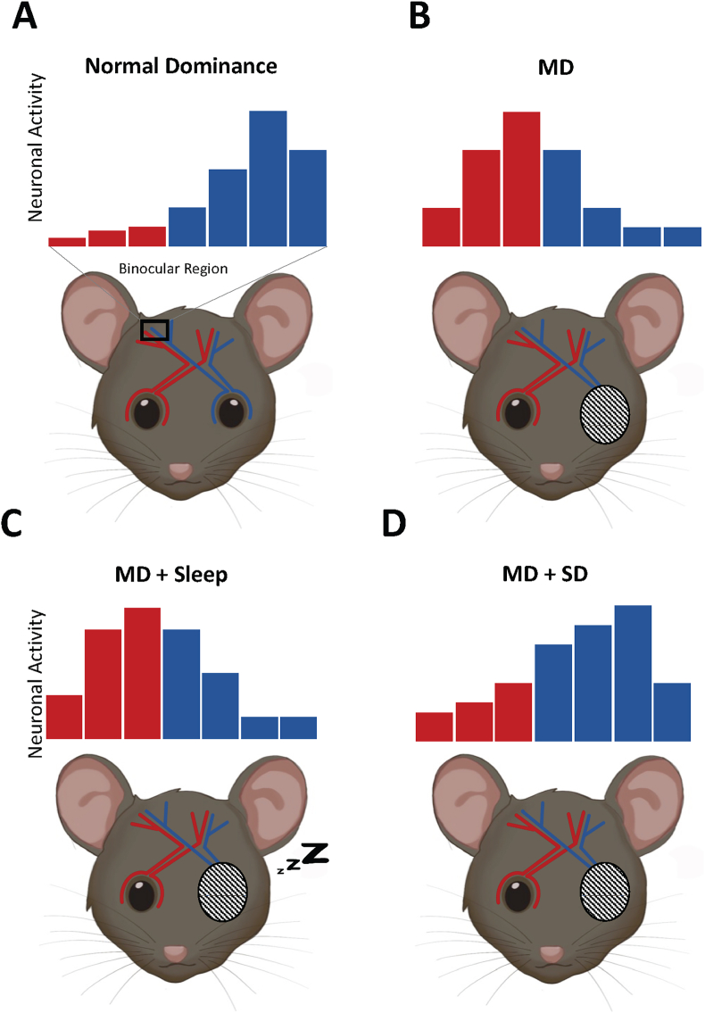
Brain development relies on both experience and genetically defined programs. Time windows where certain brain circuits are particularly receptive to external stimuli, resulting in heightened plasticity, are referred to as "critical periods". Sleep is thought to be essential for normal brain development. Importantly, studies have shown that sleep enhances critical period plasticity and promotes experience-dependent synaptic pruning in the developing mammalian brain. Therefore, normal plasticity during critical periods depends on sleep. Problems falling and staying asleep occur at a higher rate in Autism Spectrum Disorder (ASD) relative to typical development. In this review, we explore the potential link between sleep, critical period plasticity, and ASD. First, we review the importance of critical period plasticity in typical development and the role of sleep in this process. Next, we summarize the evidence linking ASD with deficits in synaptic plasticity in rodent models of high-confidence ASD gene candidates. We then show that the high-confidence rodent models of ASD that show sleep deficits also display plasticity deficits. Given how important sleep is for critical period plasticity, it is essential to understand the connections between synaptic plasticity, sleep, and brain development in ASD. However, studies investigating sleep or plasticity during critical periods in ASD mouse models are lacking. Therefore, we highlight an urgent need to consider developmental trajectory in studies of sleep and plasticity in neurodevelopmental disorders.
Keywords: Autism; Critical periods of development; Sleep; Synaptic plasticity.
© 2022 Published by Elsevier Inc.
Conflict of interest statement
The authors have no financial arrangements or connections to declare.
Figures
References
-
- Aida T., Yoshida J., Nomura M., Tanimura A., Iino Y., Soma M., Bai N., Ito Y., Cui W., Aizawa H., Yanagisawa M., Nagai T., Takata N., Tanaka K.F., Takayanagi R., Kano M., Götz M., Hirase H., Tanaka K. Astroglial glutamate transporter deficiency increases synaptic excitability and leads to pathological repetitive behaviors in mice. Neuropsychopharmacol. Off. Publ. Am. Coll. Neuropsychopharmacol. 2015;40:1569–1579. doi: 10.1038/npp.2015.26. - DOI - PMC - PubMed
-
- Altafaj X., Dierssen M., Baamonde C., Martí E., Visa J., Guimerà J., Oset M., González J.R., Flórez J., Fillat C., Estivill X. Neurodevelopmental delay, motor abnormalities and cognitive deficits in transgenic mice overexpressing Dyrk1A (minibrain), a murine model of Down's syndrome. Hum. Mol. Genet. 2001;10:1915–1923. doi: 10.1093/hmg/10.18.1915. - DOI - PubMed
Grants and funding
LinkOut - more resources
Full Text Sources
