. 2023 Jan 12;67(1):3594.
doi: 10.4081/ejh.2023.3594.

Phosphorylation mutation impairs the promoting effect of spastin on neurite outgrowth without affecting its microtubule severing ability

Affiliations

Phosphorylation mutation impairs the promoting effect of spastin on neurite outgrowth without affecting its microtubule severing ability

Yunlong Zhang et al. Eur J Histochem. .

Abstract

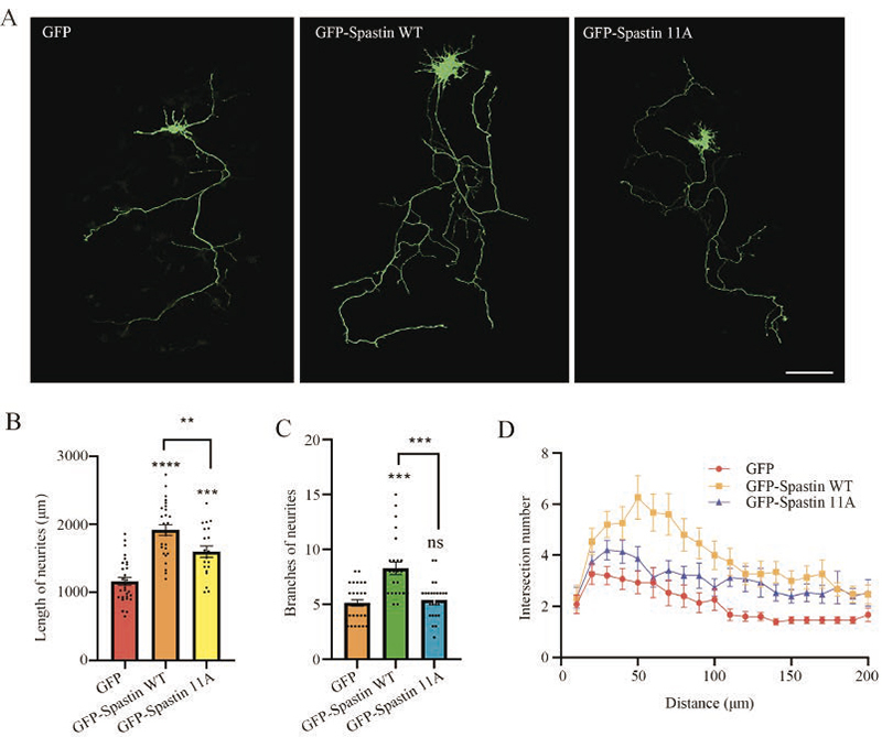
Spastin, a microtubule-severing enzyme, is known to be important for neurite outgrowth. However, the role of spastin post-translational modification, particularly its phosphorylation regulation in neuronal outgrowth, remains unclear. This study aimed to investigate the effects of eliminating spastin phosphorylation on the neurite outgrowth of rat hippocampal neurons. To accomplish this, we constructed a spastin mutant with eleven potential phosphorylation sites mutated to alanine. The phosphorylation levels of the wildtype spastin (WT) and the mutant (11A) were then detected using Phos-tag SDS-PAGE. The spastin constructs were transfected into COS7 cells for the observation of microtubule severing, and into rat hippocampal neurons for the detection of neuronal outgrowth. The results showed that compared to the spastin WT, the phosphorylation levels were significantly reduced in the spastin 11A mutant. The spastin mutant 11A impaired its ability to promote neurite length, branching, and complexity in hippocampal neurons, but did not affect its ability to sever microtubules in COS7 cells. In conclusion, the data suggest that mutations at multiple phosphorylation sites of spastin do not impair its microtubule cleavage ability in COS7 cells, but reduce its ability to promote neurite outgrowth in rat hippocampal neurons.


Figures

Figure 1.
Figure 1.
Design of construction of point mutation of potential phosphorylation sites in spastin. A) Diagram of the search for 11 spastin phosphorylation sites in the PhosphsitePlus database. B) GFP, GFP-spastin WT (wild-type), GFP-spastin 11A (11 amino acids mutated) PCR electrophoresis results. C) Comparison of gene sequences of spastin WT and 11A.
Figure 2.
Figure 2.
The phosphorylation levels between spastin WT and the mutated form 11A were examined. A) Overexpression of GFP, GFP-spastin, and GFP-spastin 11A in COS7 cells at 24 h, and cell lysates were collected and subjected to Western blotting with GFP antibodies. B) The same samples as in (A) were subjected to a Phos-tag assay to detect phosphorylation shifts; compared to spastin WT, the amount of protein phosphorylated by spastin 11A was significantly reduced compared to the amount of protein that was not phosphorylated. C) Quantification of band intensities of phosphorylated relative to unphosphorylated was performed. The relative value in the control group was set to 1. Data are presented as the mean ± SEM from at least three independent experiments; *p<0.05 vs vector group.
Figure 3.
Figure 3.
The phospho-mutated form of spastin 11A does not affect the microtubule severing ability in COS7 cells. A) Different plasmids were transfected into COS-7 cells for 18 h and then immunolabeled for tubulin (white fluorescence signal) and counterstained for nuclear DNA with DAPI (blue fluorescence). B) Relative fluorescence intensity of microtubules in COS7 cells. Data are presented as mean ± SEM; ****p<0.0001; ns, not statistically significant. Scale bar: 10 μm.
Figure 4.
Figure 4.
The phospho-mutated form of spastin 11A impairs the promoting effect of hippocampal neurite outgrowth. A) DIV 3 hippocampal neurons were transfected with different plasmids for 24 h, followed by immunocytochemical staining with GFP antibody; scale bar: 100 μm. B) Neurite length statistics (including dendrites and axons). (C) Branching statistics of neurite. D) Sholl analysis of neurite within 300 μm from the cell soma. Data are expressed as mean ± SEM; **p<0.01, ***p<0.001, ****p<0.0001.

Similar articles

Cited by

References

    1. Novarino G, Fenstermaker AG, Zaki MS, Hofree M, Silhavy JL, Heiberg AD, et al. . Exome sequencing links corticospinal motor neuron disease to common neurodegenerative disorders. Science 2014;343:506-11. - PMC - PubMed
    1. Fink JK. Hereditary spastic paraplegia: clinico-pathologic features and emerging molecular mechanisms. Acta Neuropathol 2013;126:307-28. - PMC - PubMed
    1. Solowska JM, Baas PW. Hereditary spastic paraplegia SPG4: what is known and not known about the disease. Brain 2015;138:2471-84. - PMC - PubMed
    1. Zhu Z, Zhang C, Zhao G, Liu Q, Zhong P, Zhang M, et al. . Novel mutations in the SPAST gene cause hereditary spastic paraplegia. Parkinsonism Relat Disord 2019;69:125-33. - PubMed
    1. Kuo YW, Howard J. Cutting, amplifying, and aligning microtubules with severing enzymes. Trends Cell Biol 2021;31:50-61. - PMC - PubMed